What is the quickest scoring system to predict percutaneous
nephrolithotomy outcomes? A comparative study among
S.T.O.N.E score, Guy’s Stone Ccore and CROES nomogram
_______________________________________________
Fabio C. Vicentini
1, Felipe R. Serzedello
2, Kay Thomas
3, Giovanni S. Marchini
2, Fabio C. M. Torricelli
2,
Miguel Srougi
2, Eduardo Mazzucchi
21 Seção de Endourologia, Departamento de Urologia, Hospital de Clínicas, Faculdade de Medicina da
Universidade de São Paulo, SP, Brasil; 2 Faculdade de Medicina da Universidade de São Paulo, São Paulo,
SP, Brasil; 3 Stone Unit, Guy’s and St. Thomas’ National Health services foundation Hospital, London,
United Kingdom
ABSTRACT
ARTICLE
INFO
______________________________________________________________ ______________________
Objective: To compare the application time and the capacity of the nomograms to pre-dict the success of Guy’s Stone Score (GSS), S.T.O.N.E. Nephrolithometry (STONE) and Clinical Research Office of the Endourological Society nephrolithometric nomogram (CROES) of percutaneous nephrolithotomy (PCNL), evaluating the most efficient one for clinical use.
Materials and Methods: We studied 48 patients who underwent PCNL by the same surgeon between 2010 and 2011. We calculated GSS, STONE and CROES based on pre-operative non-contrast computed tomography (CT) images and clinical data. A single observer, blinded to the outcomes, reviewed all images and assigned scores. We com-pared the application time of each nomogram. We used an analysis of variance for repeated measures and multiple comparisons by the Tukey test. We compared the area under the ROC curve (AUC) of the three nomograms two by two to determine the most predictive scoring system.
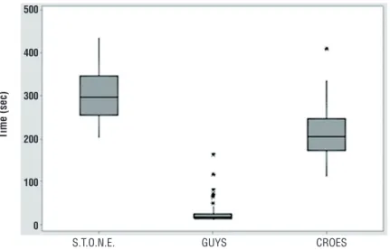
Results: The immediate success rate was 66.7% and complications occurred in 16.7% of cases. The average operative time was 122 minutes. Mean application time was significantly lower for the GSS (27.5 seconds) when compared to 300.6 seconds for STONE and 213.4 seconds for CROES (p<0.001). There was no significant difference among the GSS (AUC=0.653), STONE (AUC=0.563) and CROES (AUC=0.641) in the ability to predict immediate success of PCNL.
Conclusions: All three nomograms showed similar ability to predict success of PCNL, however the GSS was the quickest to be applied, what is an important issue for routine clinical use when counseling patients who are candidates to PCNL.
Keywords:
Nomograms; Diagnosis; Calculi
Int Braz J Urol. 2017; 43: 1102-9
_____________________ Submitted for publication: October 27, 2016
_____________________ Accepted after revision: November 21, 2016 _____________________ Published as Ahead of Print: March 08, 2017
INTRODUCTION
Nephrolithiasis is a common condition, with high prevalence and recurrence, constituting one of the most common diseases of the urina-ry tract (1). The disease affects 5% to 15% of the
stone and/or high volume stone (4-6). Despite the es-tablishment of PCNL as one of the most important methods for the treatment of kidney stones, currently there is no gold standard tool for predicting success and complications associated with this surgery (7). This is important because a scoring system could help the surgeon in planning surgical strategies, predict the success rate and complications, result in better patient counseling, and facilitate comparison of ou-tcome between the different institutions (7).
There are a few scoring systems in the li-terature which assess pre-operative parameters and predict the success rate of PCNL (8-11). They have showed to positively correlate with outcomes or complications, but comparison among them is required in order to determine the most practical and applicable one in clinical practice. Currently, three nomograms have been more extensively stu-died: Guy’s Stone Score (GSS) (8), the S.T.O.N.E. Nephrolithometry (STONE) (10) and the nomo-gram of the Clinical Research Office of the Endou-rological Society (CROES) (9). The GSS consists of a nomogram using as parameters the amount of stones, their renal location, history of spina bifida or spine injury, and the association with possible anatomical changes, as horseshoe kidney: nephro-lithiasis burden is classified in 4 degrees related to different success rates in PCNL (8). The parameters used in the STONE include stone size, distance to the skin, the degree of obstruction in the urinary tract, the number of renal calices involved, and stone density (10). Finally, the CROES uses varia-bles such as area, number and location of the sto-nes, previous treatment, staghorn stone and num-ber of cases treated per year in the institution (9). Some studies have shown that all nomo-grams correlated well with success or complica-tions (12-18) and that they have similar ability to predict surgical outcomes (19-22). Nevertheless, there are no studies assessing these nomograms in clinical practice, where time is an important fac-tor. In this study, we aim to compare the acquisi-tion times for the most used nomograms.
OBJECTIVE
Our primary goal was to compare the ap-plication time of GSS, STONE and CROES,
eva-luating which one is quickest to be applied in clinical practice.
Our secondary objective was to compare the ability of the nomograms to predict the imme-diate success rate of PCNL.
MATERIALS AND METHODS
We performed a retrospective review of medical record data from a prospectively collec-ted database. In our institution, we perform >180 PCNL/year. We analyzed patients who underwent PCNL between February, 2010 and December, 2011 at our institution by the same senior urologist (FCV) under the same technique, as previously described (22). Briefly, under general anesthesia, all patients were positioned in the supine decu-bitus with the posterior axillary line located just outside the border of the surgical Table; the flank was extended to increase the space between the last rib and the iliac crest; all csPCNLs were per-formed without boosters under the flank and all patients were maintained in the same position du-ring the entire procedure.
Tract dilation was performed with fascial dilators (numbers 10, 20 and 30Fr, sequentially) and the Amplatz® sheath was placed. Nephrosco-py was performed with a 26Fr rigid nephroscope (Karl Storz, Munich, Germany) and we used an ul-trasonic lithotripter for stone fragmentation and suction. The stone free status was verified with combined fluoroscopy and flexible nephroscopy. A 16Fr nephrostomy tube was placed at the end of the procedure in case of bleeding, residual stones, solitary kidney, suspected pelvic injury, or mul-tiple tracts. A 6Fr ureteral catheter was routinely placed; in cases of ureteropelvic junction signi-ficant edema, extensive pelvic injury, or ureteral manipulation, a double-J stent was used instead. Operative time was considered from the beginning of cystoscopy for ureteral catheter insertion to the end of nephrostomy placement.
developed for this purpose. The study protocol had ethical review board approval.
Selection criteria
Exclusion criteria comprised patients younger than 18 years old and older than 70 years old, patients with inadequate analysis by preope-rative CT (low resolution or not performed in our service) and without at least one follow-up con-sultation in a 60 days period.
Measurements
We calculate the GSS, STONE and CROES of all patients based on preoperative CT images as described by Thomas et al.(8), Okhunov et al. and Smith et al.(9), respectively. A single ob-server (FRS) reviewed all images and performed scoring according to each system. The reviewer was a 5th year medical student (of a total of 6
ye-ars), with basic knowledge in radiology, with no previous use of any of the scoring systems, who was initially trained to evaluate non-contrast CT scans by calculating the three scores for 20 different cases under supervision of two senior urologists before initiating the study. We did not use these cases for our study, just for training the observer. A concordance index among the calculations of the 3 observers for these 20 ca-ses was 0.86, showing that the reviewer has been properly trained. We analyzed all the images on the computer screen and all parameters were ac-quired through the image display program. The high concordance index allowed us to make all the analysis based on the data calculated by only one observer. The observer also had a Table with the data regarding physical examination, history of previous surgery and presence of spina bifida or not. The time for clinical data analysis was not considered, because in a clinical setting this information usually is already known by the as-sistant when analyzing the CT images, according to a regular patient evaluation.
Acquisition Time
The time required for application of no-mograms was individualized for each nomogram in each case. For this, we used a simple timer. The count began at the time the researcher started the
evaluation of the imaging exam and ended when the investigator obtained the final score of the no-mogram. First, we calculated the GSS consecutive-ly for all patients and the data inserted into an Ex-cel Table (Microsoft, California). Then, in another day, we calculated the STONE parameters also for all patients. Stone burden was estimated in mm2
using the formula, Σ (0.785 x lengthmaxx widthmax) and the value was automatically generated by the software (24). Finally, again in another day, we obtained the CROES from the same images.
We did not reuse the results of measure-ments taken for one nomogram for calculating the other one and we did all the measurements in di-fferent days in order to guarantee that the observer did not have memory of the image previously seen. Every nomogram had its proper Excel Table, with the patient´s demographic data, history of previous surgery and presence of spina bifida or not.
Definition of success
We defined success as stone fragments
≤4mm on CT scan on the first postoperative day (POD1). Stone-free rate refers to no identification of any stone fragment on the POD1 CT. Final suc-cess rate was defined as the result of the last ra-diological exam performed after all the auxiliary procedures, consisting of revision PCNL, external shockwave lithotripsy or flexible nephroscopy.
Statistical analysis
We calculate the sample size based on an expecteddifference between GSS and the others nomograms of 50% in time acquisition, with a power of 80% and a significance level of 0.05, ba-sed on the initial findings of the 20 cases studied for training the observer. With these parameters, we reached a total number of 18 patients. We stu-died 48 trying to improve the success comparison among the nomograms.
they were similar, supporting our ANOVA use for this study. To check if they were all different, we did a multiple comparison by the Tukey method, comparing the scoring systems in pairs. We genera-ted receiver operator characteristic (ROC) curves for each scoring system. We calculated the area under the curve and asymptotic 95% confidence intervals were calculated for each ROC curve. We performed all statistical analyses using SPSS 19.0 software for Windows (SPSS Inc., Chicago, USA). A p value of <0.05 was considered statistically significant.
RESULTS
Perioperative data
Demographic data are shown in the Ta-ble-1. The immediate success rate was 66.7% (29.2% of stone free and 92.4% for final success rate after a mean of 1.29 auxiliary procedures), and complications occurred in 16.7% of cases. The average operative time was122 minutes (Table-1).
Nomograms application speed
Mean application time for the GSS was 27.5±30.0 seconds, significantly shorter than the 300.6±56.5 seconds for STONE and 213.4±59.4 se-conds for CROES. There was a significant differen-ce between all groups (p <0.001) (Figure-1).
Scoring Systems Reliability
After the two by two comparison of the AUC, there was no significant difference among the GSS (AUC=0.653), STONE (AUC=0.563) and CROES (AUC=0.641) in the capacity to predict immediate success of PCNL (STONE x GSS: p= .445; STONE x CROES: p=0.513; GSS x CROES: p=0.912). Figure-2: shows the percentage of suc-cess by groups of scores. Figure-3: shows the AUC and ROC curves for each of the scoring systems. All scoring systems demonstrated similar accu-racy.
DISCUSSION
Instruments that aim to classify the sur-gical risk and estimate the percentage of success involve risk scales, nomograms, probability Tables and analysis by regression trees. They are very
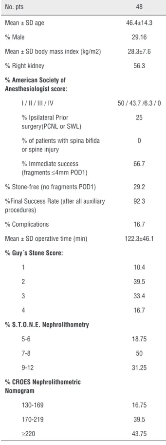
Table 1 – Baseline characteristic of study patients.
No. pts 48
Mean ± SD age 46.4±14.3
% Male 29.16
Mean ± SD body mass index (kg/m2) 28.3±7.6
% Right kidney 56.3
% American Society of Anesthesiologist score:
I / II / III / IV 50 / 43.7 /6.3 / 0
% Ipsilateral Prior surgery(PCNL or SWL)
25
% of patients with spina bifida or spine injury
0
% Immediate success (fragments ≤4mm POD1)
66.7
% Stone-free (no fragments POD1) 29.2
%Final Success Rate (after all auxiliary procedures)
92.3
% Complications 16.7
Mean ± SD operative time (min) 122.3±46.1
% Guy´s Stone Score:
1 10.4
2 39.5
3 33.4
4 16.7
% S.T.O.N.E. Nephrolithometry
5-6 18.75
7-8 50
9-12 31.25
% CROES Nephrolithometric Nomogram
130-169 16.75
170-219 39.5
Figure 1 – Time of application of the nomograms (in seconds).
Figure 2 – Percentage of success by groups of scores. useful because they help the surgeon in planning surgical strategies, result in better counselling of the patient and allow the comparison of outcomes between the experiences of different institutions.
The three nomograms evaluated in this study, GSS (8), STONE (10) and CROES (9) have been recently proposed as tools to predict success in PCNL. They use measurable and qualitative pa-rameters, acquired from preoperative imaging stu-dies and medical history. Despite the
heterogenei-ty between nomograms, the three aim to classify patients into groups with different graduations of success in PCNL.
We have demonstrated that the nomogra-ms were not significantly different in regards to the ability to predict success from PCNL. In the ROC curve analysis for the three scoring systems, we found that the Area Under the Curve (AUC) of CROES and GSS were similar (0.641 and 0.653, respectively), while the STONE was lower (0.563).
S.T.O.N.E. GUYS CROES
S.T.O.N.E. GUY’S
Time (sec)
Percentage of success
5 - 6 7 - 8 9 -12 1 2 3 4 130-169 170-219
>= 220 500
400
300
200
100
0
100 90 80 70 60 50 40 30 20 10 0
However, the two by two comparison between them revealed no significant difference (STONE x GSS: p=0.445; STONE x CROES: p=0.513; GSS x CROES: p=0.912). Other studies have compared the three nomograms and the results are similar to ours. In a study involving 246 patients, Labadie and Okhunov (19) found the area under the curve of GSS, CROES and STONE were 0.634; 0.671 and 0.670, respectively and also demonstrated that the nomograms were not significantly different. Noureldin at al. (20) also showed similar AUC be-tween GSS and STONE. Sfoungaristos et al. (21) could demonstrate the correlation between higher complexity according to the three nomograms and use of fluoroscopy. It is important to note that the value of the AUC is relatively low, suggesting a low capacity of predicting success. In our point of view, the use of AUC actually does not reflect the real benefit of using any of these nomogra-ms. When we evaluate the different groups cre-ated by the nomograms, we can clearly see that these groups are different among them regarding peri and postoperative data, according previous reports (10, 11, 13-15, 18). The information obtai-ned is critical when counselling the patient about the expectations for the surgery. Moreover, in a group of urologists used to the nomograms, when one says that is going to operate a patient with
a GSS 4, everyone knows the difficulties that are expected, including the anesthesiologist and staff room, facilitating the operative planning. This is exactly what we observe in our institution, where we have been using the GSS for the last years. Similar effect can be expected if the team is used to another nomogram.
Withington et al. (22) in a recent systema-tic review of the literature could not firmly recom-mend one nomogram over the others, but found that the quality of evidence supporting validation of the GSS was marginally superior. If all scoring system are good for predicting outcomes and are similar among then in their capacity of doing that, which one should be used in a daily basis? The applicability of a nomogram depends on how easy it is to be used in a clinical setting. Hence, time used for calculating the score is an important fac-tor when considering routine use. For that reason, we decided to study the application time for each nomogram. To the best of our knowledge, this is the first study to address this issue. In our stu-dy, we could verify that the GSS, a visual method that requires no measurement, was the fastest to be calculated. There was a large difference betwe-en application times of scoring systems, and GSS was the fastest, with an average of 27.5 seconds followed by CROES with 213.4 seconds and STO-Figure 3 – ROC curve for STONE score, Guy´s score, CROES nomogram and reference.
Sensitivity
1- Specificity
1,0
0,9
0,8
0,7
0,6
0,5
0,4
0,3
0,2
0,1
0,0
0,0 0,1 0,2 0,3 0,4 0,5 0,6 0,7 0,8 0,9 1,0
Stone
Guy’s
NE with 300.6 seconds (p<0.0001). With these findings, it has been demonstrated that the GSS could became the most practical nomogram to be used for predicting outcomes for PCNL. This new information could be useful for urologists who want to start using a nomogram but are unsure which one to choose. If all nomograms have si-milar ability to predict success, then choosing the quickest seems logical.
Our study is not without limitations. Being retrospective is a weakness, but as we evaluated basically CT scans and the clinical data was pros-pectively acquired, we believe this characteristic does not compromise the results. The number of patients is relatively small compared to other re-cent larger multire-center series, but the results rela-ted to predicting success were similar to the others studies and the analyzed cohort was large enough to identify statistically significant differences be-tween the nomograms in regards to the acquisition time. In addition, we performed a standardized pre- and postoperative evaluation of all patients with CT scan, increasing the reliability of the outcome assessment. Only one observer did all the measu-res, what could cause some bias. However, this ob-server was previously trained by two experienced urologists and as we had a high concordance index among then, we believe that one evaluator would be adequate. All patients underwent surgery per-formed by the same experienced surgeon using the same technique, reducing the potential biases in PCNL outcomes. Finally, the success evaluation was very early and rigorous, but we believe this was the better moment to have adequate and standardi-zed evaluation for all patients. Certainly, this early evaluation causes a relatively low success rate, ho-wever as our final success rate after all secondary procedures was 92.3% the difference among the groups created by the nomograms probably would not be significant. Considering this, we believed that the immediate success evaluation with CT scan would provide the best information for comparison.
In our study, we found that all three no-mograms showed similar ability to predict success of PCNL but the GSS was quicker to use than the others. Maybe these nomograms can be automated, making them easier to use, but at present this is not available. The relative low AUC of the three
no-mograms calls attention for necessity of continuing development and improvement of these tools.
CONCLUSIONS
All three nomograms showed similar abi-lity to predict success of PCNL, however the GSS was the quickest to be applied, what is an impor-tant issue for routine clinical use when counseling patients who are candidates to PCNL.
CONFLICT OF INTEREST
None declared.
REFERENCES
1. Fwu CW, Eggers PW, Kimmel PL, Kusek JW, Kirkali Z. Emergency department visits, use of imaging, and drugs for urolithiasis have increased in the United States. Kidney Int. 2013;83:479-86.
2. Sakhaee K. Pharmacology of stone disease. Adv Chronic Kidney Dis. 2009;16:30-8.
3. Moe OW. Kidney stones: pathophysiology and medical management. Lancet. 2006;367:333-44.
4. Yuhico MP, Ko R. The current status of percutaneous nephrolithotomy in the management of kidney stones. Minerva Urol Nefrol. 2008;60:159-75.
5. Preminger GM, Tiselius HG, Assimos DG, Alken P, Buck C, Gallucci M, et al. 2007 guideline for the management of ureteral calculi. J Urol. 2007;178:2418-34.
6. Türk C, Petřík A, Sarica K, Seitz C, Skolarikos A, Straub M, et al. EAU Guidelines on Interventional Treatment for Urolithiasis. Eur Urol. 2016;69:475-82.
7. Vernez SL, Okhunov Z, Motamedinia P, Bird V, Okeke Z, Smith A. Nephrolithometric Scoring Systems to Predict Outcomes of Percutaneous Nephrolithotomy. Rev Urol. 2016;18:15-27. 8. Thomas K, Smith NC, Hegarty N, Glass JM. The Guy’s
stone score--grading the complexity of percutaneous nephrolithotomy procedures. Urology. 2011;78:277-81. 9. Smith A, Averch TD, Shahrour K, Opondo D, Daels FP,
Labate G, et al. A nephrolithometric nomogram to predict treatment success of percutaneous nephrolithotomy. J Urol. 2013;190:149-56.
11. Jeong CW, Jung JW, Cha WH, Lee BK, Lee S, Jeong SJ, et al. Seoul National University Renal Stone Complexity Score for Predicting Stone-Free Rate after Percutaneous Nephrolithotomy. PLoS One. 2013;8:e65888.
12. Vicentini FC, Marchini GS, Mazzucchi E, Claro JF, Srougi M. Utility of the Guy’s stone score based on computed tomographic scan findings for predicting percutaneous nephrolithotomy outcomes. Urology. 2014;83:1248-53. 13. Mandal S, Goel A, Kathpalia R, Sankhwar S, Singh V, Sinha
RJ, et al. Prospective evaluation of complications using the modified Clavien grading system, and of success rates of percutaneous nephrolithotomy using Guy’s Stone Score: A single-center experience. Indian J Urol. 2012;28:392-8. 14. Ingimarsson JP, Dagrosa LM, Hyams ES, Pais VM Jr. External
validation of a preoperative renal stone grading system: reproducibility and inter-rater concordance of the Guy’s stone score using preoperative computed tomography and rigorous postoperative stone-free criteria. Urology. 2014;83:45-9. 15. Akhavein A, Henriksen C, Syed J, Bird VG. Prediction of single
procedure success rate using S.T.O.N.E. nephrolithometry surgical classification system with strict criteria for surgical outcome. Urology. 2015;85:69-73.
16. Sfoungaristos S, Lorber A, Gofrit ON, Yutkin V, Landau EH, Pode D, et al. External Validation and Predictive Accuracy Assessment of Guy’s Stone Score as a Preoperative Tool for Estimating Percutaneous Nephrolithotomy Outcomes. J Endourol. 2015;29:1131-5.
17. Sfoungaristos S, Gofrit ON, Yutkin V, Landau EH, Pode D, Duvdevani M. External Validation of CROES Nephrolithometry as a Preoperative Predictive System for Percutaneous Nephrolithotomy Outcomes. J Urol. 2016;195:372-6. 18. Bozkurt IH, Aydogdu O, Yonguc T, Yarimoglu S, Sen V,
Gunlusoy B, et al. Comparison of Guy and Clinical Research Office of the Endourological Society Nephrolithometry Scoring Systems for Predicting Stone-Free Status and Complication Rates After Percutaneous Nephrolithotomy: A Single Center Study with 437 Cases. J Endourol. 2015;29:1006-10.
19. Labadie K, Okhunov Z, Akhavein A, Moreira DM, Moreno-Palacios J, Del Junco M, et al. Evaluation and comparison of urolithiasis scoring systems used in percutaneous kidney stone surgery. J Urol. 2015;193:154-9.
20. Noureldin YA, Elkoushy MA, Andonian S. Which is better? Guy’s versus S.T.O.N.E. nephrolithometry scoring systems in predicting stone-free status post-percutaneous nephrolithotomy. World J Urol. 2015;33:1821-5.
21. Sfoungaristos S, Gofrit ON, Yutkin V, Landau EH, Pode D, Duvdevani M. Evaluating Parameters Affecting Fluoroscopy Time During Percutaneous Nephrolithotomy: Focus on the Predictive Role of Guy’s, S.T.O.N.E., and CROES Scoring Systems. J Endourol. 2015;29:1366-70.
22. Withington J, Armitage J, Finch W, Wiseman O, Glass J, Burgess N. Assessment of Stone Complexity for PCNL: A Systematic Review of the Literature, How Best Can We Record Stone Complexity in PCNL? J Endourol. 2016;30:13-23.
23. de la Rosette JJ, Opondo D, Daels FP, Giusti G, Serrano A, Kandasami SV, et al. Categorisation of complications and validation of the Clavien score for percutaneous nephrolithotomy. Eur Urol. 2012;62:246-55.
24. Tiselius HG, Andersson A. Stone burden in an average Swedish population of stone formers requiring active stone removal: how can the stone size be estimated in the clinical routine? Eur Urol. 2003;43:275-81.