UNIVERSIDADE FEDERAL DO CEARÁ
FACULDADE DE FARMÁCIA, ODONTOLOGIA E ENFERMAGEM PROGRAMA DE PÓS-GRADUAÇÃO EM ODONTOLOGIA
JULIANA PAIVA MARQUES LIMA
ESTUDO IN SITU DO EFEITO ANTIMICROBIANO DA TERAPIA FOTODINÂMICA EM LESÕES DE CÁRIE DENTINÁRIA
JULIANA PAIVA MARQUES LIMA
ESTUDO IN SITU DO EFEITO ANTIMICROBIANO DA TERAPIA FOTODINÂMICA EM LESÕES DE CÁRIE DENTINÁRIA
Dissertação apresentada ao Programa de Pós- Graduação em Odontologia da Faculdade de Farmácia, Odontologia e Enfermagem da Universidade Federal do Ceará como um dos requisitos para a obtenção do Título de Mestre em Odontologia.
Área de concentração: Clínica Odontológica Orientadora: Profa. Dra. Iriana Carla Junqueira Zanin
Co-Orientadora: Profa. Dra. Lidiany Karla Azevedo Rodrigues
Ficha catalográfica L698e Lima, Juliana Paiva Marques
Estudo in situ do efeito antimicrobiano da terapia fotodinâmica em lesões de cárie dentinária/ Juliana Paiva Marques Lima. – Fortaleza, 2009.
52 f. : il.
Orientador: Profa. Dra. Iriana Carla Junqueira Zanin
Dissertação (Mestrado) – Universidade Federal do Ceará. Faculdade de Farmácia, Odontologia e Enfermagem. Programa de Pós-Graduação em Odontologia.
1. Cárie Dentária 2. Fotoquimioterapia 3. Análise Microbiológica I. Zanin, Iriana Carla Junqueira (orient.) II. Título.
JULIANA PAIVA MARQUES LIMA
ESTUDO IN SITU DO EFEITO ANTIMICROBIANO DA TERAPIA FOTODINÂMICA EM LESÕES DE CÁRIE DENTINÁRIA
Dissertação apresentada à Coordenação do Programa de Pós-graduação em Odontologia da Universidade Federal do Ceará como requisito parcial para obtenção do Título de Mestre em Odontologia. Área de concentração: Clínica Odontológica
Aprovada em: ___/ ___/___
BANCA EXAMINADORA
_____________________________________ Profa. Dra. Iriana Carla Junqueira Zanin (Orientadora)
Universidade Federal do Ceará- Campus Sobral
_______________________________________ Profa. Dra. Nádia Accioly Pinto Nogueira
Universidade Federal do Ceará- UFC
A Deus,
por tudo que tenho e que sou ...
Aos meus pais, Adriano e Darcy, exemplo de
amor e respeito e por terem me ensinado
sempre a importância da perseverança na
busca pelos objetivos
À minha irmã, Adriana, pelo auxílio. Minha
eterna amiga.
Ao Renan, que sempre me estimula a cada
nova etapa, estando sempre ao meu lado e
acreditando na concretização de meus
AGRADECIMENTOS ESPECIAIS
À Profa. Dra. Iriana Carla Junqueira Zanin, minha orientadora, por seus
ensinamentos, por suas palavras de apoio e amizade e por sua maneira tranqüila de
transmitir seus conhecimentos e me acalmar nos momentos mais difíceis.
À Profa. Dra. Lidiany karla Azevedo Rodrigues, minha co-orientadora, presente,
simplesmente, em todos os momentos. Também pelo exemplo de professora e pesquisadora.
À grande amiga, Mary Anne Sampaio de Melo, pela parceria em todas as etapas do
AGRADECIMENTOS
À Universidade Federal do Ceará na pessoa do seu Reitor Jesualdo Pereira Farias.
À Faculdade de Farmácia, Odontologia e Enfermagem, na pessoa de sua diretora Profa. Dra. Neiva Fracenely Cunha Vieira.
À coordenadora do curso de Odontologia Prof. Dra. Maria Eneide Leitão de Almeida.
Ao Prof. Dr. Sérgio Lima Santiago, coordenador do curso de mestrado da UFC, pela competente e exemplar função de coordenador.
Ao Prof. Dr. Jaime Aparecido Cury por sua grandiosa contribuição nos estudos em cariologia.
Aos meus colegas da turma de mestrado, pela amizade, companheirismo e pela boa convivência.
Às minhas amigas Fátima Maria Cavalcante Borges, Rosane Pontes de Souza, Suyane Maria Luna Cruz de Vasconcelos, Maria Denise Rodrigues de Moraes e Daniela
da Silva Bezerra,agradeço pelo empenho em cada momento, inclusive nas tardes de sábado no laboratório de pesquisa. Agradecimento especial a Alrieta Henrique Teixeira por seu esforço em minha pesquisa e por viajar para a realização desta. Obrigada, também, por seu acolhimento em sua casa em 15 ininterruptosdias.
À equipe do laboratório de microbiologia de Sobral, Ruliglesio Rocha, Emanuela de Lima Rebouças, Eliane Doas Santos Pereira, por o esforço de nos acompanhar em todos os
momentos da pesquisa desenvolvida na cidade.
Aos alunos de iniciação científica, Diego Martins de Paula e João Paulo Saraiva Wenceslau,pela presteza e ajuda indispensáveis em todas as etapas da pesquisa.
À CAPES pela concessão de bolsa de auxílio financeiro.
Aos professores do curso de mestrado pela contribuição ao meu aprendizado.
Ao Prof. Dr. José Jeová Siebra Neto, primeiro coordenador do curso de mestrado em Odontologia da UFC pela dedicação na concretização deste curso e pelos ensinamentos que me repassou no decorrer do curso.
Aos funcionários de mestrado Germano Mahlmann Muniz Filho e Lúcia Ribeiro Marques Lustosa, pela constante disponibilidade.
À bibliotecária, Rosane Costa, da Biblioteca das Ciências de Saúde da UFC, pela correção das referências bibliográficas.
Ao técnico em prótese dental do curso de Odontologia, Antônio Carlos de Oliveira Filho, pela confecção dos dispositivos intra-orais.
À amiga, Raquel Saraiva Rolim, meus agradecimentos pela disponibilidade e paciência na elaboração dos esquemas gráficos.
Meus sinceros agradecimentos aos voluntários desta pesquisa.
Aos meus familiares e amigos pela compreensão às muitas horas em que tive ausente para a dedicação ao estudo.
A todos que de maneira direta ou indireta ajudaram na elaboração desta pesquisa.
“O Senhor é minha fortaleza e meu escudo;
nele confia meu coração”
RESUMO
Terapia fotodinâmica é um conceito de tratamento sugerido pela literatura como uma potencial terapia capaz de proporcionar inativação microbiana. Desta forma, este estudo avaliou a ação da terapia fotodinâmica sobre lesões de cárie dentinária, mediante um delineamento in situ de única fase. Durante 14 dias, 20 voluntários utilizaram dispositivos intra-orais palatinos, contendo, cada um, seis blocos de dentina humana. Todos os voluntários foram instruídos a gotejar sobre os blocos uma solução de sacarose a 40% dez vezes ao dia, simulando um desafio cariogênico e a utilizar dentifrício fluoretado três vezes ao dia. No final deste período clínico, os blocos foram randomicamente alocados em um dos seguintes grupos: sem fotossensibilizador e luz (F-L-); com fotossensibilizador e sem luz (F+L-); sem fotossensibilizador e com luz com densidade de energia de 47J/cm2(F-L+47); sem fotossensibilizador e com luz com densidade de energia de 94J/cm2 (F-L+94); com fotossensibilizador e irradiados com densidade de energia de 47J/cm2(F+L+47); com fotossensibilizador e irradiados com densidade de energia de 94J/cm2(F+L+94). O fotossensibilizador de escolha foi azul de orto-toluidina na concentração de 100 g/mL e a irradiação originada de um diodo emissor de luz (LED) com comprimento de onda predominante em 638,8nm. Amostras de dentina cariada de cada bloco foram coletadas antes e após os tratamentos e analisadas para contagem de microorganismos totais, estreptococos totais, estreptococos grupo mutans e lactobacilos. Os dados das contagens foram transformados em log10, os valores de log redução foram obtidos e as diferenças estatísticas
identificadas através dos testes ANOVA One way e Tukey Kramer (p<0,05). Em ambas as análises, foram observadas diferenças estatisticamente significativas entre as contagens microbiológicas antes e depois dos tratamentos nos grupos (F-L+94), (F+L+47), (F+L+94) para todos os microrganismos testados. Concluiu-se que a terapia fotodinâmica foi efetiva na morte microbiana nos parâmetros testados e que o tempo de exposição da dentina à luz de 10 minutos, também, promoveu inativação microbiana.
ABSTRACT
Photodynamic therapy is a concept of treatment, suggested by the literature as a potential therapy capable of inactivating microbial. Thus, this study assessed the efficacy of photodynamic therapy on injuries of dentine caries by a in situ design of single phase. During 14 days, 20 volunteers wore intra-oral palatal devices containing six human dental dentine slabs. The volunteers wore asked to drop a 40% sucrose solution onto the slabs ten times per day, in order to simulate a cariogenic challenge and to use fluoride dentifrice three times per day. At the end of the experimental period the slabs were randomly allocated into one of the following groups: without sensitizer and light (S-L-); with sensitizer and without light (S+L-); without sensitizer and irradiated with 47J/cm2 energy density (S-L+47); without sensitizer and irradiated with 94J/cm2 energy density (S-L+94); with sensitizer and irradiated with 47J/cm2 energy density (S+L+47); and with sensitizer and irradiated with 94J/cm2 energy density (S+L+94). The sensitizer of choice was the toluidine blue O at 100 g/mL concentration and radiation originated from a light-emitting diode (LED) with a 638,8 nm predominant wavelength. Before and after the treatments, dentine samples were collected and analyzed to figure out the total microorganisms, total streptococci, mutans streptococci and lactobacilli. The data of the counts were transformed into log10.The values of log reduction was achieved
and the statistical differences identified by ANOVA One way and Tukey Kramer tests (p<0,05). In both analyses, statistically significant difference between the microbial counts before and after treatment in groups (S-L+47), (S+L+94), (S+L+94) for all microorganisms tested are founded. It was concluded that PACT in the tested parameters was effective in promoting microbial death and that the exposure time of dentine to 10 minutes of light was also effective in microbial inactivation.
SUMÁRIO
1 INTRODUÇÃO GERAL... 12
2 PROPOSIÇÃO... 15
3 CAPÍTULOS... 16
4 CONCLUSÃO GERAL... 36
REFERÊNCIAS... 37
APÊNDICES... 44
1 INTRODUÇÃO GERAL
Os primeiros relatos da utilização da terapia fotodinâmica (TFD) datam do início do século IXX, quando Oscar Raab, um estudante alemão de medicina, reportou o conceito de indução de morte celular através da interação de agentes químicos e luz, constatando que a toxidade do hidrocloreto de acridina contra Paramecia caudatum foi dependente da quantidade de luz incidida (RAAB, 1900) A TFD, tornou-se uma alternativa promissora para o tratamento do câncer e outras doenças há duas décadas, tendo progressivamente sido bem estabelecida e recebido aprovação para uso clínico em muitos países (HAMBLIN; HASAN, 2004; PLAETZER et al., 2003). Mais recentemente, esta terapia passou a ser pesquisada e utilizada com o propósito de redução microbiana, recebendo o nome de terapia antimicrobiana fotodinâmica (MALIK; HANANIA; NITZAN, 1990).
O mecanismo de ação da TFD tanto na medicina oncológica quanto na microbiologia se baseia em uma reação fotoquímica não térmica que requer a presença de um fotossensibilizador (em geral um corante), oxigênio e luz visível (MACROBERT; BOWN; PHILLIPS, 1989). A terapia consiste na irradiação de luz visível em um comprimento de onda complementar ao fotossensibilizador, levando a excitação da molécula e sua passagem para um estado singleto. Nessa forma, a molécula apresenta um tempo de vida bastante limitado, passando, então, para um estado tripleto, que apesar de ter baixa energia, tem um tempo de vida considerado relativamente longo (10-9segundos) (RYTER; TYRREL, 1997). Nesse período, aumenta a probabilidade do fotossensibilizador transferir energia para outras moléculas e seguir um dos dois caminhos denominados como fotoprocesso do tipo I e tipo II (FUCHS; THIELE, 1997; YAVARI, 2006). No primeiro processo, há interação do fotossensibilizador com moléculas vizinhas ou átomos de hidrogênio havendo produção de radicais livres, íons superperóxidos e radicais hidroxila. No fotoprocesso do tipo II, há uma interação direta do fotossensibilizador com o oxigênio molecular, gerando oxigênio singleto (1O2) (BRANCALEON; MOSELEY, 2002; PLAETZER et al., 2003; CASTANO;
Em microbiologia, a terapia antimicrobiana fotodinâmica passou a receber atenção especial por apresentar vantagens em relação ao tratamento com agentes antimicrobianos convencionais, tais como: o confinamento do efeito ao local da lesão pela aplicação tópica do fotossensibilizador e a irradiação restrita à área de interesse, oferecendo baixo risco a outras células do hospedeiro (HAMBLIN; HASAN, 2004; ZANIN et al., 2005); dano ou morte bacteriana obtido em curto período de tempo, reduzindo a possibilidade de surgimento de resistência microbiana (WAINWRIGHT et al., 2003); e, finalmente, a inexistência de reações sistêmicas, mesmo após repetido uso (BACKHAUS et al., 2007).
Para que a terapia antimicrobiana fotodinâmica exerça algum efeito na célula bacteriana, a luz deve ser absorvida por um ou mais de seus constituintes. Embora algumas bactérias apresentem componentes capazes de absorver parte do espectro de luz visível, a maioria das espécies encontradas na cavidade oral não apresenta esses componentes, de modo que o uso de fotossensibilizadores exógenos, que atraia para si a luz, é fundamental ao sucesso dessa terapia (WILSON; DOBSON; HARVEY, 1992).
Uma grande variedade de corantes naturais e sintéticos tem sido desenvolvida e testada com relação às propriedades farmacológicas e de absorção à luz. (PRATES et al., 2006; PELOI et al., 2008 ). Um largo número de fotossensibilizadores tem sido testado in vitro contra microorganismos orais, incluindo os derivados fenotiazínicos, como azul de metileno e azul de orto toluidina (ZANIN et al., 2005); os derivados da fitalacionina, como fitalacionina dissulfonado alumínio (AlPcS2) e fitalacionina catiônica com Zn(II) (WILSON
et al., 1995); os derivados das clorinas (WILSON, 2004); além de rosa bengal (PAULINO et al., 2005), verde de malaquita (PRATES et al., 2006), e porfirinas (HAMBLIN; HASAN, 2004).
Assim como o fotossensibilizador, a escolha da fonte de luz é um importante fator para o êxito da terapia fotodinâmica. A fim de otimizar o tratamento, é essencial que a emissão de luz coincida com o pico de absorção do fotossensibilizador escolhido (FISCHER et al., 1998). Embora lasers convencionais sejam tradicionalmente utilizados, recentemente o uso dos LEDs tem se intensificado, uma vez que, por não apresentarem boa colimação e coerência, resultam em bandas de emissão de luz mais largas, favorecendo, assim, a complementaridade com o fotossensibilizador (ZANIN et al., 2006; PELOI et al., 2008; GIUSTI et al., 2008).
semicondutores pequenos e portáteis e por apresentarem baixo custo em comparação a outras fontes de luz (ZANIN et al., 2005; KONOPKA; GOSLINSKI, 2007).
A ação antimicrobiana de lasers ou LEDs, associados a fotossensibilizadores específicos sobre bactérias crescidas em cultura planctônica, já está bem documentada na literatura (WILSON et al., 1993; BURNS; WILSON; PEARSON, 1994; SOUKOS et al., 1998; WILLIAMS et al., 2003; MATEVKI et al., 2003; PAULINO et al., 2005). Da mesma forma, os efeitos desse tratamento, sobre biofilmes bacterianos organizados, já foram estabelecidos. (WOOD et al., 1999; O’NEILL; HOPE; WILSON, 2002; GAD et al., 2004; ZANIN et al., 2005, 2006; WOOD et al., 2006, METCALF et al., 2006). No entanto, ainda não há estudos que demonstrem o efeito antimicrobiano da terapia fotodinâmica em lesões de cárie dentinária humana.
A cárie dentária é uma doença crônica caracterizada pela destruição progressiva e localizada do dente (MARSH; MARTIN, 1992). Após a desmineralização do esmalte, a lesão pode progredir para a dentina, sendo caracterizada por duas camadas distintas. A camada mais externa, denominada de dentina infectada, consiste de uma área amolecida, amarelada e não é passível de remineralização. A camada mais interna, denominada dentina contaminada, é escura e endurecida, apresentando uma menor contaminação bacteriana e sendo, portanto, passível de remineralização. Kidd, Ricketts e Beighton (1996) propuseram que somente a remoção da dentina infectada seria necessária, se as bactérias da dentina contaminada fossem removidas ou mortas. Contudo a distinção entre essas zonas é extremamente crítica, de modo que os tratamentos atuais envolvem a remoção total da dentina infectada e contaminada, removendo, desnecessariamente, a dentina passível de remineralização que se encontra sobre a polpa, aumentando o risco de uma exposição pulpar e comprometendo o prognóstico do dente. Desse modo, a utilização de um tratamento alternativo complementar à remoção mecânica e capaz de matar bactérias in situ, poderia diminuir a quantidade de tecido dentinário a ser removido (BURNS; WILSON; PEARSON, 1995).
2 PROPOSIÇÃO
3 CAPÍTULOS
Esta dissertação está baseada no Artigo 46 do Regimento Interno do Programa de Pós- Graduação em Odontologia da Universidade Federal do Ceará, que regulamenta o formato alternativo para dissertações de Mestrado e permite a inserção de artigos científicos de autoria e co-autoria do candidato (Anexo A). Por se tratarem de pesquisas envolvendo seres humanos, ou parte deles, o projeto de pesquisa deste trabalho foi submetido à apreciação do Comitê de Ética em Pesquisa da Faculdade de Medicina da Universidade Federal do Ceará, tendo sido aprovado sob protocolo nº 143/06 (Anexo B). Assim sendo, esta dissertação de mestrado é composta de um capítulo que contém um artigo submetido para a publicação no periódico “European Journal of Oral Science”, conforme descrito abaixo (Anexo C), previamente analisado e corrigido por um corretor da língua inglesa (Anexo D):
Capítulo 1In situ study of the antimicrobial effect of photodynamic therapy in dentine caries lesions
In situ study of the antimicrobial effect of photodynamic therapy in dentine caries
lesions
Juliana Paiva Marques Lima1 Mary Anne Sampaio de Melo1 Fátima Maria Carvalho Borges1 Alrieta Henrique Teixeira2 Carolina Steiner-Oliveira3 Marinês Nobre dos Santos3
Lidiany Karla Azevedo Rodrigues1 Iriana Carla Junqueira Zanin2
1
Faculty of Pharmacy, Dentistry and Nursing, Federal University of Ceará, Fortaleza, CE, Brazil.
2
Faculty of Dentistry, Federal University of Ceará, Sobral, CE, Brazil.
3
PiracicabaDental School, State University of Campinas, Piracicaba, SP, Brazil.
Running Title –Lethal photosensitization in carious dentine.
Lima JPM*, Melo MAS, Borges FMC, Teixeira AH, Steiner-Oliveira C, Nobre-dos-Santos M, Rodrigues LKA, Zanin ICJ
In situ study of the antimicrobial effect of photodynamic therapy in dentine caries
lesions
Eur J Oral Sci
Abstract : Photodynamic antimicrobial therapy (PACT) promotes bacterial death due to
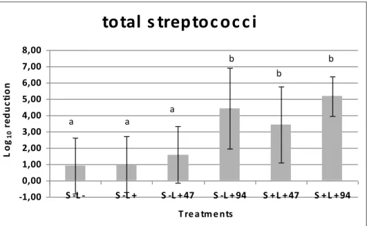
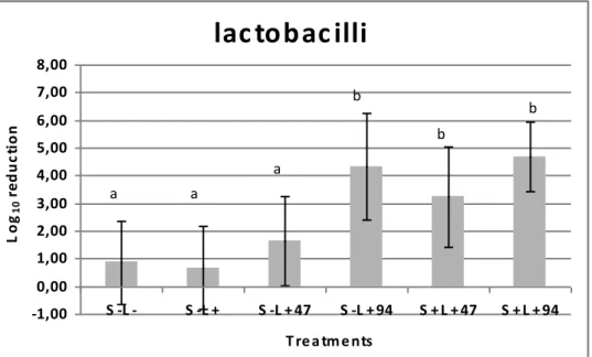
photosensitisation of microbial components. This study evaluated the effect of PACT on dentine caries produced in situ. Over the course of 14 days, 20 volunteers wore intra-oral devices containing human dentine slabs that received a 40% sucrose solution, 10 times/day. Afterwards, the antimicrobial effect of toluidine blue O associated with 47 or 94 J.cm-2 of a light emitting diode (LED) was evaluated. Before and after the treatments, dentine samples were analysed with regard to the total microorganisms, total streptococci, mutans streptococci and lactobacilli. Logreductions obtained for the several treatments were statistically analysed by one-way ANOVA and Tukey-Kramer test (p<0.05). Significant reductions were observed for PACT with both tested energy densities, with the following values observed for 47 and 94 J.cm-2: for total streptococci, 3.45 and 5.18; for mutans streptococci, 3.08 and 4.16; for lactobacilli, 3.24 and 4.66; and for total microorganisms, 4.29 and 5.43, respectively. The control using only irradiation with 94 J.cm-2 was also effective against all bacteria. Concluding, PACT was effective in killing oral microorganisms present in dentine caries produced in situ and may be a useful technique for eliminating bacteria from dentine carious lesions prior to restoration.
Keywords: Dentine Caries, Photochemotherapy, Microbiological Analysis.
INTRODUCTION
Dental caries is a chronic invasive disease involving demineralisation of the tooth followed by destruction of the organic phase of the dentine (1). During the dentine caries process, the outer layer of the lesion is characterised by a softened and wet dentine that is highly infected by bacteria. The inner layer, known as affected dentine, is frequently less contaminated with bacteria and is usually susceptible to remineralisation. However, the clinical distinction of these two regions is extremely difficult, and usually current methods of treating dentine lesions involve the removal of both dentine layers, which, in deep cavities, can result in pulp exposure (2).
KIDD et al. (1996) (3) proposed that the removal of only the softened and wet dentine is necessary for successful treatment, and that the effective sealing of a cavity with a restorative material is sufficient to render residual bacteria dormant. However, since the most restorative materials currently available are ineffective in promoting long-term sealing of the dental cavity, an effective way of disinfecting the dentine tissue is highly desirable before performing the treatment (4).
In this context, photodynamic antimicrobial therapy (PACT) may emerge as a suitable treatment because of its ability to kill bacteria in situ. The photodynamic process is based on a two-step protocol, where target cells are selectively loaded with a sensitiser followed by irradiation with a light of complementary wavelength (the maximum absorption of the sensitiser), resulting in the production of cytotoxic products, including singlet oxygen and free radicals (5, 6). These products are capable of damaging essential components of the cells or modifying metabolic activities in an irreversible way, which may result in cell death (7, 8, 9, 10, 11).
throughout dentine structures, the purpose of this study was to evaluate the antimicrobial effect of PACT in dentine carious lesions produced in situ.
MATERIALS AND METHODS
Study Population and Ethical Aspects
This study protocol was approved by the Research and Ethics Committee of the Federal University of Ceara Medical School (protocol # 143/2006). 20 healthy adults (14 females and 6 males), aged 19-36 yr, able to comply with the experimental protocol, participated in this study. They were not admitted to the study if any of the following conditions were present: active caries lesions, use of any antibiotics within the past 6 months prior to the study, or the use of antimicrobial dentifrice. 28 adult volunteers were invited to participate; 4 of them used antibiotics, and 3 refused to participate. Thus, 21 volunteers initiated the study, but one was unable to comply with the experimental protocol and was excluded.
Experimental Design
A simple-blind in situ design was conducted in one phase of 14 d, during which 20 volunteers wore palatal devices containing 6 human dental dentine slabs. At the end of the clinical phase, the slabs were randomly allocated into one of the following treatments as observed in figure 1: without sensitiser and light (S-L-), with sensitiser and without light (S+L-), without sensitiser and irradiated with 47 J.cm-2 (S-L+47), without sensitiser and irradiated with 94 J. cm-2 (S-L+94), with sensitiser and irradiated with 47 J.cm-2 (S+L+47), and with sensitiser and irradiated with 94 J.cm-2 (S+L+94).
Specimen Preparation
assessed by microscope examination at 40x magnification to ensure complete enamel removal. Thus, the fragments were fixed in acrylic devices and cut using a water-cooled diamond saw and a cutting machine (IsoMetTM Low Speed Saw, Buehler, Lake Bluff, IL, USA) to obtain dentine slabs (5 × 5 × 2 mm³). The occlusal dentine face was used once only; the remaining surfaces of the slabs were protected with resistant acid varnish (Colorama-CEIL, São Paulo, SP, Brazil). Afterwards, the specimens were polished using three different silicon carbide waterproof papers (300, 600 and 1200 grit) as well as polishing cloths with 1-µm diamond paste (Buehler, Lake Bluff, IL, USA). Microhardness measurements were performed in a hardness tester (Shimadzu HMV-2000 Shimadzu Corporation, Kyoto, Japan) using a Vickers diamond under a 50 g load for 10 s to select dentine slabs with similar hardness (50 to 70 Vickers Hardness Number). Since 40 dentine slabs were excluded due to higher hardness numbers, 120 dentine slabs were selected. Finally, the slabs were autoclaved (121ºC 15 min) (34) and stored in 100% humidity until being inserted into the palatal appliances (Figure 2A).
Palatal appliance preparation
For each subject, an acrylic palatal device was fabricated, in which 6 cavities (6 × 6 × 3 mm³) were prepared on the left and right sides; one slab was attached with wax in each cavity (35). In order to allow biofilm accumulation, and to protect it from mechanical disturbance, a plastic mesh was positioned on the acrylic resin, leaving a 1-mm space from the slab surface (35, 36, 37).
Intraoral phase
(40, 41). Before replacing the palatal appliance in the mouth, a 5-min waiting time was standardised for sucrose diffusion into the dental biofilm (Figure 2B).
The dentifrice treatment was performed 3 times a day, after mealtimes when volunteers’ habitually performed their oral hygiene. The appliances were extra-orally brushed, except the slab area, and volunteers were asked to brush carefully over the covering meshes, to avoid disturbing the biofilm. All volunteers consumed fluoridated water (0.70 mg F.l-1), and no restriction was made with regard to the volunteers’ dietary habits.
Microbiological analyses
The microbiological analyses of carious dentine were performed on the 14th d of the intraoral phase, immediately before and after performing the treatments. Around 12 h after the last sucrose solution application, the volunteers stopped wearing the intraoral device. After removing the plastic mesh, the biofilm formed on dentine slabs was removed with a scalpel, lamina #15C, and the dentine carious tissue was exposed. The baseline dentine sample was collected from half of each slab using a #5 carbide bur in a low-speed drill (Labordental, São Paulo, SP, Brazil), as described by KIDD et al. (1995) (42) (Figure 2C). The dentine samples were analytically weighed using pre-weighed microcentrifuge tubes containing the bur, to which 0.9% NaCl solution was added (1 ml.mg-1 dentine). In order to detach the bacterial cells, the tubes were agitated for 1 min in a Disrupter Genie Cell Disruptor (Precision Solutions, Rice Lake, WI, USA) after receiving three sterile glass beads (0.1-mm diameter). Afterwards, the suspension was serially diluted (1:10, 1:100, 1:1000, 1:10000, 1:100000 and 1:1000000) in 0.9% NaCl solution. In order to assess microorganism viability, samples were plated in triplicate in mitis salivarius agar (MSA agar) containing 15% sucrose to determine total streptococci (TS); mitis salivarius agar plus 0.2 bacitracin ml-1 (MSB agar) to determine mutans streptococci (MS) (43); Rogosa agar supplemented with 0.13 % glacial acetic acid to assess the number of lactobacilli (LB) (44); and blood agar to determine total microorganisms (TM) (Figure 2D). The plates were incubated for 48 h at 37°C in a partial atmosphere of 10% CO2. Representative colonies of MS, TS, LB and TM were counted using a colony counter,
Photosensitiser and light sources
Toluidine blue O (TBO) was obtained from Sigma Chemicals (Sigma-Aldrich Foundation, Poole, UK), dissolved in deionised water (100 µg.ml-1) and stored in the dark. The light source used was a red light-emitting diode (LED; Laserbeam, Rio de Janeiro, RJ, Brazil), with a spectrum of emission ranging from 620 to 660 nm and a 638.8 nm predominant wavelength. The light was distributed by a fibre optic cable with a 9.5 mm spot. Irradiation was performed with a focused beam maintained at a 2.0 mm distance from the dentine slab. A power meter Lasermate (Coherent Inc, Santa Clara, CA, USA) was used to measure the power of the LED device; an output of 40 mW was used.
Photodynamic therapy
Following the first dentine sample collection (baseline value for tissue contamination verification), PACT was performed using a random distribution of slabs into treatments. The groups that received sensitiser (S+L-; S+L+47; S+L+94) were maintained in contact with 5 µ L of TBO solution during a pre-irradiation time of 5 min in the dark. Other groups (L-; S-L+47; S-L+94) received an equal volume of sterile 0.9% NaCl solution during the same period of time. Irradiated groups (S-L+47; S-L+94; S+L+47; S+L+94) received LED light for 5 (47 J.cm-2) or 10 min (94 J. cm-2), while groups that did not receive light (S-L-; S+L-) were submitted to a 10 min waiting period in order to simulate the irradiation conditions (Figure 2C). The distribution of treatments on the palatal device was determined randomly; one option can be observed in figure 1. After each treatment, a second dentine sample was collected from another intact half of each slab and was microbiologically processed as previously described.
Statistical analysis
In order to assess the effect of the treatments, the variable log reduction promoted by each treatment was analysed. The normality distribution of the data and equality of variances were checked by using the Kolmogorov-Smirnov and Levene tests, respectively. The log reduction results were calculated by subtraction of the initial values from the final values of CFU.mg-1 of dentin after being transformed by Log10. These data were analysed by a one-way
software BioStat 2007 Professional (AnalystSoft Robust business solutions company, Vancouver, Canada) was used.
RESULTS
The log reductions found after PACT on tested microorganism growth in situ can be seen in figures 3-6. Results show that neither incubation with TBO alone nor irradiation with 47 J.cm-2, in absence of sensitiser, had a significant effect on the viability of these microorganisms, when compared to the control group (S-L-). On the other hand, irradiation with an LED device for 10 min, which resulted in an energy density of 94 J.cm-2, had a significant effect on bacterial reduction in dentine caries even in the absence of sensitiser. Also, the association of TBO and LED resulted in a significant decrease in the viability of total streptococci (p<0.0004), mutans streptococci (p<0.0223), lactobacilli (p<0.0092) and total microorganisms (p<0.0004).
DISCUSSION
Dental caries may be a disease well suited to photodynamic antimicrobial therapy. Caries is often a localised infection, so the sensitiser may be applied to the lesion by means of a syringe; light can then be delivered via an optical fibre. If bacteria within carious lesions could be eradicated by photosensitisation in vivo, there would be beneficial consequences for dental health. Dentine tissue could be better preserved, thereby making patient treatment easier for the dentist and more comfortable for patients, by enabling lesions to be restored with minimal tissue removal, and improving the long-term prognosis for the repaired tooth (19).
Sensitisers as well are essential elements in PACT; several studies have demonstrated the efficacy of a range of sensitisers in the elimination of oral bacteria (1, 19, 45). TBO is an attractive option due to its accessible cost and intense absorption in the red spectrum (>600 nm) (46). Also, TBO association with different light sources at a specific wavelength seems to be effective in producing microbiological reductions in planktonic cultures (27), scrapping plaque (1) and intact biofilms (24).
Observation of parameters used to kill bacteria in culture suspensions demonstrates that PACT is more effective at eliminating cells in planktonic cultures than in biofilms. When considering the use of photodynamic therapy on intact biofilms, it is also important to remember that bacteria in such a state are far less susceptible to antimicrobial agents because of the polymeric matrix, lower growth rate, metabolic activity and specific gene expression (47). In carious tissue, the treatment is difficult because bacteria are protected by a dentinal structure, which may limit the penetration of both the sensitiser and light (4). Moreover, the interaction of light with dentine is complex due to refraction and reflection at the surface of the peritubular and intertubular dentine. Also, it has been previously demostrated that bacterial death is lower when the microorganisms are irradiated with light passing through demineralised dentine slices and that there is no direct relationship between the extent of dentine demineralisation and the degree of bacterial death after PACT (14). Consequently, BURNS et al. (1995) (14) have suggested that there is a finite distance between the dentine and light source beyond which the irradiation will be not effective, even in highly demineralised dentine.
Although some in vitro studies simulated the effect of PACT on caries lesions using demineralised slices of dentine, (14) bacteria embedded in a collagen matrix (4, 14), bovine dentine (32), and ex vivo carious dentine (4), this is one of the first studies verifying the antimicrobial effect of photodynamic therapy on human dentine caries produced in conditions very close to those found in vivo. In situ models of caries involve the use of devices that create conditions that simulate the process of dental caries, serving as a link between the clinical uncontrolled situation and the highly controlled laboratory experiments. The model aims to simulate what occurs in the natural process of caries and also to provide information in a short period of time without causing damage to the natural teeth of volunteers (48). In this way, this model may be considered a proper tool for testing the effect of PACT on microorganisms involved in the dentine caries process.
dentine (38, 39, 49). Although the majority of in situ experiments have used a 20% sucrose solution 8 times/d (41, 50, 51), in this study the authors used a 40% sucrose solution 10 times/day since the demineralisation process seems to be more evident with carbohydrate consumption frequency equal to or higher than 6 times/d (41). A 40% sucrose solution was used in the present research, according to AIRES et al. (2006) (39), who demonstrated that this sucrose concentration is effective in producing extracellular polysaccharide on enamel slabs. However, when producing caries lesions on dentine slabs, AIRES et al. (2008) (49) used a sucrose concentration of 10%, which was different from that used in this study. This can be justified due to the necessity of obtaining a softened and drilled dentine in order to perform appropriate dentine harvest and microbiological analyses. In addition, fluoride from the toothpaste used by volunteers during all intraoral periods could decrease the progression of dentine caries. The use of fluoride-containing dentifrice was included in this experimental model, since over 95% of all dentifrices sold in the U.S., Brazil and Western Europe contain fluoride (52, 53, 54).
The results of this study show that photodynamic therapy was effective in significantly reducing all examined microorganisms at both energy densities tested. To our knowledge, there is no other in situ study testing PACT in dentine caries lesions. Thus, our results confirmed those found in a previous in vitro study performed by our research group, which used the same light source and sensitiser, concluding that photodynamic antimicrobial therapy was effective against mutans streptococci using energy densities of 47 and 94 J.cm-2 (data not published). Our work also agrees with that carried out by GIUSTI et al. (2008) (32), which demonstrated that the use of LED in association with TBO was effective in bacterial reduction of S. mutans and L. acidophilus in carious bovine dentine; a 400 mW LED device was used in an in vitro protocol. Furthermore, the authors used an unusual method for calculating the antimicrobial effect.
polysaccharide matrix and water. However, in carious dentine, the exposure to LED irradiation longer than 5 min may have caused drying of the substrate due to faster water evaporation, thus resulting in decreased bacterial viability. Consequently, some cytotoxicity from the light source might have occurred, since no effects on bacterial viabilty were obtained in the control group, which was subjected to an air-exposure time of 10 min. Nevertheless, to avoid dentine dryness and for clinical convenience, it is essential and desirable that shorter exposure times to light be achieved, while still ensuring effective killing of bacteria in the oral cavity. An alternative scenario for future research is the use of more powerful light sources because the LED currently used has a maximum output power of 40 mW. In this way, with the increase of power, the energy dose applied to bacteria could be achieved with lower exposure times, representing an advantage for clinical use.
In conclusion, our results demonstrate that the association of TBO and a red LED, with energy densities of 47 and 94 J.cm-2, was effective in reducing the viability of a large range of bacterial growth in conditions very similar to those found in the oral cavity in vivo. Although the results of this study are encouraging, the delineation of adequate parameters, especially related to irradiation time, is essential for clinical use of photodynamic antimicrobial therapy in disinfecting carious dentine.
Acknowledgments
REFERENCES
1. WILSON M, BURNS T, PRATTEN J, PEARSON GJ. Bacteria in supragingival plaque samples can be killed by low-power laser light in the presence of a photosensitizer. J Appl Bacteriol 1995; 78: 569-574.
2. BURNS T, WILSON M, PEARSON GJ. Killing of cariogenic bacteria by light from a gallium aluminium arsenide diode laser. J Den Res 1994; 22: 273-278.
3. KIDD EAM, RICKETTS DNT, BEIGHTON D. Criteria for caries removal at the enamel-dentine junction: A clinical and microbiological study. B. Dent J 1996; 180: 287-291.
4. WILLIAMS JA, PEARSON GJ, COLLES MJ, WILSON M. The photo-activated antibacterial action of toluidina blue O in a collagen matrix and carious dentine. Caries Res 2004; 38: 530-536.
5. KÜBLER AC. Photodynamic therapy. Med Laser Appl 2005; 20: 37-45.
6. WILSON BC, PATTERSON SM. The physics, biophysics and technology of photodynamic therapy. Phy Med Biol 2008; 53: 61-109.
7. MACROBERT AJ, BOWN SG, PHILLIPS D. What are the ideal properties of a photosensitizer? In: Photosensitizing Compounds: Their Chemistry, Biology and Clinical Use. Chichester: Wiley 1989: 4-16.
8. MALIK Z, HANANIA J, NITZAN Y. Bactericidal effects of photoactivated porphyrins--an alternative approach to antimicrobial drugs. J Photochem Photobiol B 1990; 5: 281-293.
9. PLAETZER K, KIESSLICH T, VERWANGER T, KRAMMER B. The modes of cell death induced by PDT: An Overview. Med Laser Appl 2003; 18: 7-19.
10.HAMBLIN MR, HASAN T. Photodynamic therapy: a new antimicrobial approach to infectious disease? Photochem Photobial Sci 2004; 3: 436-450.
11.PLAETZER K, KRAMMER B, BERLANDA J, BERR F, KIESSLICH, T. Photophysics and photochemistry of photodynamic therapy: fundamental aspects. Lasers Med Sci 2008: in press.
12.DOBSON J, WILSON M. Sensitization of oral bacteria in biofilms to killing by light from a low-power laser. Archs Oral Biol 1992; 37: 883-887.
13.GAD, F, ZAHRA, T, HASAN, T, HAMBLIN, MR. Effects of growth phase and extracellular slime on photodynamic inactivation of gram-positive pathogenic bacteria. Antimicrob. Agents Chemother 2004; 48: 2173-2178.
14.BURNS T, WILSON M, PEARSON GJ. Effect of dentine and collagen on the lethal photosensitization of Streptococcus mutans. Caries Res 1995; 29: 192-197.
15.WILSON M, YIANNI C. Killing of methicillin-resistant Staphylococcus aureus by low-power laser light. J Med Microbiol 1995; 42: 62-6.
16.GRIFFITHS MA, WREN BW, WILSON M. Killing of methicillin-resistant Staphylococcus aureus in vitro using aluminium disulphonated phthalocyanine, a light-activated antimicrobial agent. J. Antimicrob Chemother 1997; 40: 873-876. 17.ROVALDI CR, PIEVSKY NA, SOLE PM, FRIEDEN DM, ROTHSTEIN,
SPACCIAPOLI P. Photoactive porphirin derivative with broad-sprectrum activity against oral pathogens in vitro. Antimicrob Agents Chemother 2000; 44: 3364-3367. 18.CHABRIER-ROSELLO Y, FOSTER TH, PEREZ-NAZARIO N, MITRA S,
HAIDARIS CG. Sensitivity of Candida albicans germ tubes and biofilms to photofrin-mediated phototoxicity. Antimicrob Agents Chemother 2005; 49: 4288-4295.
19.WILSON M. Lethal photosensitization of oral bacteria and its potential application in the photodynamic therapy of oral infections. Photoch Photobiol Sci 2004; 3: 412-8. 20.PAULINO TP, RIBEIRO KF, THEDEI JR G, TEDESCO AC, CIANCAGLINI P.
Use of hand held photopolymerizer to photoinactivate Streptococcus mutans. Arch Oral Biol 2005; 50: 353-359.
21.BHATTI M, MACROBERT A, MEGHJI S, HENDERSON B, WILSON M. Effect of dosimetric and physiological factors on the lethal photosensitization of Porphyromonas gingivalis in vitro. Photochem Photobiol 1997; 65: 1026-1031. 22.MATEVKI D, WEERSINK R, TENENBAUM HC, WILSON B, ELLEN RP,
LÉPINE G. Lethal photosensitization of periodontal pathogens by a red-filtered Xenon lamp in vitro. J Periodont Res 2003; 38: 428-435.
24.ZANIN IC, LOBO MM, RODRIGUES LK, PIMENTA LA, HOFLING J F, GONCALVES RB. Photosensitization of in vitro biofilms by toluidine blue O combined with a light-emitting diode. Eur J Oral Sci 2006; 114: 64-69.
25.SOUKOS NS, XIMENEZ-FYVIE LA, HAMBLIN MR, SOCRANSKY SS, HASAN T. Targeted antimicrobial photochemotherapy. Antimicrob Agents Chemother 1998; 42: 2595-2601.
26.WILLIAMS JA, PEARSON GJ, COLLES M J, WILSON M. The effect of variable energy input from a novel light source on the photoactivated bactericidal action of toluidine blue O on Streptococcus Mutans. Caries Res 2003; 37: 190-193.
27.WILLIAMS JA, PEARSON GJ, COLLES MJ. Antibacterial action of photoactivated didinfection {PAD} used on endodontic bacteria in planktonic suspension and in artificial and human root canals. J Dent Res 2006; 34: 363-71.
28.WOOD S, NATTRESS B, KIRKHAM J, SHORE R, BROOKES S, GRIFFITHS J, ROBINSON C. An in vitro study of the use of photodynamic therapy for the treatment of natural oral plaque biofilms formed in vivo. J Photochem Photobiol B 1999; 50: 1-7.
29. O’NEILL JF, HOPE C, WILSON M. Oral bacteria in multi-species biofilms can be killed by red light in the presence of toluidine blue. Lasers Surg Med 2002; 31: 86-90. 30.WOOD S, METCALF D, DEVINE D, ROBINSON C. Erythrosine is a potential photosensitizer for the photodynamic therapy of oral plaque biofilms. J Antimicrob Chemother 2006a; 57: 680-684.
31.WOOD S, METCALF D, DEVINE D, ROBINSON C. Enhancement of erythrosine-mediated photodynamic therapy of Streptococcus mutans biofilms by light fractionation. J Antimicrol Chemother 2006b; 58: 190-192.
32.GIUSTI JSM, SANTOS-PINTO L, PIZZOLITO AC, HELMERSON K, CARVALHO-FILHO E, KURACHI C, BAGNATO VS. Antimicrobial photodynamic action on dentin using a light-emitting diode light source. Photomed Laser Surg 2008; 26: 279-285.
34.YAMAMOTO K, ARAI K, FUKAZAWA K, FUKUI K, NAGAMATSU K, KATO K. Effect of plaque fluoride released from a glass-ionomer cement on enamel remineralization in situ. Caries Res 2005; 2: 157-60.
35.HARA AT, QUEIROZ CS, PAES LEME AF, SERRA MC, CURY JA. Caries Progression and inhibition in Human and Bovine Root Dentine in situ. Caries Res 2003; 37: 339-344.
36.BENELLI EM, SERRA MC, RODRIGUES JR AL, CURY JA. In situ anticariogenic potential of glass ionomer cement. Caries Res 1993; 27: 280-284.
37.CURY JA; REBELLO MA; DEL BEL CURY AA. In situ relationship between sucrose exposure and the composition of dental plaque. Caries Res 1997; 31: 356-360.
38.CURY J A; REBELO MAB, DEL BEL CURY AA, DERBYSHIRE MTVC, TABCHOURY CPM. Biochemical composition and cariogenicity of dental plaque formed in the presence of sucrose or glucose and fructose. Caries Res 2000; 34: 491-497.
39.AIRES CP, TABCHOURY CP, DEL BEL CURY AA, KOO H, CURY JA. Effect of sucrose concentration on dental biofilm formed in situ and on enamel demineralization. Caries Res 2006; 40: 28-32.
40.DUGGAL MS, TOUMBA KJ, AMAECHI BT, KOWASH MB, HIGHAM SM. Enamel demineralization in situ with various frequencies of carbohydrate consumption with and without fluoride toothpaste. J Dent Res 2001; 80: 1721-1724. 41.CCAHUANA-VÁSQUEZ RA, TABCHOURY CP, TENUTA LM, DEL BEL
CURY AA, VALE GC, CURY JA. Effect of frequency of sucrose exposure on dental biofilm composition and enamel demineralization in the presence of fluoride. Caries Res 2007; 41: 9-15.
42.KIDD EA, JOYSTON-BECHAL S, BEIGHTON D. Marginal ditching and staining as a predictor of secondary caries around amalgam restorations: a clinical and microbiological study. J Dent Res 1995; 74: 1206-1211.
43.GOLD OG, JORDAN HV, VAN HOUTE J. A selective medium for Streptococcus mutans. Arch Oral Biol 1973; 18: 1357-64.
44.ROGOSA M, MITCHELL JA, WISEMAN RF. A selective medium for the isolation and enumeration of oral lactobacilli. J Dent Res 1951; 30: 682-689.
and periodontal disease. J. Appl. Bacteriol 1993; 75: 299-306.
46.JORI G, COPPELLOTTI O. Inactivation of pathogenic microorganisms by photodynamic techniques: mechanistic aspects and perspective applications. Anti-Infective Agents in Medicinal Chemistry 2007; 6: 119-131.
47.COSTERTON JW, STEWART PS, GREENBERG EP. Bacterial biofilms: a common cause of persistent infections. Sci 1999; 284: 1318-1322.
48.ZERO DT. In situ caries model. Adv Dent Res 1995; 9: 214-230.
49.AIRES CP, DEL BEL CURY AA, TENUTA LM, KLEIN ML, KOO H, DUARTE S, CURY JA. Effect of starch and sucrose on dental biofilm formation and on root dentine demineralization. Caries Res 2008; 42: 380-386.
50.RODRIGUES LK, NOBRE- DOS-SANTOS M, FEATHERSTONE JD. In situ mineral loss inhibition by co2 laser and fluoride. J Dent Res 2006; 85: 617-621.
51.SOUSA RP, ZANIN IC, LIMA JP, MELO MA, BELTRÃO HC, RODRIGUES LK. In situ effects of restorative materials on dental biofilm and enamel demineralisation. Caries Res 2008: in press.
52.CURY JA, TENUTA LM, RIBEIRO CC, PAES LEME AF. The importance of fluoride dentifrices to the current dental caries prevalence in Brazil. Braz Dent J 2004; 15: 167-174.
53.ZERO DT. Dentifrices, mouthwashes, and remineralization/caries arrestment strategies. BMC Oral Health 2006; 6: 1-13.
54.MULLEN J. History of water fluoridation. Br Dent J 2005; 8: 1-4.
Figure 1: Representation of the distribution of treatments on the palatal device. Slabs
were randomly allocated into one of the following groups: (S-L-), (S+L-), (S-L+47), (S-L+94), (S+L+47), (S+L+94).
Figure 2. (A) Dentine slabs prepared from human sound third molars stored in 0.01%
Figure 3 Effects of the treatments (S-L-), (S+L-), (S-L+47), (S-L+94), (S+L+47),
(S+L+94) on the viabilities of total streptococci in dentine caries lesions. Data represent the Log reduction and error bars represent standard deviations.
Figure 4 Effects of the treatments (S-L-), (S+L-), (S-L+47), (S-L+94), (S+L+47),
(S+L+94) on the viabilities of mutans streptococci in dentine caries lesions. Data represent the Log reduction and error bars represent standard deviations.
to tal s trep to c o c c i
-1,00 0,00 1,00 2,00 3,00 4,00 5,00 6,00 7,00 8,00
S -L - S -L + S -L + 47 S -L + 94 S + L + 47 S + L + 94
T re a tm e nts
L o g1 0 r e d u c ti o n a b a a b b
mu tan s s trep to c o c c i
-1,00 0,00 1,00 2,00 3,00 4,00 5,00 6,00 7,00 8,00
S -L - S -L + S -L + 47 S -L + 94 S + L + 47 S + L + 94
T re a tm e nts
L o g 1 0 r e d u c ti o n a a a, b
Figure 5 Effects of the treatments (S-L-), (S+L-), (S-L+47), (S-L+94), (S+L+47),
(S+L+94) on the viabilities of lactobacilli in dentine caries lesions. Data represent the Log reduction and error bars represent standard deviations.
Figure 6 Effects of the treatments (S-L-), (S+L-), (S-L+47), (S-L+94), (S+L+47),
(S+L+94) on the viabilities of total microorganisms in dentine caries lesions. Data represent the Log reduction and error bars represent standard deviations.
lac to b ac illi
-1,00 0,00 1,00 2,00 3,00 4,00 5,00 6,00 7,00 8,00
S -L - S -L + S -L + 47 S -L + 94 S + L + 47 S + L + 94
T re a tm e nts
L o g1 0 re d u c ti o n a a a b b b
to tal mic ro o rg an is ms
-1,00 0,00 1,00 2,00 3,00 4,00 5,00 6,00 7,00 8,00
S -L - S -L + S -L + 47 S -L + 94 S + L + 47 S + L + 94 T re a tm e nts
4 CONCLUSÃO GERAL
Nas condições desse estudo in situ, a terapia antimicrobiana fotodinâmica foi efetiva na redução microbiana em lesões de cárie dentinária.
A terapia antimicrobiana fotodinâmica, utilizando uma fonte de luz LED (620-660 nm) na presença de azul de orto-toluidina, mostrou-se efetiva na redução de microorganismos totais, estreptococcos totais, estreptococos do grupo mutans e lactobacilos presentes em lesões de cárie dentinária produzidas in situ.
REFERÊNCIAS
AIRES, C. P.; DEL BEL CURY, A. A.; TENUTA, L. M.; KLEIN, M. L.; KOO, H.; DUARTE, S.; CURY, J. A. Effect of starch and sucrose on dental biofilm formation and on root dentine demineralization. Caries Res., v. 42, n. 5, p. 380-386, 2008.
AIRES, C. P.; TABCHOURY, C. P.; DEL BEL CURY, A. A.; KOO, H.; CURY, J. A. Effect of sucrose concentration on dental biofilm formed in situ and on enamel demineralization. Caries Res., v. 40, n. 1, p. 28-32, 2006.
BACKHAUS, R.; TRASSIEIRA, M.; VILLA, M.; VERA DONOSCO, C. D.; CRUZ, J. F. Terapia fotodinámica em El cáncer de próstata localizado. Actas. Urol. Esp., v. 31, n. 6, p. 633-641, jun. 2007.
BENELLI, E.M.; SERRA, M.C.; RODRIGUES JR, A.L.; CURY, J.A. In situ anticariogenic potential of glass ionomer cement. Caries Res., v. 27, n. 4, p. 280-284, 1993.
BHATTI, M.; MACROBERT, A.; MEGHJI, S.; HENDERSON, B.; WILSON, M. Effect of dosimetric and physiological factors on the lethal photosensitization of Porphyromonas gingivalis in vitro. Photochem. Photobiol., v. 65, n. 6, p. 1026-1031, June 1997.
BRANCALEON, L.; MOSELEY, H. Lasers and non-lasers light sources for photodynamic therapy. Lasers Med. Sci., v. 17, n.3, p. 173-186, 2002.
BURNS, T.; WILSON, M.; PEARSON, G. J. Effect of dentine and collagen on the lethal photosensitization of Streptococcus mutans. Caries Res., v. 29, n. 3, p. 192-197, 1995.
BURNS, T.; WILSON, M.; PEARSON, G. J. Killing of cariogenic bacteria by light from a gallium aluminium arsenide diode laser. J. Dent. Res., v. 22, n. 5, p. 273-278, Oct. 1994.
CASTANO, A. P.; DEMINOVA, T. N.; HAMBLIN, M. R. Mechanisms in photodynamic therapy: part one-photosensitizers, photochemistry and cellular localization. Photodiagn. Photodyn. Ther., v. 1, n. 4, p. 279-293, Dec. 2004.
CHABRIER-ROSELLO, Y.; FOSTER, T. H.; PEREZ-NAZARIO, N.; MITRA, S.; HAIDARIS, C. G. Sensitivity of Candida albicans germ tubes and biofilms to photofrin-mediated phototoxicity. Antimicrob. Agents Chemother., v. 49, n. 10, p. 4288-4295, Oct. 2005.
COSTERTON, J. W.; STEWART, P. S.; GREENBERG, E. P. Bacterial biofilms: a common cause of persistent infections. Science, v. 284, n. 5418, p. 1318-1322, May 1999.
CURY, J. A.; REBELO, M. A. B.; DEL BEL CURY, A. A.; DERBYSHIRE, M. T. V. C.; TABCHOURY, C. P. M. Biochemical composition and cariogenicity of dental plaque formed in the presence of sucrose or glucose and fructose. Caries Res., v. 34, n. 6, p. 491-497, Nov./Dec. 2000.
CURY, J. A.; REBELLO, M. A.; DEL BEL CURY, A. A. In situ relationship between sucrose exposure and the composition of dental plaque. Caries Res., v. 31, n. 5, p. 356-360, 1997.
CURY, J. A.; TENUTA, L. M.; RIBEIRO, C. C.; PAES LEME, A.F. The importance of fluoride dentifrices to the current dental caries prevalence in Brazil. Braz. Dent. J., v.15, p. 167-174, 2004.
DOBSON, J.; WILSON, M. Sensitization of oral bacteria in biofilms to killing by light from a low-power laser. Arch. Oral. Biol., v.37, n.11, p. 883-887, Nov. 1992.
DUGGAL, M. S.; TOUMBA, K. J.; AMAECHI, B. T.; KOWASH, M. B.; HIGHAM, S. M. Enamel demineralization in situ with various frequencies of carbohydrate consumption with and without fluoride toothpaste. J. Dent. Res., v. 80, n. 8, p.1721-1724, Aug. 2001.
FISCHER, F.; GRASCHEW, G.; SINN, H. J.; MAIER-BORST, W.; LORENZ, W. J.; SCHLAG, P. M. A chemical dosimeter for the determination of the photodynamic activity of photosensitizers. Clin. Chim. Acta, v. 274, n. 1, p. 89-104, June 1998.
FUCHS, J.; THIELE, J. The role of oxygen in cutaneous photodynamic therapy. Free Radic. Biol. Med., v. 24, n. 5, p. 835-847, Mar. 1998.
GILMOUR, A. S. M.; EDMUNDS, D. H.; NEWCOMBE, R.G. Prevalence and depth of artificial caries-like lesions adjacent to cavities prepared in roots and restored with a glass ionomer or a dentin-bonded composite material. J. Dent. Res., v. 76, n. 12, p. 1854-1861, Dec. 1997.
GIUSTI, J. S. M.; SANTOS-PINTO, L.; PIZZOLITO, A. C.; HELMERSON K.; CARVALHO-FILHO, E.; KURACHI, C.; BAGNATO, V. S. Antimicrobial photodynamic action on dentin using a light-emitting diode light source. Photomed. Laser Surg., v. 26, n. 4, p.279-285, Aug. 2008.
GOLD, O. G.; JORDAN, H. V.; VAN HOUTE, J. A selective medium for Streptococcus mutans. Arch. Oral. Biol., v.18, n. 11, p.1357-1364, Nov. 1973.
GRIFFITHS, M. A.; WREN, B. W.; WILSON, M. Killing of methicillin-resistant Staphylococcus aureus in vitro using aluminium disulphonated phthalocyanine, a light-activated antimicrobial agent. J. Antimicrob. Chemother., v. 40, n. 6, p. 873-876, Dec. 1997.
HAMBLIN, M. R.; HASAN, T. Photodynamic therapy: a new antimicrobial approach to infectious disease? Photochem. Photobial. Sci., v. 3, n. 5, p. 436-450, May 2004.
HARA, A. T.; QUEIROZ, C. S.; PAES LEME, A. F.; SERRA, M. C.; CURY, J. A. Caries Progression and inhibition in Human and Bovine Root Dentine in situ. Caries Res., v. 37, n. 5, p.339-344, Sept./Oct. 2003.
JORI, G.; COPPELLOTTI, O. Inactivation of pathogenic microorganisms by photodynamic techniques: mechanistic aspects and perspective applications. Anti-Infective Agents in Medicinal Chemistry, v. 6, n. 2, p. 119-131, Apr. 2007.
KIDD, E. A. M.; RICKETTS, D. N. T.; BEIGHTON, D. Criteria for caries removal at the enamel-dentinee junction: A clinical and microbiological study. Br. Dent. J., v. 180, n. 8, p. 287-291, Apr. 1996.
KIDD, E. A.; JOYSTON-BECHAL, S.; BEIGHTON, D. Marginal ditching and staining as a predictor of secondary caries around amalgam restorations: a clinical and microbiological study. J. Dent. Res., v. 74, n. 5, p. 1206-1211, May 1995.
KÜBLER, A. C. Photodynamic therapy. Med. Laser Appl., v. 20, p. 37-45, 2005.
MACROBERT, A. J.; BOWN, S.G.; PHILLIPS, D. What are the ideal properties of a photosensitizer? In: Photosensitizing compounds: their chemistry, biology and clinical use. Chichester: Wiley, 1989. p. 4-16.
MALIK, Z.; HANANIA, J.; NITZAN, Y. Bactericidal effects of photoactivated porphyrins--an alternative approach to porphyrins--antimicrobial drugs. J. Photochem. Photobiol. B., v. 5, n. 3/4, p. 281-293, May 1990.
MARSH, P.; MARTIN, M. Oral microbiology. 3rd ed. London: Chapman & Hall, 1992.
MATEVKI, D.; WEERSINK, R.; TENENBAUM, H. C.; WILSON, B.; ELLEN R. P.; LÉPINE, G. Lethal photosensitization of periodontal pathogens by a red-filtered Xenon lamp in vitro. J. Periodontal Res., v. 38, n. 4, p. 428-435, Aug. 2003.
METCALF, D.; ROBINSON, C.; DEVINE, D.; WOOD, S. Enhancement of erythrosine-mediated photodynamic therapy of Streptococcus mutans biofilms by light fractionation. J. Antimicrob. Chemother., v. 58, n. 1, p. 190-192, July 2006.
MITRA, S. Photodynamic therapy: biophysical mechanisms and molecular responses. 2004. PhD Thesis (Doctor of Philosophy) – Department of Biochemistry and Biophysics, School of Medicine and Dentistry, University of Rochester, Rochester, NY, 2004. Disponível em: <http://www.urmc.rochester.edu/smd/Rad/foster/people/Soumya_PhDthesis.pdf>. Acesso em: 15 Jan. 2009.
MULLEN J. History of water fluoridation. Br. Dent. J., v.8, p.1-4, 2005.
O’NEILL, J. F.; HOPE C.; WILSON, M. Oral bacteria in multi-species biofilms can be killed by red light in the presence of toluidine blue. Lasers Surg. Med., v. 31, n. 2, p. 86-90, 2002.
PAULINO, T. P.; RIBEIRO, K. F.; THEDEI JR, G.; TEDESCO, A. C.; CIANCAGLINI, P. Use of hand held photopolymerizer to photoinactivate Streptococcus mutans. Arch. Oral Biol., v. 50, n. 3, p. 353-359, Mar. 2005.
PLAETZER, K.; KIESSLICH, T.; VERWANGER, T.; KRAMMER, B. The modes of cell death induced by PDT: An Overview. Med. Laser Appl., v. 18, n. 1, p. 7-19, Apr. 2003.
PLAETZER, K.; KRAMMER, B.; BERLANDA, J.; BERR, F.; KIESSLICH, T. Photophysics and photochemistry of photodynamic therapy: fundamental aspects. Lasers Med. Sci., Feb. 2008.
PRATES, R. A.; YAMADA JR, A. M.; SUZUKI, L. C.; HASHIMOTO, M. C. E.; CAI, S.; GOUW-SOARES, S.; GOMES, L.; RIBEIRO, M. S. Bactericidal effect of malachite Green and red laser on actinobacillusactinomycetemcomitans. J. Photochem. Photobiol. B, v. 86, n. 1, p. 1-7, Jan. 2006.
RAAB, V, O. Ueber die wirkung fluorescirender stoffe auf infosurien. Archiv. f. klin. Med., v. 39, p.524-546, 1900.
RODRIGUES, L. K.; NOBRE- DOS-SANTOS, M.; FEATHERSTONE, J. D. In situ mineral loss inhibition by co2 laser and fluoride. J. Dent. Res., v. 85, n. 7, p. 617-621, July 2006.
ROGOSA, M.; MITCHELL, J. A.; WISEMAN, R. F. A selective medium for the isolation and enumeration of oral lactobacilli. J. Dent. Res., v. 30, n. 5, p. 682-689, Oct. 1951.
ROVALDI, C. R.; PIEVSKY, N. A.; SOLE, P. M.; FRIEDEN, D. M.; ROTHSTEIN., SPACCIAPOLI, P. Photoactive porphirin derivative with broad-sprectrum activity against oral pathogens in vitro. Antimicrob. Agents Chemother. v. 44, n. 12, p. 3364-3367, Dec. 2000.
RYTER, S.T.; TYRRELL, M. Singlet molecular oxygen (1O2); A possible effector of
eukaryotic gene expression. Free Radic. Biol. Med., v. 24, n. 9, p. 1520-1534, June 1998.
SOUKOS, N. S.; SOCRANSKY, S. S.; MULHOLLAND, S. E.; LEE, S.; DOUKAS, A. G. Photomechanical drug delivery into bacterial biofilms. Pharm. Res., v. 17, n. 4, p. 405-409, Apr. 2003.
SOUKOS, N. S.; XIMENEZ-FYVIE, L. A.; HAMBLIN, M. R, SOCRANSKY, S. S.; HASAN, T. Targeted antimicrobial photochemotherapy. Antimicrob. Agents Chemother., v. 42, n. 10, p. 2595-2601, Oct. 1998.
WAINWRIGHT, M. Photoantimicrobials - a PACT against resistance and infection. Drugs Fut., v. 29, n. 1, p. 85, 2004.
WAINWRIGHT, M. Photodynamic antimicrobial chemotherapy (PACT). J. Antimicrob. Chemother., v. 42, n. 1, p. 13-28, July 1998.
WILLIAMS, J. A.; PEARSON, G. J.; COLLES M. J.; WILSON, M. The effect of variable energy input from a novel light source on the photoactivated bactericidal action of toluidine blue O on Streptococcus Mutans. Caries Res., v. 37, n. 3, p. 190-193, May/June 2003.
WILLIAMS, J. A.; PEARSON, G. J.; COLLES M. J.; WILSON, M. The photo-activated antibacterial action of toluidina blue O in a collagen matrix and carious dentine. Caries Res., v. 38, n. 6, p. 530-536, Nov./Dec. 2004.
WILLIAMS, J. A.; PEARSON, G. J.; COLLES, M. J. Antibacterial action of photoactivated didinfection {PAD} used on endodontic bacteria in planktonic suspension and in artificial and human root canals. J. Dent., v. 34, n. 6, p. 363-371, July 2006.
WILSON, B. C.; PATTERSON, S. M. The physics, biophysics and technology of photodynamic therapy. Phys. Med. Biol., v. 53, n. 8, p. R61-109, May 2008.
WILSON, M. Lethal photosensitization of oral bacteria and its potential application in the photodynamic therapy of oral infections. Photochem. Photobiol. Sci., v. 3, n. 5, p. 412-418, May 2004.
WILSON, M. Photolysis of oral bacteria and its potential use in the treatment of caries and periodontal disease. J. Appl. Bacteriol., v. 75, n. 4, p. 299-306, Oct. 1993.
WILSON, M.; BURNS, T.; PRATTEN, J.; PEARSON, G. J. Bacteria in supragingival plaque samples can be killed by low-power laser light in the presence of a photosensitizer. J. Appl. Bacteriol., v. 78, n. 5, p. 569-574, May 1995.
WILSON, M.; DOBSON, J.; HARVEY, W. Sensitization of oral bacteria to killing by low-power laser radiation. Curr. Microbiol., v. 25, n. 2, p. 77-81, Aug. 1992.
WOOD, S.; METCALF, D.; DEVINE, D.; ROBINSON, C. Erythrosine is a potential photosensitizer for the photodynamic therapy of oral plaque biofilms. J. Antimicrob. Chemother., v. 57, n. 4, p. 680-684, Apr. 2006.
WOOD, S.; NATTRESS, B.; KIRKHAM, J.; SHORE, R.; BROOKES, S.; GRIFFITHS, J.; ROBINSON, C. An in vitro study of the use of photodynamic therapy for the treatment of natural oral plaque biofilms formed in vivo. J. Photochem. Photobiol. B., v. 50, n. 1, p. 1-7, May 1999.
YAMAMOTO, K.; ARAI, K.; FUKAZAWA, K.; FUKUI, K.; NAGAMATSU, K., KATO, K.; NAKAGAKI, H.; ROBINSON, C. Effect of plaque fluoride released from a glass-ionomer cement on enamel remineralization in situ. Caries Res., v. 39, n. 2, p.157-160, Mar./Apr. 2005.
YAVARI, N. Optical spectroscopy for tissue diagnostics and treatment control. 2006. (Doctoral Thesis) – Department of Physics and Technology, the University of Bergen, Bergen, 2006.
ZANIN, I. C. J.; GONCALVES, R. B.; BRUGNERA-JR, A.; HOPE, C. K., PRATTEN, J. Susceptibility of Streptococcus mutans biofilms to photodynamic therapy: an in vitro study. J. Antimicrob. Chemother., v. 56, n. 2, p. 324-330, Aug. 2005.
ZANIN, I. C.; LOBO, M. M.; RODRIGUES, L.K., PIMENTA, L. A.; HOFLING J. F.; GONCALVES, R. B. Photosensitization of in vitro biofilms by toluidine blue O combined with a light-emitting diode. Eur. J. Oral. Sci., v. 114, n.1, p. 64-69, Feb. 2006.
ZERO, D. T. In situ caries model. Adv. Dent. Res., v. 9, n. 3, p. 214-234, Nov. 1995.
APÊNDICE A
INFORMAÇÕES E CONSENTIMENTO PÓS-INFORMAÇÃO PARA
PARTICIPAÇÃO EM PESQUISA – PARA OS VOLUNTÁRIOS QUE DOARÃO OS
DENTES
Nome do Voluntário:________________________________________________
As informações contidas neste prontuário foram fornecidas por Juliana Paiva Marques Lima (aluna de mestrado do curso de Odontologia) e Profa. Dra. Iriana Carla Junqueira Zanin, onde você autoriza, por escrito, sua doação de dentes terceiros molares, extraídos por necessidades clínicas, após conhecer os procedimentos que serão realizados e tendo liberdade para decidir sem qualquer coação.
Título do trabalho: “Estudo in situ do efeito antimicrobiano da terapia fotodinâmica das lesões de cárie dentinária”.
Objetivos: Este estudo vai avaliar a se a terapia fotodinâmica (associação de um Laser de baixa potência e de um corante) é capaz de matar bactérias presentes na cárie dental.
Justificativa: A maioria dos estudos que avaliou o efeito antimicrobiano da terapia fotodinâmica, utilizou como fonte de luz, lasers convencionais. Recentemente, os diodos emissores de luz (LED) surgiram como fonte de luz alternativa a serem utilizados nessa terapia. Tanto os Lasers convencionais, quanto os LEDs associados a corantes, matam microrganismos presentes na cavidade bucal. Isso é conhecido como terapia fotodinâmica. LEDs iguais ao utilizado neste estudo são vendidos no Brasil e seria interessante verificar se eles, também, são capazes de matar as bactérias que causam a doença cárie. Estudos realizados em nosso laboratório demonstraram que a terapia fotodinâmica pode matar bactérias crescidas no laboratório e, agora, queremos observar se é possível matar as bactérias presentes na cárie dental.
Procedimento experimental: Para a realização desse estudo, serão selecionados terceiros molares não erupcionados (“o dente do ciso”) que não contenham fraturas e rachaduras. Os dentes serão cortados em blocos de dentina, medindo 5x5x2 mm com uma cortadeira elétrica, serão lixados e polidos. Após o polimento final, os blocos serão lavados em água corrente e mantidos em ambiente úmido até serem inseridos nos dispositivos intra-orais palatinos.
Desconforto ou riscos esperados e benefícios vinculados à pesquisa: Os voluntários que doarem os seus dentes extraídos não sofrerão nenhum risco porque os dentes utilizados serão extraídos, segundo razões clínicas e decisão de tratamento do cirurgião dentista do próprio voluntário.