Eliza Burlamaqui Klautau(a) Karina Kato Carneiro(b) Marcelo Figueiredo Lobato(b) Sissy Maria Mendes Machado(b) Mário Honorato Silva e Souza Junior(c)
(a) Department of Dental Materials, School of
Dentistry, Federal University of Pará, Belém, PA, Brazil.
(b) School of Dentistry, Federal University of
Pará, Belém, PA, Brazil.
(c) Department of Restorative Dentistry, School
of Dentistry, Federal University of Pará, Belém, PA, Brazil.
Corresponding author:
Mário Honorato Silva e Souza Junior Tv. D. Romualdo de Seixas, 156, apto. 501, Umarizal
Belém - PA - Brazil CEP: 66050-110
E-mail: [email protected]
Received for publication on Jul 13, 2010 Accepted for publication on Oct 04, 2010
Low shrinkage composite resins:
influence on sealing ability in
unfavorable C-factor cavities
Abstract: The present investigation observed the sealing ability of low shrinkage composite resins in large and deep cavities, placed and pho-tocured in one increment. Large, deep cavities (5.0 mm diameter and 2.5 mm deep) surrounded by enamel were prepared in bovine teeth, which were then divided into ive groups. Groups 1, 2, 3 and 4: acid conditioning + Adper Single Bond (3M/ESPE, St Paul, MN, USA) and restoration with Aelite LS Posterior (BISCO Inc. Schaumburg, IL, USA) (G1); Filtek Z-350 (3M/ESPE,St Paul, MN, USA) (G2); Filtek Z-350 Flow (3M/ESPE, St Paul, MN, USA) (G3); Premisa (KERR Corpora-tion, Orange, CA, USA) (G4). Group 5: Silorane Adhesive system (3M/ ESPE, St Paul, MN, USA) + restoration with Filtek Low Shrinkage Pos-terior P90 (3M/ESPE, St Paul, MN, USA). After polymerization, the teeth were immersed in 0.5% basic fuchsine solution and immediately washed. Using the Imagetool Software, the extent of dye along the mar-gins was calculated as a percentage of total perimeter. The restorations were then transversally sectioned and the depth of dye penetration was calculated in mm, using the same software. Kruskal-Wallis analysis for all groups showed no statistical differences for extent (p = 0.54) or depth (p = 0.8364) of dye penetration. According to this methodology, the so-called low shrinkage composite resins had the same sealing ability com-pared to regular and lowable nanocomposite materials.
Descriptors: Adhesives; Composite Resins; Dental Marginal Adaptation.
Introduction
Composite resin/adhesive systems have become the irst choice for direct restoration. However, in spite of the many advantages, polymer-ization shrinkage and its associated stress continues to be a concern.1
As a result of shrinkage stresses, some events such as cusps delection; debonding or enamel cracks, which may affect marginal sealing; and postoperative sensitivity,2,3,4,5,6 are prone to occur. Among the reasons
for restoration replacement are marginal staining and marginal fracture,7
alone is not enough to achieve immediate and du-rable marginal sealing.10
The restorative material is another part of the whole process, which has signiicant inluence on marginal sealing. The so-called Low Shrinkage Composite (LSC) resins are undoubtedly an inter-esting alternative to prevent marginal sealing break-down.10
Recently, a Silorane composite resin (3M/ESPE, St. Paul, MN, USA) that uses a dedicated two-step, self-etching adhesive system claims shrinkage near 1%, by volume (Filtek Silorane - Technical Proile - 3M/ESPE), lower than the 2% to 5% exhibited by some Bis-GMA composites.11 In addition to the
materials (adhesives and composites), there are other factors, such as cavity coniguration,12,13 composite
application6,13 and inishing techniques,14 that may
inluence the marginal adaptation of resin restora-tions.
In 2007, a study15 used a simple method to
deter-mine marginal adaptation of indirect restorations: a drop of a dye was poured onto the restoration sur-faces, and the dye penetration in the margins of each restoration was calculated as a percentage of the cavity perimeter. Using a similar methodology, the proposal of this in vitro study is to assess the seal-ing ability, for depth and extent, of different types of composite resins, some of them advertising low shrinkage values, in large and unfavorable C-factor cavities restored in one increment. The purpose is to observe, mainly, the inluence of the restorative materials on marginal behavior. Therefore, the hy-pothesis to be tested is that the LSC (Aelite/BISCO, Premisa/Kerr, P-90/3M-ESPE) produce less margin-al breakdown after polymerization, when placed in large and unfavorable C-factor cavities using bulk illing technique, as compared to conventional (Z-350/3M-ESPE) and Low Viscosity (Z-350 Flow/3M-ESPE) nanocomposite resins associated with con-ventional two-step etch-and-rinse adhesives.
Materials and Methods
One hundred extracted bovine incisors present-ing lat and regular buccal surfaces were selected, thoroughly cleaned and stored in distilled water. Standardized cavities with margins totally located
in enamel (5 mm diameter and 2.5 mm deep) were cut with a # 4054 wheel shaped diamond bur (KG Sorensen - São Paulo, SP, Brazil), attached to a high-speed hand piece. The cavities were inished with the same burs, but in low speed. The dimensions of the cavities were constantly checked with a digital caliper (Starret - Las Vegas, NV, USA). Following these procedures, the teeth were randomly divided into ive groups of twenty specimens each.
For groups 1, 2, 3 and 4, the cavity walls were etched with 37% phosphoric acid (Scotchbond Etchant - 3M ESPE, St. Paul, MN, USA) for 30 sec-onds, rinsed for another 30 secsec-onds, and the excess water was blot dried. Adper Single Bond (3M/ESPE, St. Paul, MN, USA) was applied, gently air dried for 5 seconds, then light activated using a curing unit Flashlight (Discus Dental, Culver City, CA, USA) for 10 seconds. When an entire shiny surface was not achieved, the application was repeated.
Group 5 used a speciic self-etch adhesive system (Silorane, 3M/ESPE, St Paul, MN, USA), compris-ing a self-etch primer and a light-cured bondcompris-ing agent that were applied independently and polymer-ized according to the manufacturer’s directions.
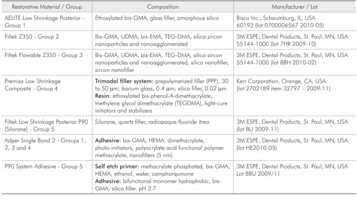
The materials used in this study, their composi-tion and the different groups are depicted in Table 1.
The cavities were bulk illed as close as possible to the margins, then light-cured for 40 s. Next, the restorations were lightly ground, wet, using 320 and 600-grit Si-C paper to remove composite overhangs.
Extent of Dye Penetration: Each specimen was immersed in a 0.5% solution of basic fuchsine for one second, removed immediately, and rinsed with running water. The excess dye on the surface of the composite and the enamel was removed using a cot-ton pallet and alcohol.
Table 1 - Overview of the different materials used in the five experimental groups.
Restorative Material / Group Composition Manufacturer / Lot
AELITE Low Shrinkage Posterior - Group 1
Ethoxylated bis-GMA, glass filler, amorphous silica Bisco Inc., Schaumburg, IL, USA 60193 (lot 0700006567 2010-05)
Filtek Z350 - Group 2 Bis-GMA, UDMA, bis-EMA, TEG-DMA, silica-zircon nanoparticles and nanoagglomerated
3M ESPE, Dental Products, St. Paul, MN, USA 55144-1000 (lot 7HR 2009-10)
Filtek Flowable Z350 - Group 3 Bis-GMA, UDMA, bis-EMA, TEG-DMA, silica-zircon nanoparticles and nanoagglomerated, silica nanofiller, zircon nanofiller
3M ESPE, Dental Products, St. Paul, MN, USA 55144-1000 (lot 8BH 2010-02)
Premisa Low Shrinkage Composite - Group 4
Trimodal filler system: prepolymerized filler (PPF), 30
to 50 µm; barium glass, 0.4 µm; silica filler, 0.02 µm
Resin: ethoxylated bis-phenol-A-dimethacrylate,
triethylene glycol dimethacrylate (TEGDMA), light-cure initiators and stabilizers
Kerr Corporation, Orange, CA, USA (lot 2702189 item 32797 – 2009-11)
Filtek Low Shrinkage Posterior P90 (Silorane) - Group 5
Silorane, quartz filler, radiopaque fluoride itreo 3M ESPE, Dental Products, St. Paul, MN, USA (lot BU 2009-11)
Adper Single Bond 2 - Groups 1, 2, 3 and 4
Adhesive: bis-GMA, HEMA, dimethacrylate,
photo-initiators, polyacrylate acid functional polymer methacrylate, nanofillers (5 nm)
3M ESPE, Dental Products, St. Paul, MN, USA (lot HE2010-05)
P90 System Adhesive - Group 5 Self etch primer: methacrylate phosphated, bis-GMA, HEMA, ethanol, water, camphorquinone
Adhesive: bifunctional monomer hydrophobic,
bis-GMA, silica filler. pH 2.7
3M ESPE, Dental Products, St. Paul, MN, USA Lot 8BU 2009/11
Hence, it was possible to calculate the extent of dye penetration as a percentage of total perimeter.
Depth of Dye Penetration: The restorations were transversally sectioned using a # 7020 dia-mond disc (KG Sorensen - São Paulo, SP, Brazil) under constant water cooling. The direction of each sectioning was guided by the areas of more evident
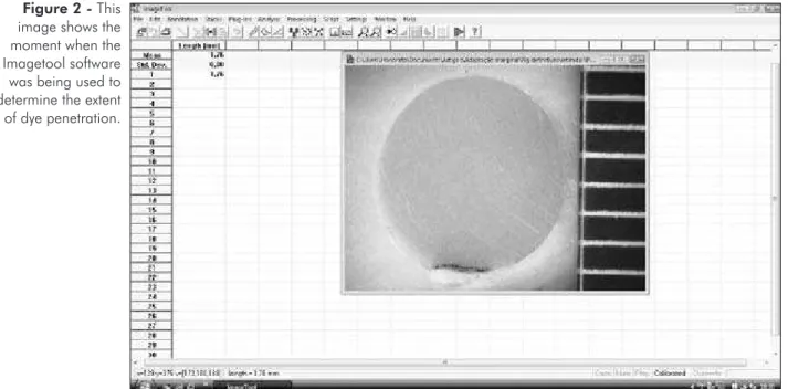
dye staining. The sectioned samples were positioned in the stereomicroscope to allow capture and trans-fer of the images to a computer (Figure 3). Depth of dye penetration was measured in millimeters, using the same software. Only the highest value of each sectioned specimen was considered for statis-tical analysis. The specimens which did not exhibit marginal staining were not sectioned, and the value Figure 2 - This
image shows the moment when the Imagetool software was being used to determine the extent of dye penetration.
0 (zero) was assigned. The Kruskal-Wallis analysis was applied to the results (P > 0.05).
Results
Marginal Adaptation - Extent of Dye Pene-tration: Table 2 summarizes the results according to the extent of dye penetration along the margins.
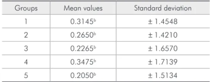
Marginal Adaptation - Depth of Dye Pene-tration: Table 3 depicts in millimeters the mean depth of dye penetration. All groups were statisti-cally similar (p = 0.8364).
Discussion
Bovine teeth have been used due to ethical con-cerns related to the use of human teeth. The advan-tages of availability, similarity in age, low risk of infection to the researcher and ease of preparation are aspects that may be considered as well. The his-tological aspects of human and bovine permanent teeth seem to be similar and no differences in bond strength values have been found.16
The marginal sealing breakdown of adhesive restorations may possibly occur as a consequence of long-term thermal and mechanical stresses; or im-mediately, during the restorative procedure itself, due to polymerization shrinkage stress. Hence, the marginal gaps in a clinically placed composite res-toration occupy between 14% and 54% of the total interface, depending on materials and techniques used.17 Several clinical methods have been suggested
to reduce polymerization stress, such as incremental layering technique,13,18,19 modulated curing
meth-ods,20,1 use of a lowable resin layer and illed
ad-hesives.13,21 Recently, some composite resin
materi-als classiied as low-shrinking composites have been commercialized; however, the material itself has not proved to be totally effective in preventing the harmful consequences of the shrinkage stresses gen-erated at the dentin-composite interface.13
Polymerization stress is a concern due to its consequences, and cavity coniguration has been strongly related to it. For instance, the study con-ducted by Loguercio et al.,22 using linear
polym-erization shrinkage measurements inside the cavi-ties, has shown that the higher the ratio of bonded to unbonded surfaces, the greater the shrinkage in the top-to-bottom direction. Thus, the restriction caused by bonding to the cavity walls would prevent the composite resin from reducing its length on the cavosurface margins during the polymerization pro-cess.
The shrinkage values presented by different types of composite resins are another point to be consid-ered. When the extent of dye penetration along the margins was determined, the Kruskal-Wallis test showed no signiicant differences (p = 0.54) among the groups. The bulk illing technique used in this study may have generated stresses on the cavity walls above the adhesive strength during polymer-ization, regardless of the type of composite used, and thus led to gap formation for all groups. From the statistical viewpoint, it could be assumed that all composite resins used presented similar shrink-age, and/or the bonding ability of the adhesive sys-tems used was not suficient to prevent the break-down of marginal sealing. However, another aspect that must be highlighted is that the difference in the stiffness of the uncured resin may impair the composite-dentin adaptation and, thus, the dimmer Table 2 - Mean values for dye penetration along the cavity
margins in millimeters. Means with the same letter are not statistically different (p = 0.54).
Groups Mean values Standard deviation
1 2.18a ± 2.29
2 3.31a ± 4.50
3 3.9a ± 3.91
4 2.61a ± 3.61
5 1.96a ± 2.84
Table 3 - Mean values for depth of dye penetration in bo-vine incisors, in millimeters. Means with the same letter are not statistically different (p = 0.8364).
Groups Mean values Standard deviation
1 0.3145b ± 1.4548
2 0.2650b ± 1.4210
3 0.2265b ± 1.6570
4 0.3475b ± 1.7139
effect caused by the thickness and transparency of the composites used.13 It is important to emphasize,
though, that the Silorane (P-90) composite was used with a self-etching adhesive system and, for the oth-er four groups, the adhesive used was the same, the etch-and-rinse Adper Single Bond. It is a common understanding that conventional etch-and-rinse adhesive systems produce higher bond strength on enamel than do self-etch ones.8,9 The self-etch
ad-hesive used in conjunction with the Silorane com-posite can be considered ultra-mild, pH 2.7;13 and,
the enamel acid-etching prior to the application of the self-etching adhesive, as recommended in some studies,8,9 was not performed. Thus, even if the
Si-lorane composite resin could reduce the shrinkage stress on cavity walls, the bond strength achieved by the self-etching system, especially on enamel mar-gins, would impair the observation of the effect of a low shrinkage restorative material.
A recent investigation,23 using micro-Raman
spectroscopy and SEM, studied the hybrid layer formed by the Silorane adhesive. A hybrid layer, comparable in thickness to two other self-etch sys-tems (G-Bond and Adhese One), was observed. To ensure adhesion to hydrated dentin, Silorane Primer contains hydrophilic monomers, whereas the Bond has hydrophobic bi-functional monomers to match the Silorane hydrophobic composite resin. Although the Bond is placed on the cured Silorane Primer sur-face prior to being cured itself, Raman spectra indi-cated an intervening zone of about 1 µm of mixed spectral intensities associated with both Silorane Primer and Bond. According to the authors, this may be due to the oxygen inhibition layer remaining on the cured Silorane Primer surface. They conclud-ed, from this particular observation, that further investigation into the bond strength of the Silorane adhesive system is necessary to assess whether this intervening zone may act as a weak link in the bonding process.
One important feature was the large standard deviation in the analysis of the extent of dye pen-etration (Table 2). It is essential to remember that each group had 20 specimens, a sample that can be considered elevated. These large standard deviation
values would be also responsible for the absence of signiicant differences. It is important to mention, however, that in addition to the interpretation of the data obtained from this applied methodology, the discussion of the related literature also shows that several factors are involved in gap formation, and that they are not clearly understood.3,10,13,19,20
Therefore, establishing a precise methodology, both to individualize the mechanisms that are responsible for gap formation and to counteract them, is still a challenge.
When the dye penetration was analyzed in depth, no signiicant differences were found as well (p = 0.8364). Nevertheless, these results need to be discussed more profoundly. Although not statistical-ly different, the mean (0.2265 mm) of the dye pene-tration for the low viscosity composite group (Filtek Flowable Z-350) was close to those observed for the other experimental groups, some of which employed low-shrinking composites. According to Hooke’s law, stress is determined by the volumetric shrink-age and the E-modulus of the material. Although low-viscosity composites generally present higher shrinkage values than high-density composites, the low E-modulus (20-25% lower) may compete with the stress development that would help to maintain the marginal seal.24,25
The polymerization rate of composites, especial-ly in deep areas, is of great importance. The depth of the cavities in this study was 2.5 mm, thus on the limit of light energy, necessary to achieve an ad-equate level of polymerization.
The incremental illing technique can be consid-ered advantageous, since the C-factor of the individ-ual layer drops and the amount of energy available at the interface increases.26 The choice of a large and
Conclusion
According to the methodology of this in vitro study, the results achieved and the statistical analy-sis performed, the tested hypotheanaly-sis must be rejected since the LSC/adhesive restorations could not pro-duce better marginal adaptation, either in extent or in depth, than the other restorative systems used in this investigation.
Acknowledgements
This study was partly supported by CNPq (Con-selho Nacional de Desenvolvimento Cientíico e Tecnológico do Brasil).
References
1. Zanchi CH, Carvalho RV, Rodrigues Júnior AS, Demarco FF, Brunetti Júnior LH. Shrinkage stress of three composites under different polymerization methods. Braz Oral Res. 2006 Apr-Jun;20(2):137-42.
2. Abbas G, Fleming GL, Harrington E, Shortall AC, Burke FJ. Cuspal movement and microleakage in premolar teeth restored with a packable composite cured in bulk or in increments. J Dent. 2003 Aug;31(6):437-44.
3. Lee M, Cho B, Son H, Um C, Lee I. Influence of cavity dimen-sion and restoration methods on cusp deflection of premolars in composite restoration. Dent Mater. 2007 Mar;23(3):288-95.
4. Pearson GL, Hegarty SM. Cusp movement of molar teeth with composite filling material in conventional and modified MOD cavities. Br Dent J. 1989 Mar;166(5):162-65.
5. Tantbirojn D, Versluis A, Pintado MR, DeLong R, Douglas W. Tooth deformation patterns in molars after composite restoration. Dent Mater. 2004 Jul;20(6):535-42.
6. Versluis A, Tantbirojn D. Theoretical considerations of con-traction stress. Compend Contin Educ Dent Suppl. 1999 Nov; (25):S24-32.quiz S73.
7. Parpaiola AR, Guimarães PS, França FMG, Basting RT. Small cross-sectional survey of composite restoration attributes asso-ciated with choices for replacement. Braz Oral Res. 2009 July-Sep;23(3):346-51.
8. De Munk J, Van Landuyt K, Peumans M, Poitevin A, Lam-brechts P, Braem M, et al. A critical review of the durability adhesion to tooth tissue: methods and results. J Dent Res. 2005 Feb;84(2):118-32
9. Van Meerbeek B, De Munck J, Yoshida Y, Inoue S, Vargas M, Vijay P, et al. Adhesion to enamel and dentin: current status and future challenges. Buonocore Memorial Lecture. Oper Dent. 2003 May-Jun;28(3):215-35.
10. Cadenaro M, Biasotto M, Scuor N, Breschi L, Davidson CL, Lenarda RD. Assessment of polymerization contraction stress of three composite resins. Dent Mater. 2008 May;24(5):681-5. 11. Raws HR, Upshaw JE. Resinas Restauradoras. In: Anusavice
KJ. Phillips Materiais Dentários. 11th ed. Saunders: Chicago; 2005. p. 375-418.
12. Carvalho RM, Perreira JC, Yoshiama M, Pashley DH. A re-view of polimerization contraction: the influence of stress development versus stress relief. Oper Dent. 1996 Jan-Feb;21(1):17-24.
13. Van Ende A, De Munk J, Mine A, Lambrechts, Van meerbeek, B. Does low-shrinking composite induce less stress at the ad-hesive interface? Dent Mater. 2010 Mar;26(3):215-22. 14. Yalçin F, Korkmaz Y, Baseren M. The effect of two
differ-ent polishing techniques on microleakage of new composites in class V restorations. J Contemp Dent Pract. 2006 Nov 1;7(5):18-25.
15. Andrade OS, Goes MF, Montes M. Marginal adaptation and microtensile bond strength of composite indirect restorations bonded to dentin treated with adhesive and low-viscosity com-posite. Dent Mater. 2007 Mar;23(3):279-87.
16. Nakamichi I, Iwaku M, Fusayama T. Bovine teeth as pos-sible substitute in the adhesion test. J Dent Res. 1983 Oct;62(10):1076-81.
17. Hannig M, Friedrichs C. Comparative in vivo and in vitro
investigation of interfacial bond variability. Oper Dent. 2001Jan-Feb;26(1):3–11.
18. He Zhengdi, Shimada Y, Tagami, J. The effects of cavity size, and incremental technique on micro-tensile bond strength of resin composite in class I cavities. Dent Mater. 2008 May;23(5):533-38.
19. Park J, Chang J, Ferracane JL, Lee IB. How should composite be layered to reduce shrinkage stress: Incremental or bulk filling? Dent Mater. 2008 Nov;24(11):1501-5.
20. Cunha JG, Alonso RCB, Pfeifer CSC, Correr-Sobrinho L, Ferracane JL, Sinhoreti MAC. Contraction stress and physical properties development of a resin-based composite irradiated using modulated curing methods at two C-factor levels. Dent Mater. 2008 Mar;24(3):392-8.
22. Loguercio AD, Reis, A, Ballester RY. Polymerization shrink-age: effects of constraint and filling technique in composite restorations. Dent Mater. 2004 Mar;20(3):236-43.
23. Santini A, Miletic V. Comparison of the hybrid layer formed by Silorane adhesive, one-step self-etch and etch and rinse systems using confocal micro Raman spectroscopy and SEM. J Dent. 2008 Sep;36(9):683-91.
24. Baroudi K, Silikas N, Watts DC. Time-dependent visco-elastic creep and recovery of flowable composites. Eur J Oral Sci 2007;115(6):517–21.
25. Braga RR, Hilton TJ, Ferracane JL. Contraction stress of flow-able composite materials and their efficacy as stress-relieving layers. J Am Dent Asssoc. 2003;134:721–8.