Evaluation of the Digisonic
®SP cochlear implant: patient outcomes
and fixation system with titanium screws
Abstract
Guilherme Machado de Carvalho1, Alexandre Caixeta Guimarães2, Fabiana Danieli3,
Lúcia Cristina Beltrame Onuki4, Jorge Rizzato Paschoal5, Walter Adriano Bianchini6, Arthur Menino Castilho7
1 MSc in Medicine, MD, ENT (Fellow in Otology. UNICAMP). 2 MD. (ENT Medical Resident. UNICAMP). 3 MSc in Bioengineering. (Specialist in Cochlear Implants). 4 Speech and Hearing Therapist. (Specialist in Cochlear Implants. UNICAMP).
5 MD, PhD, ENT. (Professor of Otorhinolaryngology. Head of the Otology Service. UNICAMP).
6 MSc in Medicine. MD, ENT. (Head and Coordinator of the Otology, Audiology, Cochlear Implant, and Implantable Hearing Aid Service. UNICAMP). 7 MD, PhD, ENT. (Head and Coordinator of the Otology, Audiology, Cochlear Implant, and Implantable Hearing Aid Service. UNICAMP).
Otology, Audiology and Implantable Ear Prostheses Ear, Nose, Throat and Head & Neck Surgery Department. E-mail: [email protected]
Send correspondence to: Dr. Guilherme Machado de Carvalho. Disciplina de Otorrinolaringologia - UNICAMP. Faculdade de Ciências Médicas. Universidade de Campinas - UNICAMP. Campinas - SP. Brasil. CEP: 13083-970. Caixa Postal: 6111.
Tel: +55 (19) 3521-7523. Fax: +55 (19) 3521-7563. E-mail: [email protected]
Paper submited to the BJORL-SGP (Publishing Management System – Brazilian Journal of Otorhinolaryngology) on August 6, 2012; and accepted on October 2, 2012. cod. 9938.
C
ochlear implants have revolutionized the way patients affected by severe hearing loss experience the world. Neurelec developed a fixation system with two titanium screws that requires no skull bone drilling.Objective: To describe the outcomes and procedure-related details of a series of patients implanted with the Digisonic® SP cochlear implant.
Method: This retrospective study analyzed patients submitted to cochlear implant placement within a period of 18 months. All patients had postlingual hearing impairment. Data was collected from patient charts and standard questionnaires answered by the surgeons in charge of carrying out the procedures.
Results: The six patients offered the Digisonic® SP cochlear implants were operated by experienced
surgeons. The procedures took 95 to 203 minutes (mean = 135’) to be completed, which is less time than what has been described for other fixation approaches. No complications were recorded and hearing improvement was satisfactory.
Conclusion: The Digisonic® SP cochlear implant developed by Neurelec offered good audiological
results for adult patients, shorter surgery time, and no surgical or postoperative complications.
ORIGINAL ARTICLE
Braz J Otorhinolaryngol. 2012;78(6):56-62.
BJORL
Keywords:
cochlear implantation, deafness,
electrodes, implanted, hearing loss
sensorineural, prostheses and implants.
.org
INTRODUCTION
Cochlear implants have revolutionized the way patients affected by severe hearing loss experience the world. Good outcomes with improved speech perception in patients of all age ranges have been observed1-3. Cochlear implants improve patient
quality-of-life even more significantly in subjects aged 65 years and older4.
The implantation procedure is safe and reliable, but complications occur in about 16% of the patients. The most frequent adverse event relates to the insertion of electrodes into the cochlea, seen in about 4% of the cases5.
Migration of internal components has been described by many authors and may lead to cochlear implant malfunction and local infection6-8. Various
implantation procedures have been described in the literature, with less invasive approaches gaining significant attention9-11.
The production of the bed on which the internal component is positioned is an important and onerous stage in the procedure, and has been the target of significant investment by cochlear implant makers12,13.
More is thus spent in the development of surgical instruments and new materials such as titanium plates, propylene mesh, GORE-TEX, absorbable materials, and others14-16.
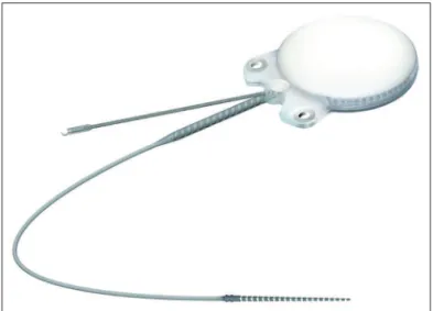
In regards to internal component fixation, Neurelec Inc. (Sophia-Antipolis, France) developed a fixation system with two titanium screws that requires no skull bone drilling17 (Figure 1).
Figure 1. Internal component with receptor, stimulator, and magnet, in a small sealed ceramic framework on a titanium base plate. Lateral
niche has two ixation screws - Digisonic® SP developed by Neurelec.
Therefore, this study aimed to describe the outcomes and procedure-related details of a series of patients implanted with the Digisonic® SP cochlear
implant and discuss the postoperative clinical and audiological findings observed in the last 18 months.
METHOD
This retrospective study was carried out in a specialized tertiary care hospital. Patients implanted with Digisonic® SP cochlear devices were assessed for
a period of 18 months.
A digital data collection protocol was designed. The following parameters were analyzed: age, gender, hearing loss etiology, time with hypacusis, side of implantation, pre and postoperative audiometric data (audiometry and speech perception testing), length of surgery, time to fixate the internal component, surgery complications, follow-up time in months. All patients in this series had post-lingual hearing loss (hearing was lost after they had developed speech and language skills).
Data was collected from patient charts, and a standard questionnaire answered by the surgeons who carried out the procedures.
The identity of the patients was not disclosed, as required by the ethical principles of the institution in which the study was conducted.
The patients
The patients selected for the study had Digisonic® SP cochlear devices implanted within
a period of 18 months. All subjects followed a preoperative protocol in which the etiology of the impairment was investigated through lab workup, genetic tests, CT and MRI scans of the ears and mastoids, psychological evaluation, and thorough speech and hearing assessment.
The device
The Digisonic® SP cochlear implant system
- made up by cochlear implant Digisonic® SP and
and a fixation system for the receiver-stimulator that uses two titanium screws and raises the stimulation rate through the “Mean Peak Interleaved Sampling” (MPIS) sound processing strategy.
Device internal component
Internal component Digisonic® SP developed
by Neurelec is shown in Figure 1.
The receiver-stimulator (RS) is made up of a convex ceramic capsule, a titanium base plate, both coated with biocompatible silicone. Digisonic®
SP is extremely compact, and both its electronic components used in signal decoding and its internal magnet are comprehended by one single structure with a diameter of 30 mm called the monoblock.
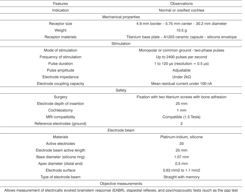
Table 1 describes the characteristics of Digiso-nic® SP.
Table 1. Digisonic® SP technical features.
Features Observations
Indication Normal or ossiied cochlea
Mechanical properties
Receptor size 4.9 mm border – 5.75 mm center – 30.2 mm diameter
Weight 10.5 g
Receptor materials Titanium base plate – A1203 ceramic capsule – silicone envelope
Stimulation
Mode of stimulation Monopolar or common ground - two-phase pulses
Frequency of stimulation Up to 2400 pulses per second
Pulse duration 1 to 120 μs (resolution = 0.5 μs)
Pulse amplitude Adjustable
Electrode impedance Under 2kΩ
Electrode coupling capacity Mean residual current under 100 nA
Safety
Surgery Fixation with two titanium screws with bone adhesion
Electrode depth of insertion 25 mm
Cochleostomy 1 mm
MRI compatibility Compatible (1.5 Tesla)
Reference electrodes (ground) 2
Electrode beam
Materials Platinum-iridium, silicone
Active electrodes 20
Electrode beam active length 25 mm
Base diameter (silicone ring) 1.07 mm
Apex diameter (distal end) 0.5 mm
Electrode surface 0.63 mm2 to 1.1 mm2
Type of electrode beam Straight with memory
Objective measurements
Allows measurement of electrically evoked brainstem response (EABR), stapedial relexes, and psychoacoustic tests (such as the gap test
etc). USB Digistim® SP diagnostic system (for Windows 98SE, ME, 2000, XP and VISTA®)
The device’s fixation is done by two 3.4 mm titanium screws bolted into two small silicone-coated titanium orifices with 5 mm in diameter positioned in the ends of the receiver-stimulator, as shown in Figures 2 and 3. The screws are driven 1.91 mm into bone tissue17.
The compact structure and the fixation screws of the Digisonic® SP allow for quicker, less invasive
surgery without the need to drill or suture bone tissue to position or fixate the implant17.
Digisonic® SP has atraumatic flexible screws
Figure 2. Digisonic® SP ixation system.
Figure 3. Digisonic® SP ixation system with two titanium screws driven
1.91 mm into the temporal bone. There is no need to drill or prepare the site. Developed by Neurelec S.A.
channel enabled by processing strategy MPIS. It is also equipped with silicone rings to facilitate insertion18.
The internal device contains a two-way telemetry system to record electrode impedance. Electrode impedance is recorded through diagnostic interface Digistim SP and software program Digistim for Windows SP® version 1.9.15, in which it is also
possible to perform other objective measurements such as electrically evoked auditory brainstem responses (EABR) and electrically evoked stapedius reflex thresholds (ESRT)18.
EABR measurement is carried out routinely during surgery to verify device function and the stimulation of peripheral auditory nerve fibers19. In
addition to that, EABR can also be used to predict the psychophysical levels to program the speech processor20, a particularly important step in the
treatment of pediatric patients. EABR is measured at the end of surgery with the patient still under general anesthesia.
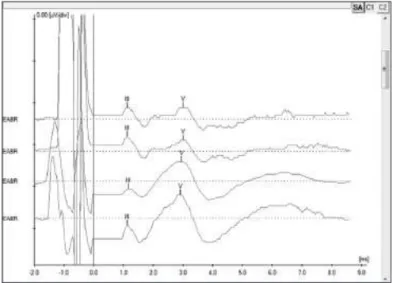
The procedure comprehends the electrical stimulation of nerve fibers through the electrodes inserted in the cochlea and the acquisition of responses through a conventional BAEP measurement device and a synchronization cord. It is possible to view waves II, III, and V, the last two being the most commonly observed; wave V is indicative of effective cochlear
Figure 4. Electrically evoked brainstem responses measured during surgery for subject 5 electrodes 19, 14, 9 and 6 using Interface Navi-gator Pro and Software AEP version 7.0.0, Biologic. Waves III and V
can be seen, showing proper nerve iber stimulation.
nerve stimulation. The absolute latencies of these waves are reduced when compared to conventional BAEP, given that the stimulation on EABR is performed directly in the cochlear nerve through the electrodes in the implant beam20 (Figure 4).
Patients using the Digisonic® SP implant can
undergo magnetic resonance imaging in scanners of up to 1.5 Tesla, if recommendations are followed18.
RESULTS
Table 1 shows the characteristics and technical details of the device used in the described procedures.
Tables 2 and 3 show general and specific data of the patients implanted with Digisonic® SP.
Table 4 shows the data on surgery-related occurrences and length of the procedure.
MRI and CT scans did not reveal radiological alterations.
EABR and ERST measurements were performed in all procedures at the end of surgery with the patients still under general anesthesia. Measurement outcomes were satisfactory and showed the device was functioning properly.
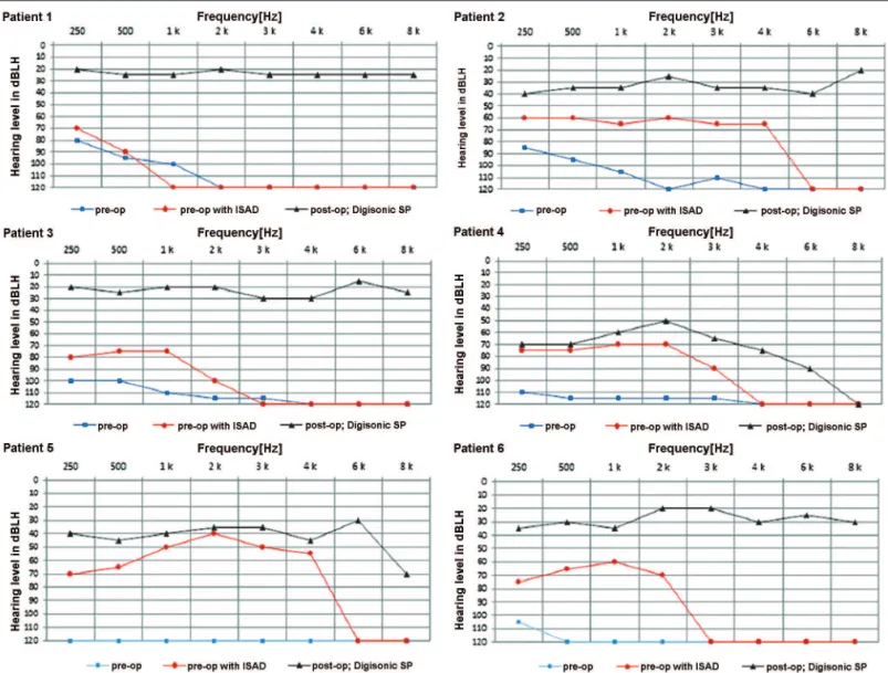
The graphs below show the pre and postoperative audiometric test results (Figure 5).
DISCUSSION
The Digisonic® SP cochlear implant developed
Table 2. General implant patient data.
Subject Gender Age* Time with dysacusis* Dysacusis etiology Date of CI Date of activation Side of CI
1 M 50 25** Infection 12/04/11 31/05/11 Left
2 F 33 21 Idiopathic (Progressive) 23/10/10 01/12/10 Right
3 F 30 10 Ototoxicidade 28/09/11 25/10/11 Left
4 F 52 15 Idiopathic 30/08/11 25/10/11 Left
5 M 20 9 Idiopathic (Progressive) 20/03/12 26/14/12 Right
6 F 26 20 Rubeola 14/12/11 02/02/12 Left
* years; ** right ear dysacusis at one year of age; hearing on left ear worsened at age 25 after meningitis; stroke at age 49 obliterated remaining
left ear hearing; M: male; F: female; CI: cochlear implantation; dates on DD/MM/YY format.
Table 3. Speciic implant patient data.
Subject Electrode insertion site Electrode type Active channels External processor Follow Up*
1 Cochleostomy Digisonic® SP All Digi SP® 15
2 Cochleostomy Digisonic® SP All Digi SP® 33
3 Cochleostomy Digisonic® SP All Digi SP® 10
4 Cochleostomy Digisonic® SP All Digi SP® 11
5 Cochleostomy Digisonic® SP All Digi SP® 4
6 Cochleostomy Digisonic® SP All Digi SP® 8
* months.
Table 4. Surgery implant patient data.
Subject Total length* Fixation time* Time saved*, ** Complications (intra and postoperative)***
1 158 3.58 30 None
2 203 5.60 30 None
3 144 4.12 30 None
4 100 4.68 30 None
5 109 3.44 30 None
6 95 6.01 30 None
* minutes; ** time to ixate internal component as reported by surgeon. The mean time to the completion of this portion of the procedure (pro
-duction of internal component niche) was calculated by the analysis of 10 random cases done within the same time period using different ixation modes (temporal bone drilling to make the niche); *** ixation errors, bleeding, injured noble structures, infection, dehiscence, internal component
migration, electrode migration, need to remove the implant, cholesteatoma, otitis media.
niche to place the cochlear implant internal component or any drilling on the patient’s skull bone. In addition to reducing the risks and complications associated with the production of the cochlear implant niche, this fixation system reduces the length of surgery.
In our study, six patients were implanted with Digisonic® SP by experienced surgeons, and surgery
length ranged from 95 and 203 minutes, with a mean length of 135 minutes. The mean length of a conventional cochlear device implantation procedure is 255 minutes in the hands of experienced surgeons using the S-shaped retroauricular incision to make the implant niche, and 200 minutes when using a small retroauricular incision and a subperiosteal pouch21.
Therefore, length of surgery in our series was shorter than that of procedures using other modes of fixation.
Studies looking into the cost of cochlear implants use a wide array of methods, and cost estimates may vary significantly depending on what is considered as part of the cost22. However, according
to the literature, the cost of a unilateral implant in a patient with post-lingual hearing loss ranges between
€ 30,026 (USD 21,018) and € 45,770 (USD 32,039), with
the device accounting for most of the cost23. In Brazil,
the cost of a Digisonic® SP is similar to the cost of a
conventional fixation device.
Figure 5. Pre and postoperative audiometric thresholds of six cochlear device implantation patients using Digisonic® SP. Note threshold impro-vement.
The patients in our series had no complications, and the level of auditory gain was satisfactory in all cases. In general terms, mean complication rates of 16% have been described in cochlear device implantation procedures5. Despite the small size of
our sample, our results were better than average.
CONCLUSION
The Digisonic® SP cochlear implant developed
by Neurelec presented good audiological outcomes in our series, shorter length of surgery, and no intra or postoperative complications.
REFERENCES
1. Yang WS, Moon IS, Kim HN, Lee WS, Lee SE, Choi JY. Delayed cochlear implantation in adults with prelingual severe-to-profound hearing loss. Otol Neurotol. 2011;32(2):223-8.
2. Budenz CL, Cosetti MK, Coelho DH, Birenbaum B, Babb J, Waltzman SB, et al. The effects of cochlear implantation on speech perception in older adults. J Am Geriatr Soc. 2011;59(3):446-53.
3. Klop WM, Briaire JJ, Stiggelbout AM, Frijns JH. Cochlear implant outcomes and quality of life in adults with prelingual deafness. Laryngoscope. 2007;117(11):1982-7.
4. Chung J, Chueng K, Shipp D, Friesen L, Chen JM, Nedzelski JM, et al. Unilateral multi-channel cochlear implantation results in significant improvement in quality of life. Otol Neurotol. 2012;33(4):566-71. 5. Brito R, Monteiro TA, Leal AF, Tsuji RK, Pinna MH, Bento RF.
Sur-gical complications in 550 consecutive cochlear implantation. Braz J Otorhinolaryngol. 2012;78(3):80-5.
6. Yun JM, Colburn MW, Antonelli PJ. Cochlear implant magnet dis-placement with minor head trauma. Otolaryngol Head Neck Surg. 2005;133(2):275-7.
7. Hoffman RA, Cohen NL. Complications of cochlear implant surgery. Ann Otol Rhinol Laryngol Suppl. 1995;166:420-2.
8. Migirov L, Muchnik C, Kaplan-Neeman R, Kronenberg J. Surgical and medical complications in paediatric cochlear im- plantation: a review of 300 cases. Cochlear Implants Int. 2006;7(4):194-201.
10. Loh C, Jiang D, Dezso A, Fitzgerald O’Connor A. Non-sutured fixa-tion of cochlear implants using a minimally-invasive approach. Clin Otolaryngol. 2008;33(3):259-61.
11. Carvalho GM, Valente JPP, Duarte ASM, Muranaka EB, Guimarães AC, Soki MN, et al. Electro acoustic stimulation of the auditory system: Unicamp’s surgical approach. Braz J Otorhinolaryngol. 2012;78(1):43-50. 12. James AL, Papsin BC. Device fixation and small incision access
for pediatric cochlear implants. Int J Pediatr Otorhinolaryngol. 2004;68(8):1017-22.
13. Dalchow CV, Werner JA. A new instrument for minimal access surgery in cochlear implantation. Otol Neurotol. 2005;26(4):678-9.
14. Lee DJ, Driver M. Cochlear implant fixation using titanium screws. Laryngoscope. 2005;115(5):910-1.
15. Davis BM, Labadie RF, McMenomey SO, Haynes DS. Cochlear implant fixation using polypropylene mesh and titanium screws. Laryngos-cope. 2004;114(12):2116-8.
16. Otto RA, Lane AP, Carrasco VN. A new technique for securing cochlear implants. Otolaryngol Head Neck Surg. 1999;120(6):897-8.
17. Guevara N, Sterkers O, Bébéar JP, Meller R, Magnan J, Mosnier I, et al. Multicenter evaluation of the Digisonic SP cochlear implant fixation system with titanium screws in 156 patients. Ann Otol Rhinol Laryngol. 2010;119(8):501-5.
18. Vincent C, Ruzza I, Vaneecloo FM, Dubrulle F. Magnetic resonance imaging with the Digisonic SP Neurelec cochlear implant. Eur Arch Otorhinolaryngol. 2008;265(9):1043-6.
19. McCormick B, Archbold S (eds). Cochlear implants for young children: the Nottingham approach to assessment and rehabilitation. 2nd ed. London: Whurr; 2003. p.162-215.
20. Truy E, Gallego S, Chanal JM, Collet L, Morgon A. Correlation between electrical auditory brainstem response and perceptual thresholds in Di-gisonic cochlear implant users. Laryngoscope. 1998;108(4 Pt 1):554-9. 21. Prager JD, Neidich MJ, Perkins JN, Meinzen-Derr J, Greinwald JH Jr.
Minimal access and standard cochlear implantation: A comparative study. Int J Pediatr Otorhinolaryngol. 2012;76(8):1102-6.
22. Nadège C, Valérie G, Laura F, Hélène DB, Vanina B, Olivier D, et al. The cost of cochlear implantation: a review of methodological considerations. Int J Otolaryngol. 2011;2011:210838.
23. Turchetti G, Bellelli S, Palla I, Berrettini S. Systematic review of the scientific literature on the economic evaluation of cochlear implants in adult patients. Acta Otorhinolaryngol Ital. 2011;31(5):319-27. 24. Turchetti G, Bellelli S, Palla I, Forli F. Systematic review of the
scien-tific literature on the economic evaluation of cochlear implants in paediatric patients. Acta Otorhinolaryngol Ital. 2011;31(5):311-8. 25. Peñaranda A, Mendieta JC, Perdomo JA, Aparicio ML, Marín LM,