Case 10940
A pitfall case of endometrial serous adenocarcinoma
Lara Delgado , Teresa Margarida Cunha 1 2
Genital (Female) Imaging Section:
2013, Jul. 10 Published:
78 year(s), female Patient:
Authors' Institution
(1) Centro Hospitalar de Lisboa Ocidental, Lisbon, Portugal
(2) Instituto Português de Oncologia de Lisboa Francisco Gentil, Lisbon, Portugal Email: [email protected]
Clinical History
A 78 year-old-woman presented with a 6 month history of metrorrhagia and pelvic pain. The past medical history was unknown. The patient was referred for a pelvic ultrasound examination and, subsequently, a pelvic magnetic resonance imaging (MRI) was performed.
Imaging Findings
A gynecological ultrasound (not showed) revealed two contiguous cystic lesions with 63 and 104 mm, with scattered internal echos. A solid component was observed as a vegetation on the the largest cyst. The uterus was not visualized.
signal intensity on T1WI, intermediate signal intensity on T2WI and presenting homogeneous enhancement after Gadolinium-DTPA administration on fat-suppressed T1WI (Fig. 3). Both ovaries were seen separate from the lesion without detectable abnormalities.
Discussion
Endometrial carcinoma is the fourth most common female cancer and the most common
malignancy of the female reproductive tract [1]. Most cases are diagnosed after menopause with highest incidence around the seventh decade of life [2].
About 80% of all endometrial carcinomas are adenocarcinomas of the endometriod type. Most of the endometriod carcinomas are well to moderately differentiated and arise on a background of endometrial hyperplasia [2].
Serous carcinoma is the most aggressive type of non-endometrioid endometrial carcinomas [2]. In postmenopausal patients atrophy related to decrease in endocervical glands results in decreased secretion of mucus and represent a frequent cause of cervical stenosis, which when severe results in hydrometra, pyometra or hematometra [3].
In this case report the patient presents with a 6 month history of metrorrhagia and pelvic pain, and is referred for ultrasound examination and subsequently for MRI.
In the presence of a pelvic dilated tubular fluid-filled (high protein or hemorrhagic content)
structure with folded configuration, incomplete septations and scattered solid vegetation, a fallopian tube or an endometrial malignant tumor should be considered as differential diagnose.
Tubal origin should be suspected in the presence of a dilated tubular adnexal structure with folded configuration separated from the ovaries, with relatively thin walls and thin longitudinally oriented folds along the interior of the tube [3]. The lesion was centrally located in the pelvis, just above the vaginal cuff between the bladder and the rectum. The inverted pear shape and the reduction in the diameter in the median zone (uterine isthmus) allow the identification of the uterus.
The MRI revealed distention of the endometrial cavity and cervical canal with hematrometra and scattered foci of solid endometrial and cervical canal vegetations, without invasion of the outer half of the myometrium.
MRI is essential for the preoperative staging of endometrial cancer because it can accurately depict the depth of myometrial invasion, which is the most important morphological prognostic factor and correlates with tumor grade, presence of lymph node metastases, and the overall patient survival [4].
Cervical and endometrial and biopsy were performed revealing a serous adenocarcinoma. Despite the aggressive behavior of the tumor, given the age of the patient three months later, total hysterectomy, bilateral pelvic adnexectomy, partial omentectomy and paraaortic and pelvic
lymphadenectomy were performed. Histological evaluation revealed a multifocal endometrial tumor with invasion of the inner half of the myometrium without lymph node involvement.
Differential Diagnosis List
Endometrial Carcinoma, Primary Fallopian Tube Carcinoma
Figures
Figure 1 Figure 1 Pelvic MR
T1-weighted image on axial plane shows a high signal intensity fluid filled structure with
folded configuration and with scaterred low signal vegetations (arrows).
© Ramalho M, Department of Radiology, Hospital Garcia de Horta, Lisboa, Portugal
Area of Interest: Genital / Reproductive system female;
Imaging Technique: MR;
Procedure: Staging;
Special Focus: Tissue characterisation;
Figure 2 Figure 2 Pelvic MR
T2-weighted image on sagittal plane shows a high signal intensity fluid filled structure with
folded configuration and a incomplete septation (arrow).
© Ramalho M, Departement of Radiology, Hospital Garcia de Horta, Lisboa, Portugal
Area of Interest: Genital / Reproductive system female;
Imaging Technique: MR;
Procedure: Staging;
T2-WI on sagittal plane shows above the vaginal cuff between the bladder and the rectum a
centrally located high signal intensity fluid filled structure with folded configuration with
scatterred intermediate signal intensity vegetations (arrow)
© Ramalho M, Departement of Radiology, Hospital Garcia de Horta, Lisboa, Portugal
Area of Interest: Genital / Reproductive system female;
Imaging Technique: MR;
Procedure: Staging;
Special Focus: Tissue characterisation;
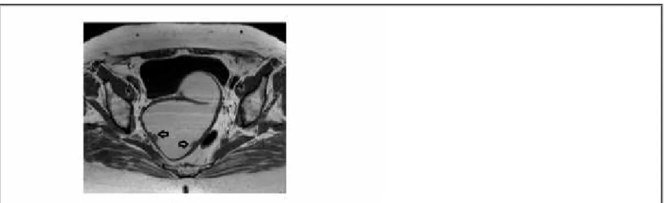
T2-weighted image on axial plane shows a high signal intensity fluid filled structure with
folded configuration and with scaterred intermediate signal vegetations.
© Ramalho M, Departement of Radiology, Hospital Garcia de Horta, Lisboa, Portugal
Area of Interest: Genital / Reproductive system female;
Imaging Technique: MR;
Imaging Technique: MR;
Procedure: Diagnostic procedure;
Special Focus: Neoplasia;
T2-weighted image on coronal plane shows a cystic lesion, with intermediate signal intensity
vegetations.
© Ramalho M, Departement of Radiology, Hospital Garcia de Horta, Lisboa, Portugal
Area of Interest: Genital / Reproductive system female;
Imaging Technique: MR;
Procedure: Diagnostic procedure;
Special Focus: Neoplasia;
Figure 3 Figure 3 Pelvic MR
Fat-suppressed T1WI after gadolinium administration on sagittal plane shows the high signal
intensity of the fluid filled structure showing either high protein or hemorrhagic contend and
the solid vegetations (arrow)
© Ramalho M, Departement of Radiology, Hospital Garcia de Horta, Lisboa, Portugal
Area of Interest: Genital / Reproductive system female;
Imaging Technique: MR;
Procedure: Staging;
Fat-suppressed T1WI after gadolinium administration on axial plane shows the enhancement
of the solid vegetations.
© Ramalho M, Departement of Radiology, Hospital Garcia de Horta, Lisboa, Portugal
Area of Interest: Genital / Reproductive system female;
Imaging Technique: MR;
Procedure: Diagnostic procedure;
Special Focus: Neoplasia;
MeSH
[A05.360.319.679.490] Endometrium
ampulla, an infundibulum, and fimbriae. Its wall consists of three histologic layers: serous, muscular, and an internal mucosal layer lined with both ciliated and secretory cells.
[C13.371.270.875.750] Endometrial Neoplasms
Tumors or cancer of ENDOMETRIUM, the mucous lining of the UTERUS. These neoplasms can be benign or malignant. Their classification and grading are based on the various cell types and the percent of undifferentiated cells.
References
[1] Sala E, Walkely S, Senior E, Lomas D (2007) MRI of Malignant Neoplasms of the Uterine Corpus and Cervix AJR Jun;188(6):1577-8
[2] Hamm B, Forstner R, Baert A, Knauth M, Sartor K (2007) MR and CT of the Female Pelvis Springer-Verlag Berlin Heidelberg (101-102)
[3] Hricak H, Akin O, Sala E, Ascher S, Levine Deborah, Reinhold C (2007) Diagnostic imaging Gynecology (3-2;8-6)
[4] Beddy P, O'Neil A, Yamamoto A, Addley H, Reinhold C, Sala E (2012) Figo Staging System for Endometrial Cancer: Added Benefits of MR Imaging Radiographics Jan-Feb;32(1):241-54
Citation
Lara Delgado , Teresa Margarida Cunha (2013, Jul. 10) 1 2