371
RADIOLOGY PAGE
Vol. 43 (2): 371-372, March - April, 2017doi: 10.1590/S1677-5538.IBJU.2015.0207
Carcinoma prostate masquerading as a hemorrhagic
pelvic cyst
_______________________________________________
Rajat Arora
1, Arun Jacob Philip George
1, Anu Eapen
2, Antony Devasia
11 Department of Urology, Christian Medical College, Vellore, Tamil Nadu, India; 2 Department of Urology,
Christian Medical College, Vellore, Tamil Nadu, India
_______________________________________________________________________________________
371
RADIOLOGY PAGE
CASE
A 53 year-old man, with untreated lower urinary tract symptoms for two years was catheterized for acute retention of urine. He had an enlarged, boggy non tender prostate.
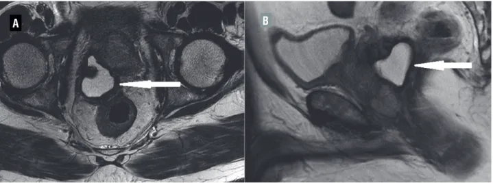
The transrectal ultrasound (TRUS) revealed a fluid filled cystic mass arising from the prostate and 200cc of hemorrhagic fluid was aspirated. There was no growth on culture of fluid and Xpert® MTB/RIF along with three acid fast bacillus smears was negative for tuberculosis. Cytology was not performed on the aspirated fluid. Magnetic resonance imaging revealed prostate volume of 52cc and a 94x77mm cystic and solid lesion (Figure-1) with seminal vesicle and rectal infiltration. There was abnormal signal intensity with T2-weighted hypointensity of the peripheral zone of the prostate. Close to the apex of the prostate, the cystic lesion merged with the right side of the base of the prostate. Above mentioned area near the apex showed restricted diffusion. His prostate specific antigen (PSA) was 910.0ng/mL. TRUS guided biopsy of the cyst wall revealed adenocarcinoma of prostate (Gleason grade: 4+4=8). The Technetium-99m methylene diphosphonate scan showed no evidence of osseous metastasis.
A B
IBJU| RADIOLOGY PAGE
372
ARTICLE INFOInt Braz J Urol. 2017; 43: 371-2
_____________________
Submitted for publication: April 11, 2015
_____________________
Accepted after revision: February 18, 2016
_____________________
Published as Ahead of Print: November 02, 2016
REFERENCES
1. Chang YH, Chuang CK, Ng KF, Liao SK. Coexistence of a hemorrhagic cyst and carcinoma in the prostate gland. Chang Gung Med J. 2005;28:264-7.
2. Chen C-H, Lin Y-H, Tzai T-S, Tsai Y-S. Prostate Cancer Associated with Hemorrhagic Cyst: Findings on Transrectal Doppler Sonography. J Med Ultrasound 2008;16:292–5.
_______________________ Correspondence address: Rajat Arora, MS Department of Urology Christian Medical College Unit - 1, CMC Hospital, Vellore Tamil Nadu, 632004, India Fax: +91 416 226-2788 E-mail: rajat_zzz@cmcvellore.ac.in
3. Khorsandi M. Cystic prostatic carcinoma. J Urol. 2002;168:2542.
4. Curran S, Akin O, Agildere AM, Zhang J, Hricak H, Rademaker J. Endorectal MRI of prostatic and periprostatic cystic lesions and their mimics. AJR Am J Roentgenol. 2007;188:1373-9. 5. Lucey BC, Kuligowska E. Radiologic management of
cysts in the abdomen and pelvis. AJR Am J Roentgenol. 2006;186:562-73.
A B
Figure 2 - (A and B): Reduction in size of cyst (arrow) after androgen deprivation therapy.
Based on the locally advanced nature of the disease, he was initiated on neoadjuvant Leuprolide acetate 22.5mg subcutaneously (luteinizing hormone releasing hormone analog) and he voided successfully without a catheter on follow-up. At three months, his PSA was 21.9ng/mL with marked reduction in the size of the cystic lesion (50x40mm) (Figure-2). He received radiotherapy (Intensity Modulated Radiation Therapy technique with image guidance) after six months of androgen deprivation. A total dose of 79.2Gy was delivered in 44 fractions and his nadir PSA was 0.04ng/mL after 18 months of follow-up. Leuprolide is being continued for three years.

