389
Rev Bras Med Esporte – Vol. 17, No 6 – Nov/Dec, 2011
Original Article EXERCISE AND SPORTS
MEDICINE CLINIC
Evaluation of Exercise-Induced Bronchospasm
Assessed by Peak Flow Meter in Obese Adolescents
Luciana Oliveira e Silva1
Patrícia Leão da Silva1
Ana Maria Oliveira Caixeta Nogueira1
Morgana Borges Silva1
Gabriela Costa Pontes Luz1
Fernanda Veruska Narciso1
Eliane Maria de Carvalho2
Nadia Carla Cheik2
1. University Center of Triangle (UNITRI) – Uberlândia, MG. 2. Federal University of Uberlândia – Physical Education College – Uberlândia, MG.
Mailing address:
Universidade Federal de Uberlândia, Instituto de Ciências Biomédicas, Faculdade de Educação Física. Faculdade de Educação Física FAEFI/UFU.
Rua Benjamin Constant, 1.286 Bairro Aparecida 38400-678 – Uberlândia, MG, Brasil
E-mail: [email protected]
ABSTRACT
Introduction: Children and adolescents who are overweight have a higher prevalence of exercise-induced bronchospasm (EIB), as compared to eutrophics. Spirometry and peak flow meter are important evaluation methods of lung function. However, the applicability of the peak expiratory flow (peak flow meter) in the detection of EIB in children and adolescents who are overweight is not known, hence the development of this research. Objectives: To evaluate and compare the onset of exercise-induced bronchospasm (EIB) in children and adolescents non-asthmatic who are overweight, evaluated by spirometry and the peak flow meter (PEF). Methods: The study included 39 volunteers above the 85th percentile (OB) and 30 normal weight (EU), with the age of 8 to 15 years. The evaluation of lung function before and after bronchial provocation test was performed by spirometry and peak flow meter, according to the protocol of Del Río-Navarro et al, (2000). The EIB was considered positive when the volunteer showed a reduction ≥ 10% of baseline FEV1 or ≥ 20% reduction in PEF PFM and/or PEFE. Results: The detection of the BIE, the prevalence of obese group was 26% measured by peak flow meter (PEFPFM) and 23% for FEV1. The time of the BIE occurred with the first 15 minutes post-exercise in both parameters: (PFEPFM) and FEV1. Conclusion: The obese volunteers presented similar time and prevalence of EIB, when evaluated by both methods of pulmonary assessment. The easy handling and low cost from this method created greater accessibility for the general population from the peak flow meter, which shows its importance as part of an educational program in the initial diagnosis of EIB in large airway caliber.
Keywords: lung function in exercise, peak expiratory flow, obesity.
INTRODUCTION
Exercise-induced bronchospasm is assessed through the behavior of the pulmonary function before and after exercise, being characterized by significant decrease of the pulmonary function(1). Symptoms such as dyspnea, dry cough and clinical signs as the presence of wheezing during or immediately after an intense physical activity (PA) characterize the exercise-induced bronchospasm (EIB)(2).
The peak of expiratory flow represents the maximal flow generated during a forced expiration, performed with maximum intensity, starting from the total pulmonary capacity(3).
EIB is diagnosed through reduction of 20% to 25% of post-exercise PEF in asthmatic individuals(4), athletes(5)and obese women the reduction may reach up to 10% of the baseline value(6). Studies observe higher prevalence of EIB onset in children with weight excess(7,8).
Among the methods used to evaluate the pulmonary function, spirometry has the aim to measure the expired air maximal quantity and velocity; however, despite the high reliability, it presents high technical complexity, since it requires good cognition and understanding from the evaluated individual, besides being an evaluation method of difficult access due to its high cost(3,9). The FEV
1
is a spirometric parameter of the beginning of the forced expiration, expresses the emptying ofthe central air way; however, it does not include the pulmonary volume in which the limitation to the air flow in the current volume occurs(9).
On the other hand, the expiratory flow meter, also known as
peak-flow meter, is a non-invasive method, of easy application and low cost which presents high correlation both to the FEV1and other results obtained through the conventional spirometry instrument(3,10). The measurement of the expired flow peak represents the maximum flow generated during a forced expiration and has as aim to determine the asthma severity, diagnose exercise-induced asthma and hyperesponsiveness of the air way, monitor the treatment and detect worsening of pulmonary function(11). The peak flow meter application is not known in the detection of EIB in non-asthmatic children and adolescents with weight excess, which justifies the development of this research.
Thus, this study had the aim to evaluate and compare the triggering of exercise-induced bronchospasm (EIB) in non-asthmatic children and adolescents with weight excess, evaluated by spirometry and the peak flow meter (PEF).
METHODS
390 Rev Bras Med Esporte – Vol. 17, No 6 – Nov/Dec, 2011 The exclusion criteria adopted were: presence of acute and
chronic pulmonary diseases, cardiopathies, diabetes, musculoskeletal deformities, pain in lower limbs, use of steroid and non-steroid anti-inflammatory medication, presence of symptomatology compatible with viral infection scenario (cold, flu) in the last six weeks and baseline pulmonary function proof (spirometry) with values of the FEV1/FVCratio (forced expiratory volume at the first second of the forced vital capacity) < 80% andof FEV1and PEF< 70% from the expected value.
This study was previously approved by the Ethics in Research Committee of the University Center of the Triangle (# 621616). The parents and legal tutors were informed on the study’s aims and subsequently were invited to sign the consent form.
Sexual maturation was assessed according to the Tannercriteria(12). Asthma was diagnosed using the questionnaire by the International
Study of Asthma and Allergies in Childhood-ISAAC(13,14), considering questions 1 and 2 for confirmation of the asthma diagnosis.
Evaluation of the pulmonary function and the bronchial challenge test
Pulmonary function was assessed with a portable spirometer EasyOne® model 2001 (zurich, Switzerland), according to recommendations by the ATS. The bronchial challenge test by exercise was performed in a room with temperature range between 22-25°C and air relative humidity below 50%. Prior to the bronchial challenge test, the volunteers rested for 15 minutes, being informed on the research procedures.
Subsequently, the nasal clip was put on the volunteers when they were told to perform a forced maximal expiration and one forced maximal inspiration following it. The flow-volume curves were observed during the test performance, being the best value of three reproducible forced expiration maneuvers selected. The FEV1 (L) parameter was assessed. The predicted values according to Knudson reference determined according to age, race, gender, weight and height were considered. Evaluation of the pulmonary function was performed pre-exercise to obtain a baseline value and two. 10, 15, 20, 25, 30 and 60 minutes after exercise, in periods similar to the ones used by Del Río,Navarroet al.(7). The test was considered positive when the volunteer presented FEV1 ≥ 10% reduction and/or PEFE (peak of expiratory flow evaluated by spirometry) ≥ 20% of its baseline value until 60 minutes post-exercise and/or clinical criteria such as: cyanosis, arrhythmia or thoracic pain. The test was performed on a treadmill on which initial velocity was 1km/h and 0% of total inclination, increasing 1.5km/h and 2,5% of inclination at every 30 seconds for two minutes until reaching 6km/h and 10% of total inclination. When the volunteers’ inclination reached the submaximal cardiac frequency (220 – age* 0.65), they should continue for four extra minutes, with the same workload.
The volunteers returned to the laboratory in 48 hours and performed the bronchial challenge test once again when the PEF(L/min) parameter was used and evaluated by the peak flow meter Health scan® Personal Best (PEFPFM) instrument. The expected values were considered according to the reference table(15). Likewise, the evaluation of the pulmonary function was performed pre-exercise to obtain a baseline value and two, 10, 15, 20, 25,
30 and 60 minutes after the exercise. The test was considered positive when the volunteer presented reduction of PEF≥ 20% than its baseline vale until 60 minutes post-exercise on treadmill(4).
STATISTICAL ANALYSIS
Normality of the collected data was verified using the Kolmogorov Smirnov test through the SPSS software, version 13.0. Significance level of 5% (p < 0.05) was considered in all tests. The prevalence variables were assessed by the independence chi-square test. Comparative analysis of the anthropometric variables, spirometric variables obtained in the pulmonary function test and age, between groups was performed through the ttest for non-paired samples. The FEV1, obtained values and pre and post-test PEF of intergroup bronchial challenge test of the volunteers, were compared with the application of the analysis of variance (two-way ANOVA) and the Tukey post hoc test.
RESULTS
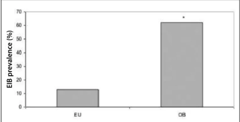
EIB prevalence when the FEV1criterion was adopted was of 62% in the case group versus 13% in the control group (p < 0.05) (figure 1).
When the prevalence of EIB was evaluated in the obese group, 23% (10 volunteers) triggered it by the FEV1, 26% (11 volunteers) triggered it by the PFEPFM (figure 2).
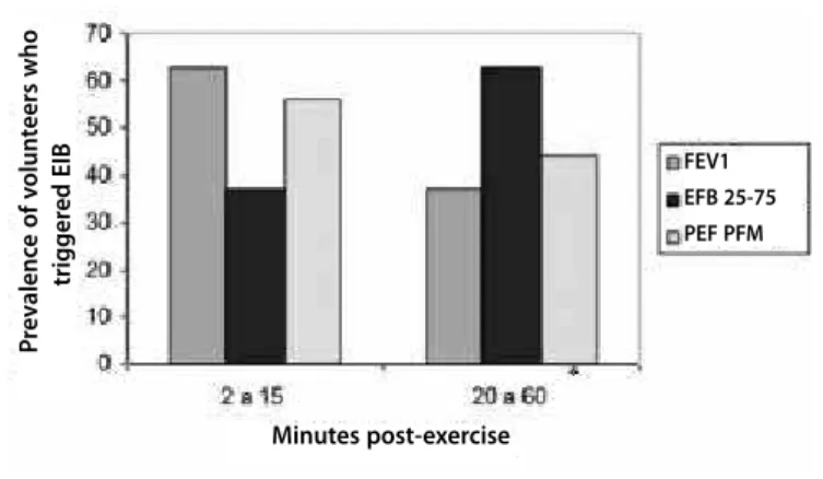
Concerning the EIB triggering time, it is observed that when the FEV1parameter and PEFPFM is evaluated, there was higher prevalence of EIB in the first 15 minutes post-exercise (figure 3).
In the intergroup analysis (EU and OB), statistically significant difference has not been found between the predicted values of baseline FEV1. However, our overweight volunteers presented lower baseline PFE values evaluated by spirometry (PEFE) (table 1).
Figure 1. Prevalence of EIB evaluated by the FEV1between groups.*p < 0.05.
EIB pr
e
v
alenc
e (%)
Figure 2. EIB prevalence in the obese group (n = 39). *p < 0.05, comparing FEV1,
forced expiratory flow on the first second and PEFPFM, peak expiratory flow measured
by the peak flow meter.
EIB pr
e
v
alenc
e (%)
391
Rev Bras Med Esporte – Vol. 17, No 6 – Nov/Dec, 2011 DISCUSSION
Obesity is considered a chronic disease and directly or indirectly related to some other pathological situations which contribute to morbimortality such as cardiovascular, osteomuscular and neoplastic diseases(16), as well as alterations of the respiratory system in its volume and pulmonary capacities(17,18). The buildup of adipose tissue is deleterious to the ventilatory function in children of both genders(19,20). Obesity directly affects the mechanics of the respiratory system changing the pulmonary volume, the airway caliber and the strength of the respiratory muscles(8).
Although alterations in the pulmonary function are found in adults, especially in severe obesity(21), in children the mechanical load of the fat deposition positively affects the pulmonary growth, which induces reduction of pulmonary function(22). Our overweight volunteers presented lower baseline PEFE values, which is according to the findings by Ulgeret al.(8).
The bronchial challenge test is performed through a maximal exertion, being an exercise of high intensity and short duration, between six and eight minutes, which allows that the heart rate (HR) is increased up to 80% of the maximum for the age, through treadmill or cycle ergometer(23). In our study the Del Río-Navarro et
al.(7) protocol which monitors the submaximal heart rate through the formula: (220 – age * 0.65) was used. The protocol on treadmill was due to the higher metabolic demand when compared to the cycle ergometer, since it generates higher cardiac and ventilator stress, being useful in the evaluation of the exercise-induced bronchoconstriction(24). It is worth mentioning that four volunteers
o four sample interrupted the test for having interpreted the load imposed as maximal effort due to the discomfort caused by the peripheral muscular fatigue.
Among the evaluation methods of the pulmonary function, spirometry has the aim to measure the maximal quantity and velocity of expired air, and despite its high reliability it is a clinical evaluation method of difficult access due to its high cost(3,9). On the other hand, the expired flow meter, also known as peak-flow meter, is a non-invasive method of easy application and low cost(3,10). In the EIB detection, 26% of our volunteers of the obese group had EIB triggering evaluated by the peak flow meter. The PEF is an effort-dependent expiratory parameter which reflects the big airways caliber and can be used with an indirect index of the expiratory strength(9).
Since the EIB prevalence by the PEFPFM is similar to the one found by the FEV1 (23%) in the obese group, our findings demonstrate the importance of this instrument as intrinsic part of an educational program as an alternative of initial diagnosis for EIB in airways of big caliber; however, the PEFPFM should not completely substitute the traditional spirometry, but can help in the early detection of airway obstruction(25).
When the EIB triggering times are assessed by the FEV1and PEFPFM parameters, higher prevalence in the first 15 minutes after exercise is observed. We believe that the EIB triggering time is related to the size of the airways affected, being the FEV1and PEF more directly related to the central airways(26,27).
The EIB causes are not well-evidenced(28), but there are currently hypotheses which explain the EIB triggering.
In obese individuals, the hyper responsiveness to exercise may be explained by three hypotheses. The first one is supported by the differences in anatomy of the lungs and airways, due to the possible effect in the compromising of the pulmonary growth, and consequently, in reducing the pulmonary function by the deposition of adipose tissue in the thoracic and abdominal regions(29).
In the second hypothesis, the increase of adipose tissue in the abdominal and thoracic region causes reduction of the functional residual capacity(19,29), which directly acts on the straight muscle of the airways which excessively shortens. The fact that obese individuals breathe ore rapidly and superficially, predisposes them to increase of airway responsiveness and is justified by the fact that the pulmonary is the greatest determinant of the airway diameter(29).
The third hypothesis is that obesity is related to a chronic inflammatory process, in which systemic inflammation characterized by increase of the circulation of leucocytes and increase of the cytokines, cytokines receptors, chemokines and proteins of acute phase concentrations released by the adipose tissue occurs. It is particularly interesting that obesity increases the TNFα seric concentration, which when connecting to the receptors placed in the straight muscles of the airways, promotes bronchoconstriction. In addition to these factors, there are others which when altering their concentrations may affect the airways function leading to their hyper responsiveness, which include: leptin, adiponectin and PAI-1(18,29,30). It is believed that in obese individuals an association between these hypotheses possibly occurs, which predisposes them to more remarkable EIB triggering.
Table 1. Spirometric variables of the baseline pulmonary function testof both groups
Characteristics EU N = 30
OB N = 39
FVCpredicted (%) 94 ± 12 97 ± 16
FEV1predicted (%) 99 ± 13 98 ± 14
FEV1/FVC (%) 103 ± 7 100 ± 9
FVC, forced vital capacity; FEV1, forced expired volume on the first secondPEF, peak expiratory flow. Values presented
in MA ± SD. *p < 0.05.
Figure 3. Most prevalent times of the EIB triggering by the different adopted criteria. FEV1, forced expiratory volume on the first second; PEF25-75%, expiratory flow between
25-75% of the FVC; PEFPFM, peak of expiratory flow by the peak flow meter.* p < 0.05,
comparing the three parameters.
P
re
v
alenc
e of v
olun
teers who
trigger
ed EIB
Minutes post-exercise
FEV1 EFB 25-75
392 Rev Bras Med Esporte – Vol. 17, No 6 – Nov/Dec, 2011 CONCLUSION
Important results to the clinical practice of the professionals from the health field are present in this study, such as the use of the peak flow meter as an important instrument in the detection and initial diagnosis of EIB in central airways, in order to provide a suitable evaluation and previous guidance about physical exercise practice for obese children and adolescents, besides enabling the investigation of possible associated factors such as respiratory discomfort and dropout from physical activity programs.
Possibly, distinct etiologies are related to the EIB; this fact
corroborates the importance for original research in order to elucidate the complex physiopathological mechanisms related to the central and peripheral airways in the triggering of EIB in overweight children and adolescents.
Acknowledgement
To the Development Organs FAPEMIG (#: APQ-01102-09) and CAPES.
All authors have declared there is not any potential conflict of interests concerning this article.
REFERENCES
1. Anderson SD, Daviskas E. The mechanism of exercise-induced asthma is... J Allergy Clin Immunol 2000;106:453-9.
2. Tan RA, Spector SL. Exercise-induced asthma: diagnosis and management. Ann Allergy Asthma Immunol 2002;89:226-36.
3. Fonseca ACCF, Fonseca MTM, Rodrigues MESM, Lasmar LMLBF, Camargos PAM. Pico do fluxo expiratório no acompanhamento de crianças asmáticas. J Pediatr 2006;82:465-9.
4. Marostica PJC. Broncoespasmo induzido pelo exercício na infância. Revista HCPA 2000;20:28-36.
5. Uçok K, Dane S, Gökbel H, Akar S. Prevalence of exercise-induced bronchospasm in long distance runners trained in cold weather. Lung 2004;182:265-70.
6. Matteoni SPC, Júnior CRB, Teixeira LR. Efeito de um Programa de Condicionamento Físico no Broncoespasmo induzido Pelo Exercício em Mulheres obesas. Rev Bras Med Esporte 2009;15:190-4.
7. Del Río-Navarro BE, Cisneros-Rivero MG, Berber-Eslava A, Espínola-Reyna G, Sienra-Monge JJL. Exercise induced bronchospasm in asthmatic and non-asthmatic obese children. Allergol et Immunopathol 2000;28:5-11.
8. Ulger z, Demir E, Tanaç R, Gökşen D, Gülen F, Darcan S,et al. The effect of Childhood obesity on respiratory function tests and airway hyperresponsiveness. Turk J Pediatr 2006;48:43-5.
9. Sociedade Brasileira de Pneumologia e Tisiologia. Diretrizes para teste de função pulmonar. J Pneumol 2002;28:S(3).
10. Menezes AMB, Victora CG, Horta BL, Rigatto M. Valores de referência para o pico de fluxo expiratório em adultos acima de 40 anos, Pelotas-RS. J Pneumol 1995;21:119-22.
11. Rodrigues JC, Cardieri JMA, Bussamra MHCF, Nakaie CMAN, Almeida MB, Filho LVFS, et al. Provas de função pulmonar em crianças e adolescentes. In: Pereira, C.A.C; Neder, J.A. Diretrizes para testes de função pulmonar. J Pneumol 2002;28(Supl. 3):207-21.
12. Tanner JM, Whitehouse RH. Clinical longitudinal standards for height, weight, height velocity, weight velocity, and stages of puberty. Arch Dis Child 1976;62:57-62.
13. Asher MI, Keil U, Anderson HR, Beasly R, Crane S, Martinez F, et al. International Study Protocol: International study of asthma and allergies in childhood (ISAAC): rationale and methods. Eur Respir J 1995;8:483-91.
14. Solé D, Naspitz CK. Epidemiologia da asma: Estudo ISAAC (International Study of Asthma and Allergies in Childhood. J Invest Allergol Clin Immunol 1998;21:38-45.
15. Godfrey S, Kamburoff PL, Nairn JR. Spirometry lung volumes and airway resistance in normal children aged 5 to 18 years. Br J Dis Chest 1970;64:15-24.
16. Cabrera MAS, Jacob Filho W. Obesidade em idosos: Prevalência, distribuição e associação com hábitos e co-morbidades. Arq Bras Endocrinol Metabol 2001;45:494-501.
17. Casali CCC. Efeito do treinamento muscular inspiratório sobre a função muscular e pulmonar após gas-troplastia em obesos. [dissertação]. Uberlândia, MG: Mestrado em Fisioterapia do Centro Universitário do Triângulo; 2005.
18. Poulain M, Doucet M, Major GC, Drapeau V, Sériès F, Boulet LP, et al. The effect of obesity on chronic respiratory diseases: pathophysiology and therapeutic strategies. CMAJ 2006;74:1293-9.
19. Li AM, Chan D, Wong E, Yin J, Nelson E, Fok T. The effects of obesity on pulmonary function. Archives of Disease in Childhood 2003;88:361-3.
20. Delorey DS, Wyrick BL, Babb TG. Mild-to-moderate obesity: implications for respiratory mechanics at rest and during exercise in young men. Int J Obes 2005;29:1039-47.
21. Ferretti AMD, Giampiccolo PMD, Cavalli AMD, Milic-Emili JMD, Tantucci CMD. Expiratory flow limitationn and orthopnea in massively obese subjects. Chest 2001;119:1401-8.
22. Shore SA, Fredberg JJ. Obesity, smooth muscle, and airway hiperresponsiveness. J Allergy Clin Immunol 2005;115:925-7.
23. Dalamón RS. Broncoespasmo inducido por ejercicio: un desafío diagnóstico. Arch Arg Pediatr 2004;102:163-4.
24. Neder JA, Nery LE. Fisiologia Clínica do Exercício. Teoria e Prática. 1a. ed. São Paulo: Artes Médicas, 2003. 481p.
25. Jain P, Kavuru MS, Emerman CL, Ahmad M. Utility of peak expiratory flow monitoring. Chest 1998;114:861-76.
26. Godoy I. Pico de fluxo expiratório (PFE): mitos e verdades. 2005 Disponível em: http://www.emv.fmb.unesp. br/material_estudo/pneumologia/pico_de_flu/clin_med_picodeflu.asp.
27. Hegewald MJ, Lefor MJ, Jensen RL, Crapo RO, Kritchevsky SB, Haggerty CL, et al. Peak Expiratory Flow Variability and FEV1 Are Poorly Correlated in an Elderly Population. Chest 2007;131:1494-9.
28. Rosas MA, Perez J, Blandon V, del Rio B, Sienra M, Juan JL. Broncoespasmo inducido pelo ejercicio: diagnóstico y manejo. Revista Alergia México 2004;51:85-93.
29. Shore SA, Fredberg JJ. Obesity, smooth muscle, and airway hiperresponsiveness. J Allergy Clin Immunol 2005;115:925-7.