LETTERS TO THE EDITORS
New insights into cortisol
levels in PTSD
Rev Bras Psiquiatr 2016;38:176 doi:10.1590/1516-4446-2015-1795
The article ‘‘Relationship of cortisol, norepinephrine, and epinephrine levels with war-induced posttraumatic stress disorder in fathers and their offspring’’ by Yahyavi et al.1 provided interesting data on the neuroendocrinology of post-traumatic stress disorder (PTSD). The mechanisms that underlie the associations of cortisol levels with traumatic exposures and PTSD are still not well under-stood and, as stated by the authors, findings are far from consistent.
Similarly to the findings of Yahyavi et al., in a sample of individuals exposed to trauma during the preceding 5 years, patients with PTSD had neither an increase nor a decrease in mean urinary cortisol levels.2Conversely, in a recently published study, Wingenfeld et al.3reported decreased cortisol values in outpatients with PTSD recruited from two Veterans Affairs medical centers. Furthermore, researchers have reported lower cortisol levels in the acute aftermath of trauma in patients who later developed PTSD.4
It seems that inadequate glucocorticoid release follow-ing stress not only delays recovery by disruptfollow-ing biological homeostasis in the short run but can also interfere with the processing or interpretation of stressful information, resulting in long-term disruptions in memory integration.5
Consistent with these findings is the fact that a single dose of hydrocortisone administered in the acute after-math of trauma produced recovery while promoting enhanced synaptic plasticity and connectivity in the secondary prevention of PTSD.5In this sense, it remains unclear whether deregulation of the HPA axis, leading to low peritraumatic levels of cortisol, endures post-traumatically.
Another critical issue in this matter has to do with changes in cortisol levels secondary to pharmacological treatment of PTSD (e.g., sertraline), an important aspect not addressed by the authors.6 Finally, it is worth mentioning that PTSD is characterized by the presence of four symptom clusters (intrusion, avoidance, negative alterations in cognitions and mood, and alterations in arousal and reactivity) instead of the three mentioned in the paper.
Gabriela de Moraes Costa Departamento de Neuropsiquiatria, Universidade Federal de Santa Maria (UFSM), Santa Maria, RS, Brazil
Submitted Aug 11 2015, accepted Feb 12 2016.
Disclosure
The author reports no conflicts of interest.
References
1 Yahyavi ST, Zarghami M, Naghshvar F, Danesh A. Relationship of cortisol, norepinephrine, and epinephrine levels with war-induced posttraumatic stress disorder in fathers and their offspring. Rev Bras Psiquiatr. 2015;37:93-8.
2 Young EA, Breslau N. Cortisol and catecholamines in posttraumatic stress disorder: an epidemiologic community study. Arch Gen Psychiatry. 2004;61:394-401.
3 Wingenfeld K, Whooley MA, Neylan TC, Otte C, Cohen BE. Effect of current and lifetime posttraumatic stress disorder on 24-h urinary catecholamines and cortisol: results from the Mind Your Heart Study. Psychoneuroendocrinology. 2015;52:83-91.
4 Witteveen AB, Huizink AC, Slottje P, Bramsen I, Smid T, van der Ploeg HM. Associations of cortisol with posttraumatic stress symptoms and negative life events: a study of police officers and firefighters. Psychoneuroendocrinology. 2010;35:1113-8.
5 Zohar J, Yahalom H, Kozlovsky N, Cwikel-Hamzany S, Matar MA, Kaplan Z, et al. High dose hydrocortisone immediately after trauma may alter the trajectory of PTSD: interplay between clinical and animal studies. Eur Neuropsychopharmacol. 2011;21:796-809. 6 Pacella ML, Feeny N, Zoellner L, Delahanty DL. The impact of
PTSD treatment on the cortisol awakening response. Depress Anxiety. 2014;31:862-9.
Clozapine-induced
esophagitis at therapeutic
dose: a case report
Rev Bras Psiquiatr. 2016;38:176–177 doi:10.1590/1516-4446-2015-1787
The pharmacological profile of clozapine, which is often described as a ‘‘broad spectrum’’ antagonist, is differ-ent from that of other antipsychotics. It is thought that the side effects reported with clozapine may be attributed in part to its anti-serotonin action together with its anti-a-adrenergic, anticholinergic, and
antihis-taminic effects.1
A 58-year-old chronic schizophrenic patient had been treated with haloperidol for over 10 years. Haloperidol was eventually replaced with a combination of antipsy-chotics (olanzapine/zuclopenthixol and later aripiprazole/ zuclopenthixol). Given the persistence of major psychotic symptoms, clozapine was gradually introduced in combination with the following treatments: aripiprazole (10 mg/d), zopiclone (7.5 mg/day, as needed), tropate-pine (15 mg/day), and lorazepam (7.5 mg/d). During the first month of treatment, the dose of clozapine was gradually increased to 275 mg/d (at the end of 1 month). However, various side effects were observed, leading to the increase in tropatepine (30 mg/d) for extrapyramidal side effects and to the introduction of macrogol 4,000 (30 g/d) for constipation, heptaminol (563.4 mg/d) for orthostatic hypotension, and paracetamol (3 to 4 g/d) for headaches.
After a second month of treatment the patient continued to describe somatic complaints, including nausea. Aripiprazole was stopped, and tropatepine was gradually reduced to 10 mg/d. In the meantime, clozapine was gradually increased based on good hematologic Revista Brasileira de Psiquiatria. 2016;38
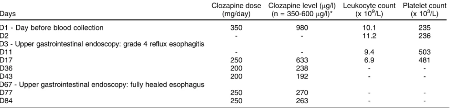
Table 1 Summary of changes on dosage and plasma clozapine levels following clozapine intoxication with available leucocyte and platelet counts
Days
Clozapine dose (mg/day)
Clozapine level (mg/l) (n = 350-600mg/l)*
Leukocyte count (x 109/L)
Platelet count (x 103/L)
D1 - Day before blood collection 350 980 10.1 235
D2 - - 11.2 236
D3 - Upper gastrointestinal endoscopy: grade 4 reflux esophagitis
D11 - - 9.4 503
D17 250 633 6.9 481
D36 200 238 -
-D43 200 192 -
-D67 - Upper gastrointestinal endoscopy: fully healed esophagus
D77 250 270 -
-D84 250 263 -
-tolerance since the start of treatment. Nefopam was also introduced for the management of hemorrhoid pain as needed, at 20 mg/d (po). After an additional 35 days of treatment with clozapine at 350 mg/d, one night the patient had hematemesis and diffuse abdominal pain. The next morning, she experienced vomiting and complained of epigastric pain. The patient was treated with domper-idone (30 mg/d) and metoclopramide (20 mg/d, po, as needed). Clinical examination revealed reflux esophagitis (grade 4) associated with a hiatal hernia (4 cm). A blood test showed plasma clozapine levels of 980mg/l
(labora-tory alert level is plasma concentration41,000mg/L).2In
this context, pantoprazole was introduced at a dosage of 20 mg/d, whereas domperidone was stopped after 1 week. Clozapine was gradually reduced to 200 mg/d. After 15 days, gastroesophageal symptoms had dimin-ished and clozapine levels had fallen to 633 mg/L
(therapeutic reference range = 350-600 mg/L).2 About
2 months after esophagitis, a gastroscopic control was performed, demonstrating complete esophageal healing. Table 1 synthesizes the clozapine dose changes following intoxication along with the plasma levels of clozapine.
Some disorders affecting the gastrointestinal tract, including cases of esophagitis with hiatal hernias, have been reported with the use of clozapine, but very few have been published.3 These gastrointestinal distur-bances appear in connection with hypomotility and changes in digestive secretion induced by clozapine, as a result of its antiserotoninergic and anticholinergic properties.3 Anticholinergic medications are often
impli-cated in exacerbating gastroesophageal reflux disease by decreasing lower esophageal sphincter pressure and consequently causing or aggravating heartburn, but such conditions were not identified in our patient before the occurrence of esophagitis.
In our patient, the combined use of clozapine, tropatepine, and aripiprazole (stopped a week before the adverse effect but with a long half-life) may have enhanced the effects of clozapine by contributing to anticholinergic and antiserotoninergic effects respectively (nefopam was only administered on 2 days: 4 and 24 days before esophagitis). However, the dramatic increase in clozapine blood concentration seems suffi-cient to account for the adverse effect. To the best of our knowledge, this is the first case of clozapine-induced
severe esophagitis correlated to a measured-level of plasma clozapine to be published in the literature.
Herve´ Javelot,1Bruno Michel,2Divya Kumar,3
Brigitte Audibert3 1Clinical Pharmacy Service, Mental Health Establishment (EPSAN),
Brumath, France.2Faculte´ de Pharmacie, Laboratoire HuManiS (EA 7308), Service Pharmacie - CHU de Strasbourg, Strasbourg, France.3Service G06, EPSAN, Brumath, France
Submitted Jul 31 2015, accepted Nov 09 2015.
Disclosure
The authors report no conflicts of interest.
References
1 Naheed M, Green B. Focus on clozapine. Curr Med Res Opin. 2001;17:223-9.
2 Hiemke C, Baumann P, Bergemann N, Conca A, Dietmaier O, Egberts K, et al. AGNP consensus guidelines for therapeutic drug monitoring in psychiatry: update 2011. Pharmacopsychiatry. 2011;44:195-235.
3 Laker MK, Cookson JC. Reflux oesophagitis and clozapine. Int Clin Psychopharmacol. 1997;12:37-9.
Circadian rhythm
disturbances and
conversion to psychosis
in ultra high-risk youth
Rev Bras Psiquiatr. 2016;38:177–179doi:10.1590/1516-4446-2015-1859
Our group has recently showed that individuals in at risk mental states for psychosis and bipolar disorder (BD) have a poorer quality of sleep, start their daily activity later, and have a fragmented circadian rhythm compared with age and gender-matched healthy controls.1,2Another study shows that adolescents at high risk of developing psychosis are more prone to nocturnal awakening, which reduces sleep efficiency.3Nevertheless, the relationship