SUMMARY
Objective: To evaluate the prevalence of actinic skin lesions in patients with basal cell car-cinoma of the head. Methods: A case-control study was carried out. Cases were patients with primary, solid basal cell carcinoma of the head, less than two centimeters in diam-eter; and as controls, patients with other dermatoses. Constitutional and behavioral vari-ables were analyzed, as well as actinic lesions. Results: One hundred twenty cases and 360 controls were evaluated. Facial milia (OR = 2.3), leukoderma punctata of the upper limbs (OR = 2.9), and cutis rhomboidalis nuchae (OR = 1.8) were associated with neoplasms re-gardless of other variables, suggesting a risk phenotype. here was also association with light hair and eye color phenotypes, family genetics, and cumulative sun exposure. Sun-burn, smoking, and alcoholism were not identiied as risk factors. he use of sunscreens showed no evidence of protection; however, the control group consisted of dermatology patients who are oten prescribed sunscreens. Conclusion: Actinic lesions were more prevalent in patients with solid basal cell carcinoma of the head than in controls, espe-cially milia, cutis rhomboidalis nuchae, and leukoderma punctata, regardless of other known risk factors.
Keywords: Head and neckneoplasias; basal cell carcinoma; epidemiology; case-control studies; UV rays; risk factors.
©2012 Elsevier Editora Ltda. All rights reserved.
Study conducted at the Departament of Dermatology, School of Medicine of Botucatu, Universidade Estadual Paulista (UNESP), Botucatu, SP, Brazil
Submitted on: 09/27/2011
Approved on: 12/19/2011
Correspondence to:
Hélio Amante Miot Departamento de Dermatologia e Radioterapia, SN Campus Universitário CEP: 18618-000 Botucatu, SP, Brazil Phone/Fax: +55 (14) 3882-4922 heliomiot@fmb.unesp.br
Conlict of interest: None.
Prevalence of actinic skin lesions in patients with basal cell carcinoma
of the head: a case-control study
VALQUÍRIA PESSOA CHINEM1, HÉLIO AMANTE MIOT2
INTRODUCTION
Basal cell carcinoma (BCC) is the most common neopla-sia in humans, especially among fair-skinned individuals. It constitutes 70% to 80% of malignant skin tumors and represents an important demand on and cost to the health
system1,2. According to a German sample, throughout life,
the cumulative risk of its development in the Caucasian
population is over 30%3. he expected incidence in 2011
in Brazil is approximately 120,000 cases1. Over the past
50 years, there was an increase in the incidence in several
countries, including the involvement of younger patients4,5.
his neoplasia has a low degree of malignancy and mortality due to the typical slow growth and reduced po-tential to metastasize, in addition to its early diagnosis, as it is preferentially located in areas exposed to sunlight. However, its morbidity is high due to local invasion, tis-sue destruction, and the possibility of recurrence,
afect-ing patients’ quality of life4,6-8.
In contrast, a smaller fraction of malignant neoplasms of the skin (20-30%) consists mainly of squamous cell car-cinoma (SCC) and melanomas, which are aggressive forms, with clear metastatic potential. Whereas the SCC favors sun-exposed and mucosal areas, melanomas also occurs in less irradiated areas such as the back, upper thighs, and
extremities, which can lead to diagnostic delay1.
Ultraviolet radiation (UVR) is the main environmen-tal risk factor associated with the genesis of the BCC, as shown by a higher frequency of lesions in sun-exposed ar-eas. In addition to the immunosuppressive action on the skin, compromising the local antitumor monitoring activ-ity2-4,9, UVB radiation generates mutagenic photoproducts
in the DNA, which promotes mutations in genes such as PTCH and p53.In turn, UVA radiation has mainly indi-rect efects by generating cytotoxic and mutagenic free radicals9-12.
he most important constitutional risk factors are: fair skin (diiculty to tan and predisposition to sunburns), light-colored eyes and hair, family history of BCC, and freckles in childhood. Noteworthy behavioral factors are: professional activity unprotected from UVR, rural
activi-ties, and sunburns in youth7,13.
Although most authors point to a greater role of inter-mittent sun exposure, especially in youth, in the genesis of the BCC, some recent studies have suggested that nodular
BCC would be more related to chronic sun exposure14,15.
As their genesis is associated with exposure to UVR, chronic actinic skin lesions may be indicators of the risk for the development of BCC. Skin lesions such as actinic keratosis, solar lentigines, solar elastosis, and facial tel-angiectasias are commonly found in patients with this cancer, but have not been systematically studied, nor considered regarding the other epidemiological markers of risk13,16-18.
Early diagnosis is a key strategy to improve prognosis, and reduce sequelae (local and therapeutic) and costs for the health system. he identiication of risk phenotypes can be favorable in public health interventions aiming at primary and secondary prevention.
he authors aimed to evaluate the prevalence of chronic actinic skin lesions in patients with solid, pri-mary BCC of the head, and compared them with patients without skin cancer, in order to identify skin phenotypes associated with the risk of BCC.
METHODS
his was a case-control study involving patients older than 40 years of age, of both genders, treated at the De-partment of Dermatology of the Medical School of UN-ESP - Botucatu, from March 2007 to December 2009.
he cases were deined as patients with solid, primary BCC (conirmed by histopathological analysis), less than two centimeters in diameter, located on the head, diag-nosed less than a year before the interview. Controls were patients with other benign dermatoses, interviewed at a proportion of three respondents for each case, in a non-paired manner, to allow risk estimation and stratiied analysis of constitutional variables such as gender, age,
and skin type (Fitzpatrick classiication)19.
Patients presenting with evidence of immunosup-pression (medication-induced or associated with chronic diseases with signiicant reduction in immune response), genetic syndromes predisposing to cancer, difuse skin dermatoses, skin type VI, presenting more than one BCC lesion or any other skin neoplasm at any time were excluded.
he demographic and behavioral information were obtained from standardized forms, and the actinic skin lesions were examined by a dermatologist. Systematic pa-tient sampling was not used, thus including all those who were consecutively available.
he main dependent variable was the appearance of sporadic, solid BCC, less than two centimeters in diam-eter, located on the head. he independent variables were age, gender, skin type, constitutional characteristics, sun-burns, photoaging measurement, dermatoses associated with chronic sun exposure, smoking, alcohol consump-tion, family occurrence of BCC, sun exposure related to occupational or leisure activity, and use of sunscreen.
Initially, a bivariate analysis was performed to esti-mate associations between the primary variables in each group. Categorical variables were expressed as the per-centage of occurrence and compared by the chi-square tests or the chi-square test for trend, for ordinal charac-teristic. Continuous and discrete quantitative variables were represented by their means or medians, and
the distribution was nonparametric. Normality of distri-butions was estimated by the Shapiro-Wilk’s test.
Subsequently, the independent variables were adjusted from a non-conditional multiple logistic regression model of hierarchical structure to control confounding factors. he progressive inclusion of variables into the model,
ac-cording to each hierarchical level, occurred with p < 0.2520-22.
he hierarchical model of analysis to control con-founding factors showed, in the irst level (forced entry variables): gender, age, and skin type. At the second level, other constitutional variables included: light-colored eyes, light-colored hair, freckles in childhood, family history of BCC. In the third level, behavioral and exposure variables were included: rural activities, working in the sun, num-ber of hours working in the sun (expressed by the product of hours/days and years of exposure = h/d x year), leisure activities in the sun, number of hours during leisure activi-ties in the sun (expressed by the product of hours/week and years of exposure = h/wk x year), smoking (expressed as the product of packs/day and years of smoking = packs/d x year), alcoholism, use of sunscreen in the past 10 years (stratiied as: never, when leaving the house/going to the beach, every day). In the last level, chronic actinic lesions were included: wrinkles, facial and upper limb actinic kera-toses, telangiectasias, leukoderma punctata, facial milia, acne scars, comedones, facial and upper limb lentigines,
poikiloderma of Civatte, cutis rhomboidalis nuchae, stellar
scars of the upper limbs, Bateman’s purpura, actinic cheili-tis, and palmar keratosis.
he groups were further analyzed and stratiied by gender, skin types, and extremes of age tertiles. he addi-tive efects of the interaction between variables were esti-mated by assessing the modiication of the overall efect of
the combination of variables20,22.
he association measures were represented as odds ra-tios, at a 95% conidence interval (CI), and a two-tailed p-value of 0.05 was considered signiicant.
Data were tabulated in MS Excel® 2003, and analyzed using the Statistical Package for the Social Sciences (SPSS)
sotware version 17.023.
he sampling was estimated based on a pretest with 50 cases and 150 controls, and calculated for a model of non-conditional multiple logistic regression, according to the
estimation of variables for the inal model24,25.
he project was approved by the institution’s ethics and research committee (protocol number 331/2007). Ater all interviews, the patients signed the free and informed con-sent form.
RESULTS
A total of 120 cases and 360 controls were evaluated. he main demographic data are shown in Table 1. here was a direct association of BCC with fair-skin phenotypes, older
age, predisposing family genetics, and work activity in the sun; 95% of the controls were exposed for less than 400 h/d x year. Smoking, regular alcohol consumption, sunscreen use, leisure activities in the sun, and sunburns were not risk factors for this sample.
Cases and controls mainly came from the city of Botucatu (45% and 41%) and surrounding regions (98% and 91%). No patients of Asian origin were interviewed and none of the cases or controls used tanning beds or phototherapy.
Among controls, 98 diferent dermatological diagno-ses were identiied, of which the main ones (> 2% of diag-noses) were onychomycosis, contact dermatitis, urticarial rash, melanocytic nevi, seborrheic keratoses, actinic
kera-tosis, pyoderma, discoid lupus erythematosus, tinea pedis,
stasis eczema, psoriasis, which are in agreement with the Brazilian dermatological outpatient sample for this age group26.
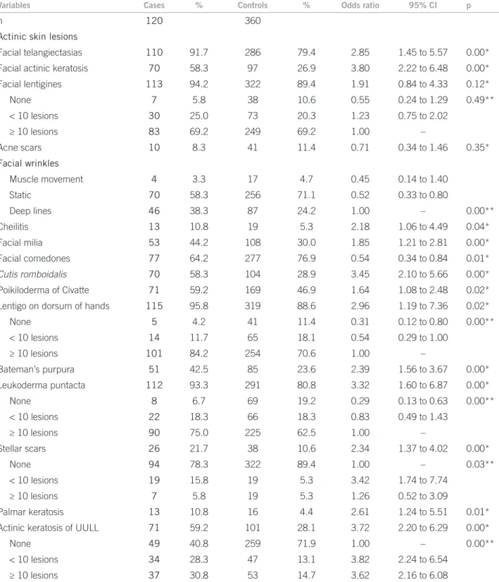
Table 2 speciies the frequency of chronic actinic le-sions between the groups, showing a direct association between BCC and the skin lesions studied, except for len-tigines and facial acne scars.
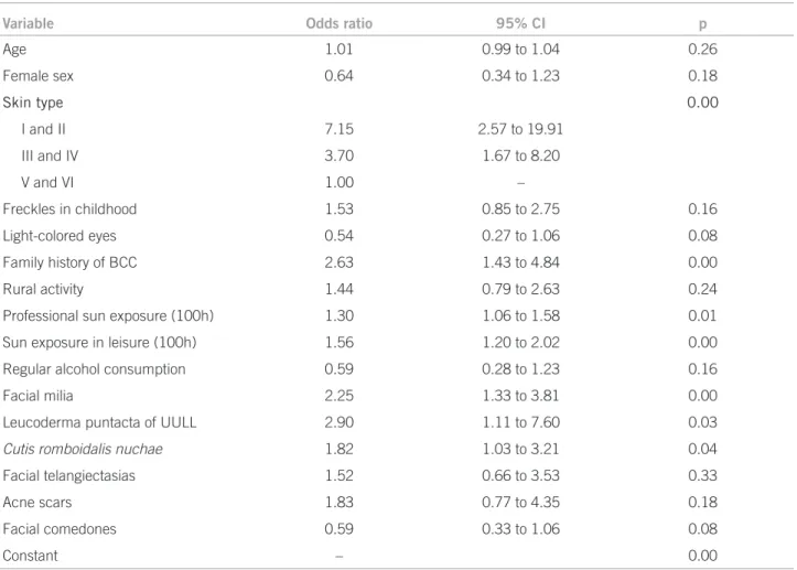
Table 3 shows the inal reduced hierarchical logistic model of the prevalence of actinic lesions. Milia,
leuko-derma punctata of the upper limbs, and cutis rhomboidalis
nuchae were associated with the BCC, regardless of other covariates.
No signiicant interactions were identiied between the independent variables, and there was no data loss in the sample. he inal model showed no outliers (Zres > 3).
Female cases had lighter skin types, reported less al-cohol consumption, and had higher sun exposure during work and leisure time than their controls of the same sex. Family history was more frequent (OR = 2.38) among cas-es, and skin lesions indicative of risk in this subgroup were milia (OR = 2.16) and stellar scars (OR = 2.55).
Male patients had the following skin lesions as indic-ative of risk: milia (OR = 3.85), acne scars (OR = 8.29),
leukoderma punctata (OR = 3.71), and cutis
rhomboida-lis nuchae (OR = 4 17). Family history was more frequent among cases (OR = 23.83), which also showed lighter phe-notypes, reported higher sun exposure during work and leisure periods, and more freckles in childhood in com-parison to their controls. he presence of facial comedones showed a protective efect in these patients (OR = 0.28).
Overall, the women in the study worked fewer hours exposed to the sun as compared to men (average: 91.82
versus 182.94 h, p < 0.05), and underwent less sun
expo-sure during leiexpo-sure time than men (average: 31.68 versus
53.33 h, p < 0.05).
Variables Cases % Controls % Odds
ratio 95% CI p
n 120 360
Constitutional and biological variables
Female sex 76 63.3 241 66.9 0.85 0.55 to 1.31 0.47*
Skin type
I 4 3.3 6 1.7 13.00 1.94 to 87.14 0.01**
II 42 35.0 67 18.6 12.22 2.80 to 53.29
III 61 50.8 150 41.7 7.93 1.86 to 33.87
IV 11 9.2 98 27.2 2.19 0.46 to 10.33
V 2 1.7 39 10.8 1.00 -
Light-colored hair 32 26.7 59 16.4 1.86 1.14 to 3.03 0.01*
Light-colored eyes 48 40.0 105 29.2 1.62 1.05 to 2.49 0.03*
Freckles in childhood 36 30.0 59 16.4 2.19 1.36 to 3.51 0.00*
Family history of BCC 31 25.8 46 12.8 2.38 1.35 to 3.53 0.00*
Median age (1st-3rd quartiles) years 64 (54-72) 56 (48-66) 1.04 1.02 to 1.06 0.00#
Behavioral and exposure variables
Sun exposure at work 105 87.5 263 73.1 2.58 1.43 to 4.65 0.00*
Sun exposure at work - median
(1st-3rd quartiles) hours/day x years 112.5 (24-350) 45 (5-150) 1.00 1.00 to 1.01 0.00 #
Rural activity 58 48.3 101 28.1 2.40 1.57 to 3.67 0.00*
Leisure in the sun 42 35.0 102 28.3 1.36 0.88 to 2.11 0.17*
Sun exposure during leisure activities -
median (1st-3rd quartiles) hours/week x years 0 (0-38) 0 (0-15) 1.00 1.00 to 1.01 0.21 #
Painful sunburn 54 45.0 157 43.6 1.06 0.70 to 1.60 0.79*
Sunburn with blisters 23 19.2 71 19.7 0.97 0.57 to 1.63 0.89*
Smoking 49 40.8 176 48.9 0.72 0.48 to 1.10 0.13*
Tobacco load - median
(1st-3rd quartiles) packs/days x years 0 (0-21) 0 (0-21) 1.00 1.00 to 1.01 0.36 #
Regular alcohol consumption 18 15.0 81 22.5 0.61 0.34 to 1.06 0.08*
Sunscreen use for > 10 years
Never 102 85.0 289 80.3 1.00 - 0.20**
Leaving the house/on the beach 8 6.3 26 7.2 0.87 0.38 to 1.99
Everyday 10 8.3 45 12.5 0.63 0.31 to 1.30
*Chi-square test; **Chi-square trend test; #Mann-Withney test.
Table 1 - Analysis of constitutional and behavioral demographic variables between the groups
he injury most oten associated with risk of BCC was fa-cial telangiectasia (OR = 12.20).
Compared to controls with skin types IV and V, cases with darker skin types underwent higher sun exposure during leisure time. he age of the cases, which was old-er than controls (OR = 1.14), and the refold-erence to family history (OR = 39.99) must be emphasized. he skin
le-sion most oten associated was cutis rhomboidalis nuchae
(OR = 8.32).
Variables Cases % Controls % Odds ratio 95% CI p
n 120 360
Actinic skin lesions
Facial telangiectasias 110 91.7 286 79.4 2.85 1.45 to 5.57 0.00*
Facial actinic keratosis 70 58.3 97 26.9 3.80 2.22 to 6.48 0.00*
Facial lentigines 113 94.2 322 89.4 1.91 0.84 to 4.33 0.12*
None 7 5.8 38 10.6 0.55 0.24 to 1.29 0.49**
< 10 lesions 30 25.0 73 20.3 1.23 0.75 to 2.02
≥ 10 lesions 83 69.2 249 69.2 1.00 –
Acne scars 10 8.3 41 11.4 0.71 0.34 to 1.46 0.35*
Facial wrinkles
Muscle movement 4 3.3 17 4.7 0.45 0.14 to 1.40
Static 70 58.3 256 71.1 0.52 0.33 to 0.80
Deep lines 46 38.3 87 24.2 1.00 – 0.00**
Cheilitis 13 10.8 19 5.3 2.18 1.06 to 4.49 0.04*
Facial milia 53 44.2 108 30.0 1.85 1.21 to 2.81 0.00*
Facial comedones 77 64.2 277 76.9 0.54 0.34 to 0.84 0.01*
Cutis romboidalis 70 58.3 104 28.9 3.45 2.10 to 5.66 0.00*
Poikiloderma of Civatte 71 59.2 169 46.9 1.64 1.08 to 2.48 0.02*
Lentigo on dorsum of hands 115 95.8 319 88.6 2.96 1.19 to 7.36 0.02*
None 5 4.2 41 11.4 0.31 0.12 to 0.80 0.00**
< 10 lesions 14 11.7 65 18.1 0.54 0.29 to 1.00
≥ 10 lesions 101 84.2 254 70.6 1.00 –
Bateman’s purpura 51 42.5 85 23.6 2.39 1.56 to 3.67 0.00*
Leukoderma puntacta 112 93.3 291 80.8 3.32 1.60 to 6.87 0.00*
None 8 6.7 69 19.2 0.29 0.13 to 0.63 0.00**
< 10 lesions 22 18.3 66 18.3 0.83 0.49 to 1.43
≥ 10 lesions 90 75.0 225 62.5 1.00 –
Stellar scars 26 21.7 38 10.6 2.34 1.37 to 4.02 0.00*
None 94 78.3 322 89.4 1.00 – 0.03**
< 10 lesions 19 15.8 19 5.3 3.42 1.74 to 7.74
≥ 10 lesions 7 5.8 19 5.3 1.26 0.52 to 3.09
Palmar keratosis 13 10.8 16 4.4 2.61 1.24 to 5.51 0.01*
Actinic keratosis of UULL 71 59.2 101 28.1 3.72 2.20 to 6.29 0.00*
None 49 40.8 259 71.9 1.00 – 0.00**
< 10 lesions 34 28.3 47 13.1 3.82 2.24 to 6.54
≥ 10 lesions 37 30.8 53 14.7 3.62 2.16 to 6.08
UULL, upper limbs; *Chi-square test; **Chi-square trend test.
Table 2 – Bivariate analysis of prevalence of chronic actinic lesions between the groups
he cases in the oldest tertile (> 78 years), when com-pared with older controls, showed lighter phenotypes and higher sun exposure during work. he associated actinic
lesions in this subgroup were milia (OR = 3.63), cutis
rhomboidalis nuchae (OR = 4.73), leukoderma punctata (OR = 6.80), and actinic cheilitis (OR = 6.35).
DISCUSSION
he chronic actinic lesions were more prevalent among patients with solid BCC on the head than in the controls of the studied population. Especially, facial milia,
leuko-derma punctata of the upper limbs, and cutis
Variable Odds ratio 95% CI p
Age 1.01 0.99 to 1.04 0.26
Female sex 0.64 0.34 to 1.23 0.18
Skin type 0.00
I and II 7.15 2.57 to 19.91
III and IV 3.70 1.67 to 8.20
V and VI 1.00 –
Freckles in childhood 1.53 0.85 to 2.75 0.16
Light-colored eyes 0.54 0.27 to 1.06 0.08
Family history of BCC 2.63 1.43 to 4.84 0.00
Rural activity 1.44 0.79 to 2.63 0.24
Professional sun exposure (100h) 1.30 1.06 to 1.58 0.01
Sun exposure in leisure (100h) 1.56 1.20 to 2.02 0.00
Regular alcohol consumption 0.59 0.28 to 1.23 0.16
Facial milia 2.25 1.33 to 3.81 0.00
Leucoderma puntacta of UULL 2.90 1.11 to 7.60 0.03
Cutis romboidalis nuchae 1.82 1.03 to 3.21 0.04
Facial telangiectasias 1.52 0.66 to 3.53 0.33
Acne scars 1.83 0.77 to 4.35 0.18
Facial comedones 0.59 0.33 to 1.06 0.08
Constant – 0.00
Hosmer and Lemeshow Test p = 0.35; correct classiication = 81.3%; R2Nagelkerke = 0.37.
Table 3 – Logistic model adjusted for the other risk factors
when adjusted for age, skin type, gender, family history of BCC, and number of hours of work and leisure activities in the sun.
Milia are keratinocytic cysts that are formed in the upper dermis as a result of sun exposure, burns, trauma, sweat or sebaceous gland obstruction, or recurrent cicatri-cial processes as it occurs in porphyria and epidermolysis bullosa. hey also occur in Gorlin, Bazex-Dupré-Chris-tol, and Rombo syndromes, which course with multiple BCCs4,27,28. To date, no study had investigated its
associa-tion with BCC.
Cutis rhomboidalis nuchae is a manifestation of exu-berant dermal actinic elastosis that occurs in the neck. It commonly afects fair-skinned elderly individuals who were chronically exposed to UVR. he exacerbated pro-duction of thick and disarrayed elastic ibers is due to a chronic inlammatory process, with mast cell degranula-tion and activadegranula-tion of ibroblasts that produce collagen-de-grading metalloproteinases. he association of BCC with photoaging, elastosis, and Favre-Racouchot syndrome has
been described before16,17,29. Facial wrinkles are skin
le-sions associated with elastosis, which have been identiied
as a possible protective factor in a German series30. his,
however, was not demonstrated in this sample, as the di-rect analysis showed that wrinkles represent risk factors that are proportional to their intensity.
Leukoderma punctata or idiopathic guttate hypomela-nosis is common in fair-skinned adults and occurs mainly in sun-exposed surfaces of the upper and lower limbs. It is caused by local melanocytic hypoplasia with reduced melanin pigmentation. It has been previously identiied as
a risk element for BCC ater multivariate adjustment29.
Leukoderma punctata, milia, and cutis rhomboidalis
are not neoplastic lesions, and their physiopathogenesis occurs independently from each other; in addition, they do not share other factors in the biological chain of the BCC, except chronic exposure to UVB. he concomitant susceptibility of these actinic lesions with BCC must de-rive from an individual phenotype of common risk.
Solar lentigines, actinic keratoses, actinic cheilitis, fa-cial telangiectasia, and solar elastosis were not variables independently associated with BCC in this population,
unlike other studies16,17,29-31. his discrepancy may result
analysis, such as milia, leucoderma, and cutis
rhomboida-lis. Other explanations for this diference may result from
the selection of patients with BCCs in other topographies, other histological types and multiple forms; moreover, the control individuals come from a group of adult dermatol-ogy patients, in whom a higher frequency of skin lesions
than in the normal population is to be expected14.
Prospective studies should be carried out to corrobo-rate these indings and disclose the physiopathology of individual sensitivity to BCC that predisposes the simulta-neous expression of these actinic dermatoses to the detri-ment of others.
he literature indicates lower-sensitiveness skin types and sun exposure as the most important risk factors for BCC. Some studies also report association with previous sunburns, light hair and eyes, positive family history, and
presence of actinic lesions7,16,17,31-36.
In the present study, there was a direct association of BCC with lighter skin types, load of sun exposure during professional activities, family history, and actinic lesions. here was no association with previous sunburns, smok-ing, and alcohol, and the regular use of sunscreen did not protect against BCC.
Contrary to what occurs in SCC, there is no consen-sus with regard to smoking and subsequent development
of BCC37,38. he same goes for the protection provided
by the regular use of sunscreen (in the past 10 years), and most studies also found no such association for this neoplasia29,34,39-41.
he population studied, typically coming from lower social classes, presented a report of current smoking at a proportion that was similar to that found in the state
capi-tal of São Paulo (20.4% versus 19.9%)42. However, the
regu-lar use of sunscreen, even occasionally, was reported with a lower frequency than the result obtained from the
Bra-zilian population survey (18.5% versus 38.4%), where the
rate reaches up to 53.3% in the highest-income class43. It
is noteworthy that, in this study, the estimated use of sun-screen considered the past 10 years, which underestimates the frequency compared to the national survey; however, it is more consistent with the long latency time for the
de-velopment of BCC, and in accordance with the study aim3.
In addition to the fact that the genesis of BCC could have occurred more than 10 years before the study, the sunscreen protection factor (SPF > 30), the spectrum (UVA and UVB), the amount of product used, and reap-plication frequency were not analyzed among patients who reported daily use, which can interfere with the accuracy of the conclusion about its role in preventing BCC, and therefore it should not discourage the recommendation for the regular use of the product, with broad spectrum and SPF > 30, especially in populations at risk for
UVR-induced skin cancer4.
he lack of association of BCC with a history of sun-burn in this study may be due to the exclusion of tumors of the trunk and supericial types, associated with the most intense intermittent exposure and sunburns, while solid facial tumors are associated mainly with chronic exposure14,29,44.
When the genders are analyzed separately, the
asso-ciation of risk for cutis rhomboidalis nuchae only for men
highlights the protection of long hair on women. As for stellar scars of the upper limbs only associated with wom-en, it may indicate aspects of personal protection at work, or the possibility that hair on male forearms can ofer some protection. Facial milia were lesions that indicated risk in both sexes.
Men also had acne scars as a risk factor for BCC, in disagreement with previous studies that correlated them with a protective efect, as it is referred for oily skin. he evidence that facial comedones exert a protective efect is consistent with this hypothesis, but contradicts the
ind-ings on acne scars4,45. he restricted choice of primary
BCCs of the head and the solid subtype prevents the di-rect comparison of these indings with the literature, and adequate designs to investigate this aspect should be used. here are peculiarities in the epidemiology of gen-der-related neoplasias. In the case of BCC, in addition to clothing and professional aspects, it can be suggested that sun exposure occurs daily for women, although it is little perceived during housework activities; they less oten report frequent smoking and alcohol consumption, and, moreover, there are protective factors against cancer spe-ciically related to gender that are observed in carcinogen initiation-promotion animal models, but that need to be
evaluated in humans46,47.
he cases with lighter skin types develop BCC earlier, when compared to controls. In addition to the fact that they have higher sun exposure, the importance of early recommendations regarding behavioral factors, particu-larly in this group of individuals, is emphasized. hese data are consistent with the observation of the emergence of BCCs in patients younger than 40 years, mainly among lighter skin types2,48-50.
In contrast, in darker skin types, the important inlu-ence of family genetics is emphasized, indicating suscep-tibility to BCC, regardless of protective melanin, which justiies recommendations on the risks of unprotected sun exposure, even in groups that do not burn easily. he possibility of BCC in black individuals supports this
observation51-53.
In the comparison between young cases and young controls, sun exposure during leisure time was higher among cases, but not sunburns, supporting the hypothesis that solid BCCs are not induced by sunburns, but mainly
Light skin types and positive family history of BCC were elements that were strongly associated with the de-velopment of BCC in younger individuals, mainly in those with actinic lesions and unprotected sun exposure. hese elements identify subgroups that beneit from preventive primary and secondary measures, as an increased inci-dence of BCC in individuals younger than 40 years has
been identiied17,50.
Older cases had higher number of sun exposure hours,
higher incidence of cutis rhomboidalis, actinic cheilitis,
and leukoderma punctata than controls of the same age, reinforcing the importance of chronic sun exposure as a risk factor for BCC in this group.
he limitations of this study are related to the possibil-ity of memory bias, the selection of a control group among dermatology patients - who have a higher frequency of ac-tinic lesions -, and momentary assessment of the presence of lesions. However, the sample size, the choice of three controls for each case, and the magnitude of the associa-tions found by multivariate analysis of these elements re-duce the impact on the results.
he identiication of actinic lesions as risk for BCC, despite the exclusion of patients with skin type VI, who would mainly compose the control group and would re-duce the prevalence of actinic lesions in this group, is an-other element that strengthens the results of the study.
Memory bias may occur regarding smoking, alcohol consumption, sunburns, sunscreen use, family history, freckles in childhood, light-colored hair in youth, rural activities, sun exposure in leisure and work activities; a greater reference to these factors is expected in the case group. he controls, who are outpatients, favor compliance with the study and decrease memory and information bias
when compared to healthy subjects31,54.
With the exception of family history and sun exposure during work and leisure activities, none of the other fac-tors remained signiicant in the inal risk model, minimiz-ing the inluence of these elements in the analysis. Fur-thermore, family history and hours of sun exposure are risk factors substantiated by other studies and related to other actinic lesions2,7,16,36.
he quality of information regarding the type of skin neoplasia in the family, and especially sun exposure and
habits are classically diicult to assess55,56. he authors
ad-opted the cumulative quantiication of these latter vari-ables to maximize the diferences between the groups re-garding exposure to these factors.
he accuracy of the dermatologic diagnosis of actinic lesions and its quantiication is a matter of attention re-garding the results. While there is little reproducibility in
the counting of skin lesions57, the arbitrary choice of a
cut-of cut-of 10 lesions, and the clear evidence that the absence cut-of lesions increases the accuracy of estimating the number of
lesions, was a decision considered also by other studies16,58.
Furthermore, histological lesion veriication would make the sample size unfeasible, in addition to the ethical limits of this procedure. Another confounding factor arises from the diferent classiications and choices among authors for lesions such as comedones, lentigines, cysts, and photoag-ing criteria16,17,31,59.
he choice of controls from the dermatology outpa-tient clinic of the institution beneits the homogeneity of the social and geographical origin of patients; however, it may overestimate the occurrence of actinic lesions, due to the nature of dermatological care, and modify the cur-rent reference to photoprotection by medical indications. Even so, the common social structure of groups reduces the generalization of the results to other populations and socioeconomic realities.
Still, the higher frequency of actinic lesions among cases and the maintenance of this proile ater adjustment for other covariates support the association between BCC and actinic lesions.
Subsequent comparative studies to investigate the general population should substantiate our indings, may evidence greater magnitude of these associations, and even reveal the existence of other actinic lesions associated with BCC. hey would also allow the stratiication of the analy-sis by social status and ethnic groups, which would con-tribute to the generalization of the results.
he study of risk factors for the development of BCC allows the creation of statistical risk models, the identiica-tion of individuals susceptible to tumor development, and the planning of strategies for primary prevention in these groups, such as campaigns for periodic dermatological examinations, resulting in the increase of the diagnostic index, earlier treatments, and more efective national
cam-paigns to prevent skin cancer60.
CONCLUSION
Actinic lesions were more prevalent in patients with solid basal cell carcinoma of the head than in controls,
especial-ly milia, cutis rhomboidalis nuchae and leukoderma
punc-tata, regardless of other known risk factors.
REFERENCES
1. INCA. Estimativas 2010: Incidência de câncer no Brasil. Rio de Janeiro: Minis-tério da Saúde. Secretaria de Atenção à Saúde. Instituto Nacional de Câncer. Rio de Janeiro: Coordenação de Prevenção e Vigilância de Câncer; 2009. p. 98. 2. Lear W, Dahlke E, Murray CA. Basal cell carcinoma: review of epidemiology,
pathogenesis, and associated risk factors. J Cutan Med Surg. 2007;11:19-30. 3. Roewert-Huber J, Lange-Asschenfeldt B, Stockleth E, Kerl H. Epidemiology
and aetiology of basal cell carcinoma. Br J Dermatol. 2007; 157(Suppl 2):47-51. 4. Chinem VP, Miot HA, Epidemiology of basal cell cacinoma. An Bras
Derma-tol. 2011;86:292-305.
5. Schmitt JV, Chinem VP, Marques MEA, Miot HA. Aumento da incidência de carcinoma basocelular em hospital universitário: 1999 a 2009. An Bras Derma-tol. 2011;86:375-7.
7. Rubin AI, Chen EH, Ratner D. Basal-cell carcinoma. N Engl J Med. 2005;353:2262-9.
8. Chren MM, Sahay AP, Bertenthal DS, Sen S, Landefeld CS. Quality-of-life outcomes of treatments for cutaneous basal cell carcinoma and squamous cell carcinoma. J Invest Dermatol. 2007;127:1351-7.
9. Madan V, Hoban P, Strange RC, Fryer AA, Lear JT. Genetics and risk factors for basal cell carcinoma. Br J Dermatol. 2006;154(Suppl 1):5-7.
10. Welsh MM, Karagas MR, Applebaum KM, Spencer SK, Perry AE, Nelson HH. A role for ultraviolet radiation immunosuppression in non-melanoma skin cancer as evidenced by gene-environment interactions. Carcinogenesis. 2008;29: 1950-4.
11. Donovan J. Review of the hair follicle origin hypothesis for basal cell carci-noma. Dermatol Surg. 2009;35:1311-23.
12. Leiter U, Garbe C. Epidemiology of melanoma and nonmelanoma skin cancer-the role of sunlight. Adv Exp Med Biol. 2008;624:89-103.
13. Hoban PR, Ramachandran S, Strange RC. Environment, phenotype and genet-ics: risk factors associated with BCC of the skin. Expert Rev Anticancer her. 2002;2:570-9.
14. Scrivener Y, Grosshans E, Cribier B. Variations of basal cell carcinomas ac-cording to gender, age, location and histopathological subtype. Br J Dermatol. 2002;147:41-7.
15. Pelucchi C, Di Landro A, Naldi L, La Vecchia C. Oncology Study Group of the Italian Group for Epidemiologic Research in Dermatology. (GISED). Risk factors for histological types and anatomic sites of cutaneous basal-cell carci-noma: an Italian case-control study. J Invest Dermatol. 2007;127:937-44. 16. Rocha FP, Menezes AMB, Almeida Jr HL, Tomasi E. Marcadores e fatores de
risco para queratoses actínicas e carcinomas basocelulares: um estudo de caso-controle. An Bras Dermatol. 2004;79:441-54.
17. Maia M, Proenca NG, Moraes JC. Risk factors for basal cell carcinoma: a case-control study. Rev Saúde Pública. 1995;29:27-37.
18. Richmond-Sinclair NM, Pandeya N, Williams GM, Neale RE, van der Pols JC, Green AC. Clinical signs of photodamage are associated with basal cell carcinoma multiplicity and site: a 16-year longitudinal study. Int J Cancer. 2010;127:2622-9.
19. Fitzpatrick TB. Soleil et peau. J Med Esthet. 1975;2:33-4.
20. Bagley SC, White H, Golomb BA. Logistic regression in the medical literature: standards for use and reporting, with particular attention to one medical do-main. J Clin Epidemiol. 2001;54:979-85.
21. Rahman M, Sakamoto J, Fukui T. Conditional versus unconditional logistic regression in the medical literature. J Clin Epidemiol. 2003;56:101-2. 22. Greenland S. Modeling and variable selection in epidemiologic analysis. Am J
Public Health. 1989;79:340-9.
23. SPSS 17.0 for Windows. 17th ed. Chicago (IL): SPSS Incorporation, Statistical
Package for Social Science (SPSS); 2008.
24. Ortega Calvo M, Cayuela Dominguez A. Unconditioned logistic regression and sample size: a bibliographic review . Rev Esp Salud Publica. 2002;76:85-93. 25. Demidenko E. Sample size and optimal design for logistic regression with
bi-nary interaction. Stat Med. 2008;27:36-46.
26. Sociedade Brasileira de Dermatologia SBD. Peril nosológico das consultas dermatológicas no Brasil. An Bras Dermatol. 2006;81:549-58.
27. Van Steensel MA, Jaspers NG, Steijlen PM. A case of Rombo syndrome. Br J Dermatol. 2001;144:1215-8.
28. Kidd A, Carson L, Gregory DW, Silva D, Holmes J, Dean JC, Haites N. A Scottish family with Bazex-Dupre-Christol syndrome: follicular atropho-derma, congenital hipotrichosis, and basal cell carcinoma. J Med Genet. 1996;33:493-7.
29. Gon AS. Fatores de risco para o carcinoma basocelular: estudo de casos e controles. In: Curso de pós-graduação em medicina e ciências da saúde da Universidade Estadual de Londrina. PhD. Londrina: Universidade Estadual de Londrina – UEL; 2008. p. 76.
30. Walther U, Kron M, Sander S, Sebastian G, Sander R, Peter RU, et al. Risk and protective factors for sporadic basal cell carcinoma: results of a two-centre case-control study in southern Germany. Clinical actinic elastosis may be a protective factor. Br J Dermatol. 2004;151:170-8.
31. Lascano AR, Kuznitzky R, Garay I, Ducasse C, Albertini R. Factores de riesgo para carcinoma basocelular. Estudio de casos-controles en Cordoba. Medicina (B Aires.) 2005;65:495-500.
32. Diepgen TL, Mahler V. he epidemiology of skin cancer. Br J Dermatol. 2002; 146(Suppl 61):1-6.
33. Armstrong BK, Kricker A. he epidemiology of UV induced skin cancer. J Photochem Photobiol B. 2001;63:8-18.
34. Lear JT, Tan BB, Smith AG, Jones PW, Heagerty AH, Strange RC, et al. A com-parison of risk factors for malignant melanoma, squamous cell carcinoma and basal cell carcinoma in the UK. Int J Clin Pract. 1998;52:145-9.
35. Gallagher RP, Hill GB, Bajdik CD, Fincham S, Coldman AJ, McLean DI, et al.
Sunlight exposure, pigmentary factors, and risk of nonmelanocytic skin can-cer. I. Basal cell carcinoma. Arch Dermatol. 1995;131:157-63.
36. Geller AC, Annas GD. Epidemiology of melanoma and nonmelanoma skin cancer. Semin Oncol Nurs. 2003;19:2-11.
37. De Hertog SA, Wensveen CA, Bastiaens MT, Kielich CJ, Berkhout MJ, Wes-tendorp R, et al. Relation between smoking and skin cancer. J Clin Oncol. 2001;19:231-8.
38. Freiman A, Bird G, Metelitsa Al, Brankin B, Lauzon GJ. Cutaneous efects of smoking. J Cutan Med Surg. 2004;8:415-23.
39. Okida F, Madalosso G, Souza TL, Pouza CET, Scaf A, Romit, N. Estudo da prevalência de casos de câncer da pele e análise da eicácia da proteção solar na prevenção de lesões causadas por radiação ultravioleta em uma amostra da população. An Bras Dermatol. 2001;76:403-12.
40. Sahl WJ, Glore S, Garrison P, Oakleaf K, Johnson SD. Basal cell carcinoma and lifestyle characteristics. Int J Dermatol. 1995;34:398-402.
41. van der Pols JC, Williams GM, Pandeya N, Logan V, Green AC. Prolonged prevention of squamous cell carcinoma of the skin by regular sunscreen use. Cancer Epidemiol Biomarkers Prev. 2006;15:2546-8.
42. INCA. Prevalência de tabagismo no Brasil. 2004. Rio de Janeiro: Ministério da Saúde. Secretaria de Atenção à Saúde. Rio de Janeiro: Instituto Nacional de Câncer; 2004. p. 2.
43. Lupi O, Nunes S, Gomes Neto A, et al. Doenças dermatológicas no Brasil: per-il atitudinal e epidemiológico. An Bras Dermatol. 2010;85:S1-S20. 44. Betti R, Inselvini E, Carducci M, Crosti C. Age and site prevalence of histologic
subtypes of basal cell carcinomas. Int J Dermatol. 1995;34: 174-6.
45. Friedman-Birnbaum R, Linn S, Eidlitz-Markus T, Harth Y, Cohen E. Sebor-rheic skin and acne vulgaris as protective factors against the development of basal cell epithelioma. Dermatologica. 1991;183:160-3.
46. Mancuso M, Gallo D, Leonardi S, Pierdomenico M, Pasquali E, De Stefano I, et al. Modulation of basal and squamous cell carcinoma by endogenous estrogen in mouse models of skin cancer. Carcinogenesis. 2009;30:340-7.
47. Sverko V, Sobocanec S, Balog T, Marotti T. Age and gender diferences in anti-oxidant enzyme activity: potential relationship to liver carcinogenesis in male mice. Biogerontology. 2004;5:235-42.
48. Weinstock MA. Controversies in the public health approach to keratinocyte carcinomas. Br J Dermatol. 2006;154(Suppl 1):3-4.
49. Rubin P, Mykula R, Griiths RW. Ectropion following excision of lower eyelid tumours and full thickness skin grat repair. Br J Plast Surg. 2005;58:353-60. 50. Van Hattem S, Aarts MJ, Louwman WJ, Neumann HA, Coebergh JW, Looman
CW, et al. Increase in basal cell carcinoma incidence steepest in individuals with high socioeconomic status: results of a cancer registry study in he Neth-erlands. Br J Dermatol. 2009;161:840-5.
51. Gohara MA. Skin cancer in skins of color. J Drugs Dermatol. 2008;7:441-5. 52. Gloster HM Jr, Neal K. Skin cancer in skin of color. J Am Acad Dermatol.
2006;55:741-60.
53. Jackson BA. Non melanoma skin cancer in persons of color. Semin Cutan Med Surg. 2009;28:93-5.
54. Hennekens CH, Buring JE. Epidemiology in medicine. Boston: Little, Brown and Co; 1987. v. 1.
55. Rosso S, Minarro R, Schraub S, Tumino R, Franceschi S, Zanetti R, et al. Re-producibility of skin characteristic measurements and reported sun exposure history. Int J Epidemiol. 2002;31:439-46.
56. English DR, Armstrong BK, Kricker A. Reproducibility of reported measure-ments of sun exposure in a case-control study. Cancer Epidemiol Biomarkers Prev. 1998;7:857-63.
57. Weinstock MA, Bingham SF, Cole GW, Eilers D, Naylor MF, Kalivas J, et al.
Reliability of counting actinic keratoses before and ater brief consensus dis-cussion: the VA topical tretinoin chemoprevention (VATTC) trial. Arch Der-matol. 2001;137:1055-8.
58. Foote JA, Harris RB, Giuliano AR, Roe DJ, Moon TE, Cartmel B, et al. Pre-dictors for cutaneous basal- and squamous-cell carcinoma among actinically damaged adults. Int J Cancer. 2001;95:7-11.
59. Lear JT, Harvey I, de Berker D, Strange RC, Fryer AA. Basal cell carcinoma. J R Soc Med. 1998;91:585-8.