ORIGINAL ARTICLE
SUMMARY
Objective: his study aimed to evaluate the body fat content of HIV patients accord-ing to the duration of antiretroviral therapy use (DURARV), < 1 year and ≥ 1 year. Methods: Multiple linear regression was used to investigate the association between ul-trasonographic variables of body fat compartments (BFCs) of the face, arm, subcutane-ous and visceral abdomen, and the following explanatory variables: gender, age, BMI, and DURARV. Results: Of all patients (187), 102 of them with DURARV ≥ 1 year were sufering from HIV-related lipodystrophy (HIV-LD), diagnosed through clinical ques-tionnaires. hose with DURARV < 1 year (n = 85, ≈ 46%) did not have HIV-LD. Regard-ing the visceral compartment, the diference between those with DURARV ≥ 1 year and < 1 year was 11 mm of additional fat content in those with DURARV ≥ 1 year. Women had more fat than men in all peripheral BFCs, while men had 7.2 mm more visceral fat than women, on average. Conclusion: Ultrasonography is a method capable of measur-ing the thickness of BFCs and is applicable to clinical practice to diagnose HIV-LD. Keywords: HIV-related lipodystrophy syndrome; highly active anti-retroviral therapy; cross-sectional studies; HIV infection.
©2012 Elsevier Editora Ltda. All rights reserved.
Study conducted at Hospital Universitário Gaffrée e Guinle (HUGG), Rio de Janeiro, RJ, Brazil
Submitted on: 10/30/2011 Approved on: 01/13/2012
Correspondence to: Dario Jose Hart Pontes Signorini
Rua Mariz e Barros, 775, Tijuca CEP: 20270-901 Rio de Janeiro – RJ, Brazil
Conflict of interest: None.
Differences in body fat distribution assessed by ultrasonography in
patients receiving antiretroviral drugs
DARIO JOSE HART PONTES SIGNORINI1, ANA MARIA SCHMIDTDE OLIVEIRA NETTO2, MICHELLE CARREIRA MIRANDA MONTEIRO3,
DARIO HART SIGNORINI4, CLÁUDIA TORRES CODEÇO5, FRANCISCO I. BASTOS6, SERGIO GABBAY7, MARIONDE FÁTIMA CASTRODE ANDRADE8
1 PhDin Public Health (Epidemiology), Escola de Saúde Pública Sérgio Arouca (ENSP), Fundação Oswaldo Cruz (Fiocruz); Adjunct Professor, Universidade Federal do Estado do Rio de Janeiro (UNIRIO), Rio de Janeiro, RJ, Brazil
2 Postgraduate in Endocrinology, Pontifícia Universidade Católica do Rio de Janeiro (PUC-Rio); Ultrasonographist, Instituto Brasileiro de Ultrassonografia, Rio de Janeiro, RJ, Brazil 3 Postgraduate in Endocrinology, UNIRIO; Assistant Physician, Hospital Universitário Gaffrée e Guinle (HUGG), Rio de Janeiro, RJ, Brazil
4 Medical Student, Medical School, Universidade Estácio de Sá; Graduate Student, HUGG, Rio de Janeiro, RJ, Brazil 5 PhD in Quantitative Biology; Researcher, Scientific Computing Program, Fiocruz, Rio de Janeiro, RJ, Brazil
6 PhD in Public Health at ENSP-Fiocruz; Researcher, Instituto de Comunicação e Informação Científica e Tecnológica em Saúde (ICICT), Fiocruz, Rio de Janeiro, RJ, Brazil 7 MSc in Nuclear Biosciences, Universidade Estadual do Rio de Janeiro (UERJ); Head of the Nuclear Medicine Service, Hospital Federal dos Servidores do Estado (HFSE), Rio de
Janeiro, RJ, Brazil
INTRODUCTION
he advent of highly active antiretroviral therapy (HAART) resulted in a substantial increase in survival and
improved quality of life for patients living with AIDS1,2.
However, these patients started to present chronic com-plications such as, for example, lipodystrophy and diverse
metabolism alterations3,4.
HIV-related lipodystrophy (HIV-LD) involves disor-ders of lipid metabolism and glucose and body fat redis-tribution. A typical pattern of body fat redistribution is the loss of subcutaneous fat in the face, limbs, and buttocks, called lipoatrophy. Another commonly observed pat-tern is dorsocervical and trunk fat accumulation, as well as increased abdominal visceral fat, called lipohypertro-phy. he combination of the two patterns is called mixed
lipodystrophy3,5.
he diagnosis of lipodystrophy in HIV patients is usu-ally subjective, whether it is carried out by patient
self-assessment or clinical evaluation by the physician6,7. In
order to meet the needs for an objective method capable of performing this assessment accurately and easily, sev-eral imaging methods have been suggested. Computed to-mography (CT), magnetic resonance imaging (MRI), and dual energy X-ray absorptiometry (DEXA) are the
imag-ing methods considered to be the gold standard8.
How-ever, their use increases the already high cost of treating
the patient with HIV-LD8-10. In this context, ultrasound
is a promising method, with lower cost and good patient acceptance, besides being an easily accessible, simple, and
noninvasive method11.
his article analyzed the diferences in the amount of fat in body fat compartments (face, arms, subcutaneous and visceral abdomen) analyzed through ultrasonography in HIV patients on antiretroviral therapy (HAART).
METHODS
he study sample consisted of patients with HIV on
an-tiretroviral regimen using HAART12, in accordance with
the guidelines of the Brazilian Ministry of Health, who did or did not present HIV-related lipodystrophy (HIV-LD). Eligible patients included those with recent use of HAART (<1 year) and who did not have HIV-LD, and those under treatment for a period of ≥ 1 year, with HIV-LD. All pa-tients were older than 18 years and were recruited during their visits to the outpatient clinic of a public hospital in the city of Rio de Janeiro, between November 2006 and October 2009.
he selected patients completed a form that included their demographic, clinical, and therapeutic character-istics. he information of interest were: age (in years), gender, presence or absence of signs/symptoms associ-ated with HIV/AIDS and lipodystrophy; ongoing or prior treatments, which enabled the later establishment of the
presence of AIDS-deining diseases in the previous 30 days, according to the criteria of the Centers for Disease
Control and Prevention (CDC)13; the presence or absence
of HIV-LD according to the criteria of Lichtenstein et al.6;
and the duration of antiretroviral therapy use (DURARV) (continuous variable).
he variable DURARV was further categorized into ≥ 1 year and < 1 year of antiretroviral therapy use. Weight (kg) and height (m) were measured, and the body mass
in-dex (BMI) (kg/m2) was calculated. he clinical and
thera-peutic data provided by the patients were carefully veriied in their medical records. In case of discrepancy between the information provided by the patient and that in the medical records, the information in the latter was chosen as the reference information.
Using the questionnaires for the assessment of body fat compartments (BFCs), all patients performed a self-as-sessment of their BFCs, followed by a medical evaluation of these same compartments by a single physician blinded to the results of the patient self-assessment.he purpose was to diferentiate body shape alterations in these assess-ments, such as loss of subcutaneous fat in the face, limbs
and buttocks, and fat increase in the trunk and abdomen6.
he diagnosis of HIV-LD followed the validated
diag-nostic criteria proposed by Lichtenstein et al.6, which rely
on the presence of at least one immediately perceptible sign associated with a discrete sign (detected only by care-ful examination) of alteration in the previously described body compartment; on the agreement between the medi-cal assessment and that performed by the patients on the assessed BFCs; and on the absence of any sign of AIDS-de-ining illness for at least 30 days prior to the consultation. Abdominal obesity was not considered a deining sign of HIV-LD.
he patients, at the end of clinical and ultrasonograph-ic assessments, were then divided in two groups: those with DURARV ≥ 1 year and presence of HIV-LD, and those with DURARV < 1 year and no HIV-LD.
he documents that summarized the physicians’ and the participants’ assessments were kept in a safe storage by the lead investigator and compared only ater all the ultra-sound measurements had been completed.
ULTRASONOGRAPHY
Figure 1 – Measurement of subcutaneous adipose thickness of the face. Cross-sectional plane (10MHz) of the most prominent part of the zygomatic bone, from the internal skin layer to the outer fascia of the facial musculature.
S, skin; A, subcutaneous adipose tissue; FM, facial muscle; Z, zygomatic bone; M, maxillary bone.
Figure 2 – Measurement of the subcutaneous adipose
thickness of the abdomen. Cross-sectional view (10MHz), from the internal skin layer to the outer surface of the rectus abdominis muscles.
S, skin; A, adipose subcutaneous tissue; LA, linea alba; R, rectus abdominis; ABD, abdomen; U, above umbilicus.
average = 2, edge enhanced = 0, dynamic range = 105%, reject level = 2, view area = wide, tissue = normal, trap-ezoid = of, apex = up, frame rate = fast, and power = 80. he mean time between the interview/clinical assessment and ultrasonographic measurements was three days.
he measurements of fat thickness were performed us-ing a high-frequency linear probe (10 MHz) lightly posi-tioned transversely at a perpendicular angle to the body surface with the patient lying on the table in the supine position without a pillow. he probe was positioned on three anatomical landmarks, according to the description
of the study by Martinez et al.11. On the malar region, the
positioning was on the most prominent point of the zy-gomatic bone (cheekbone). Facial fat thickness was mea-sured from the inner layer of the skin to the external fascia of the supericial facial muscle, at the point of junction of the zygomatic and maxillary bones (Figure 1).
In the brachial region, the mid-third of the right arm was placed in pronation and aligned with the body. Once the images of the humerus and the muscle had been dis-played together on the screen, the image was frozen and the measurement of fat thickness was taken from the in-ner layer of skin to the external fascia of the biceps. In the umbilical region, just above the umbilicus, fat thickness was measured from the inner layer of the skin to the up-per surface of the rectus abdominis, in the middle of the
linea alba, with the patient holding the breath (Figure 2).
Visceral fat thickness was measured in the umbilical area
with a low-frequency probe (3.5 MHz) positioned perpen-dicularly and transversely to the body surface. Visceral fat measurement was performed from the inner surface (pos-terior) of the rectus abdominis muscles to the posterior wall of the abdominal aorta, just above its bifurcation, in
accordance with the study by Radominski et al.14.
he study protocol was approved by the hospital eth-ics committee and all patients signed the informed con-sent for the ultrasonographic and clinical-epidemiological evaluation.
STATISTICALANALYSIS
Multiple linear regression was used to investigate the as-sociation between the variables of the BFCs (face, arm, subcutaneous and visceral abdominal fat), measured by ultrasonography, and the following explanatory vari-ables: gender, age, BMI, and time on antiretroviral therapy (DURARV ≥ 1 year and < 1 year). he assumptions of linear regression models were analyzed graphically and
through statistical tests15. he normality and
homoscedas-ticity test results of the residues of each model are shown in Table 1. Statistical analysis was performed using the
open source statistical package R, release 2.11.116.
RESULTS
he study sample consisted of 187 patients, mean age (± standard error) of 42 (± 0.8) years, of which 137 (≈ 73%) were males. he time of treatment showed a
S S
A A
FM
LA
ABD M
R
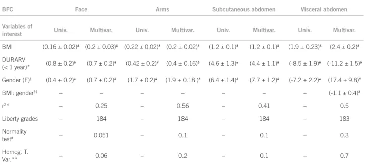
BFC Face Arms Subcutaneous abdomen Visceral abdomen
Variables of
interest Univ. Multivar. Univ. Multivar. Univ. Multivar. Univ. Multivar.
BMI (0.16 ± 0.02)δ (0.2 ± 0.03)δ (0.22 ± 0.02)δ (0.2 ± 0.02)δ (1.2 ± 0.1)δ (1.2 ± 0.1)δ (1.9 ± 0.23)δ (2.4 ± 0.2)δ
DURARV
(< 1 year)* (0.8 ± 0.2)
δ (0.7 ± 0.2)δ (0.42 ± 0.2)¥ (0.4 ± 0.16)δ (4.6 ± 1.3)α (4.4 ± 1.1)δ (-8.5 ± 1.9)δ (-11.2 ± 1.5)δ
Gender (F)§ (0.4 ± 0.2)α
(0.7 ± 0.2)δ
(1.7 ± 0.2)δ
(1.9 ± 0.18 )δ
(6.4 ± 1.4)δ
(7.7 ± 1.2)δ
(-7.2 ± 2.2)α (17.4 ± 9.8)¥
BMI: gender§§ – – – – – – – (-1.1 ± 0.4)δ
r2 // – 0.25 – 0.56 – 0.41 – 0.5
Liberty grades – 184 – 184 – 184 – 183
Normality
test# – 0.051 – 0.1 – 0.1 – 0.3
Homog. T.
Var.** – 0.06 – 0.2 – 0.1 – 0.7
BMI, body mass index (continuous variable); *duration of antiretroviral therapy use with exposure category < 1 year; §exposure category: female gender; §§interaction term between BMI and gender; //quality adjustment of the linear model; #p-value of the Kolmogorov-Smirnov test for normality; **p-value of Bartlett
test of homogeneity of variances; Univ., univariate estimation of the model; Multivar., multivariate estimation of the model ± standard deviation; BFC, body fat compartments; δ
< 0.01; α
< 0.05; ¥< 0.10.
Table 1 – Linear models of body fat compartments
bimodal distribution, and a cutof was set at one year. In the group of patients with more than 1 year of treatment (n = 85, ≈ 46%) the mean time was 3,418 (± 139) days of treatment. All these patients had HIV-LD. In patients with < 1 year of antiretroviral drug use (n = 102), the average time of treatment was 73 (± 7.2) days and there were no cases of HIV-LD. he signiicant diference in time be-tween the treatment groups (t = 24, p < 0.0001) and the in-terval of 475 days between the extremes of the two groups justiies the dichotomization of this variable used in the multivariate analysis.
he mean ultrasonographic measurements of the BFCs of face, arm, subcutaneous and visceral abdomen were re-spectively 3.43 (± 0.1) mm, 1.8 (± 0.13) mm, 13.4 (± 0.7), and 45.5 mm (± 1) mm for all patients. he univariate re-gression coeicients of the variables of interest (age, BMI, DURARV, and gender) and their signiicance values are shown in Table 1. he mean BMI of the study population
was 24.4 kg/m2 (± 0.28). here was no association between
BMI and gender (males: 24.5 (± 0.30) kg/m2); females:
24.1 (± 0.62) kg/m2, t = 0.76, p = 0.5), or between BMI
and treatment duration (group > 1 year: 24 (± 0.5) kg/m2;
group < 1 year: 25 (± 0.3) kg/m2); t = -1.713, p = 0.9). On
the other hand, patients with DURARV > 1 year were signiicantly older (49 (± 1)) than those with DURARV < 1 year (age 37 (± 0.9), t = 8.8, p < 0.0001).
In multivariate analysis, the variables BMI, gender, and time of treatment were signiicantly associated with all BFC measurements analyzed by ultrasound.
Females had 0.7 mm, 1.9 mm and 7.7 mm more fat, respectively, in the face, neck, and subcutaneous abdomen, on average, than the corresponding values of the respec-tive male compartments. As for the male visceral compart-ment, there was an increase of 7.2 mm in fat compared to the same compartment in females. he increase of one unit of BMI was associated with increased fat in all BFCs studied, and ranged from 0.2 mm in the face and arm to 1.2 mm and 2.4 mm in the subcutaneous and visceral ab-domen, respectively.
he thickness of body fat compartments was strongly associated with treatment time in both men and women. In those with DURARV < 1 year there was, respectively, an increase of fat of 0.4 mm and 0.7 mm, in the arm and face, and 4.4 mm in the subcutaneous abdominal compart-ment (Table 1). Conversely, in the visceral compartcompart-ment, patients with DURARV ≥ 1 year had on average 11 mm more visceral fat than patients with shorter treatment time (Table 1).
DISCUSSION
he Ministry of Health reported 592,914 cases of AIDS
(clinical syndrome) in Brazil, from 1980 until June 201018;
of these, 200,000 regularly receive antiretroviral
treat-ment19. In spite of the substantial increases in the quality
and length of survival of HIV patients since its irst use in
1996, this type of therapy has signiicant adverse efects20,
in addition to the diiculties of treatment compliance in the long term, which, so far, corresponds to a period that
encompasses the life of each patient with HIV/AIDS21.
he prevalence of abnormal fat redistribution in difer-ent studies is closely associated with the duration of anti-retroviral use, and other factors such as age, gender, body weight, and duration of HIV infection. Except for the last variable, of which information is inaccurate or unknown for most people with HIV, the others were studied in the
present study, as well as in other international studies3,7,8,
by regression models of fat compartments of interest. he gender of patients has a signiicant efect on changes in body composition of fat that occur during HIV infection; women, who usually have a higher proportion of fat mass when compared to men, lose disproportion-ately more fat, thus reducing the sexual diferences in body
composition of HIV patients over time22. Kotler et al.22 also
observed that the amounts of fat in subcutaneous com-partments are higher among women, when compared to men. he opposite is observed with respect to the visceral compartment: there is a smaller amount of fat in women when compared to men, and these observations were cor-roborated by the present study.
Height and weight have a strong inluence on the mass
of body cells, in relation to both lean mass and fat mass22.
As expected, a positive association between BMI and fat content of body compartments, particularly in the visceral compartment, was found. Conversely, there was no statis-tically signiicant association between BMI and length of use of antiretrovirals. his result suggests that the fat loss occurs in the same amounts in patients with diferent BMI values. In turn, the impact of this loss, when measured as loss relative to the initial weight, will be greater in those with lower BMI, as the loss of fat mass will be signiicantly higher in relation to the pre-existing fat mass.
he smaller amounts of fat measured in the peripheral compartments of patients with DURARV ≥ 1 year/HIV-LD compared with those with DURARV < 1 year/without HIV-LD, suggest a thinning of the arm and fat atrophy in the face and abdomen, possibly in response to treatment. In the latter (the stomach compartment compressible with the ingers), which is not normally measured by the clinical
diagnostic methods of HIV-LD5,23, there was a substantial
reduction of fat associated with longer time of treatment. Regarding the visceral compartment, there was a great-er amount of fat in the group receiving prolonged treat-ment (> 1 year), compared to the group that had started
treatment more recently (< 1). he visceral fat accumula-tion usually occurs in the clinical pictures of abnormal fat redistribution, but its presence alone does not deine the diagnosis of HIV-LD, as there are other diseases or disor-ders that also course with visceral obesity, such as meta-bolic syndrome. herefore, it is necessary to perform dif-ferential diagnosis before associating visceral obesity with HIV-LD.
In routine care, the diagnosis of HIV-LD is per-formed based on the self-assessment of patients and on the attending physician’s assessment (subjective informa-tion), due to the considerable advantages, such as low cost and lexibility of the language used in the question-naire, adjusted to the patient’s language skills. his
diag-nostic method was validated by Lichtenstein6, who, in a
multicenter U.S. study, evaluated 1,077 patients with HIV submitted to self-assessment and evaluation by their phy-sicians for signs of HIV-LD. Patients who had diagnostic concordance of HIV-LD in both evaluations were con-sidered as having HIV-LD.
In a study of objective deinition of lipodystrophy, in which 10 clinical and metabolic variables were used, and the variables related to body compartments were measured
by DEXA, the authors24 showed a sensitivity of 79% and a
speciicity of 80% for the diagnosis of HIV-LD. However, this method is currently restricted to health facilities with better infrastructure, as it relies on sophisticated metabolic tests and DEXA, which are not on the list of routine ex-aminations in most Brazilian hospitals. Ultrasound may represent an intermediate option between the high-tech methods, which have higher cost and lower availability in medical facilities, and methods based on subjective infor-mation only, which have lower cost and are widely avail-able, but also generally show problems with accuracy, reli-ability, and validity8.
Sectional studies with HIV patients, conducted in
Spain25,26, assessed the inter-exam reliability of ultrasound,
CT, and DEXA, when evaluating fat compartments in the subcutaneous abdomen and arm in patients with
lipoatro-phy, and found statistically signiicant correlations25,26. In
the arm, the Spearman’s rank correlation coeicient var-ied from 0.64 to 0.84, whereas in the subcutaneous abdo-men, these correlations, although weak, were statistically signiicant, and ranged from 0.34 (p-value < 0.05) to 0.40
(p = 0.001)25,26. Similarly, for the visceral fat, a good
repro-ducibility of ultrasound measurements when compared
with CT (r = 0.84) was observed26.
In another study, which measured visceral fat thickness by ultrasound in patients with HIV receiving antiretroviral
therapy versus those who had never received it, Guimarães
et al.27 also found diferences in visceral adiposity that
Most studies of inter-exam reliability that evaluated the ultrasonography for the face and clinical methods found a
signiicant correlation between them11,28,29. However, Carey
et al.30 found no signiicant association between the malar
region studied by ultrasound and other body regions stud-ied by diferent methods of measurement, such as CT and DEXA. It can be postulated that it would not be possible to establish correlation between the measurements, as the method used in this study did not maintain similar ob-servation conditions in the several procedures performed. An additional advantage of ultrasonography is the low variability of its measurements of subcutaneous
(< 5%)11,27,31 and visceral (2.6%) fat thickness when
per-formed by a single well-trained operator.
he multivariate model adjusted for ultrasonographic
data of the face had the worst quality of it (r2 = 0.25). Two
possible explanations can be proposed. First, this result suggests the need to include in these evaluations additional variables that were not studied in the present study. How-ever, no studies in the literature that used linear regression to study the diferences in body fat compartments in this population were found, so it is not possible to compare the present results with those of other studies.
he fact that the measurement of facial fat has the highest degree of technical diiculty among the proce-dures used in this study is noteworthy. Anatomical varia-tions and variability in facial shape and fat content of this body compartment require the ultrasonographist’s skill to produce an image that includes the anatomical reference points (the junction of the zygomatic and maxillary bones in the lower right eye socket, and adjacent muscle and fat tissues) (Figure 1). herefore, one possible explanation for the high variability observed in measurements of the face (and consequent low quality of it) would be the result of signiicant diferences in the form and content of the fat compartments of patients examined by ultrasound.
Simultaneously with the expansion of antiretroviral treatment provided to millions of HIV patients, there is ongoing development of new drugs with less toxic pro-iles, tending to replace irst and second-line antiretrovi-ral drugs, which are relatively inexpensive, efective, and widely prescribed, but are oten associated with metabolic disorders and HIV-LD. he economic impact of these sub-stitutions in antiretroviral programs with free and univer-sal access is likely to jeopardize the continued expansion of these programs for new patients and even the maintenance
of those already being treated32.
It is suggested that ultrasonography should be used to study the fat compartments, in parallel or in series with the clinical method of diagnosis of HIV-LD through-out through-outpatient-based treatment. his approach is similar to that which has been routinely used in relation to the monitoring of immunological and metabolic parameters
of patients in follow-up care. It is expected that ultrasound can be used as an important tool in early diagnosis of ab-normal fat redistribution, and can indicate patients for whom replacement of antiretroviral drugs closely associ-ated with HIV-LD by others with less pronounced side ef-fects is recommended.
REFERENCES
1. Signorini DJHP, Codeço CT, Carvalho MS, Campos DP, Monteiro MCM, Andrade MFC, et al. Efect of sociodemographic, clinical-prophylactic and therapeutic procedures on survival of AIDS patients assisted in a Brazilian outpatient clinic. Rev Bras Epidemiol. 2005;8(3)253-61.
2. Marconi VC, Grandits GA, Weintrob AC, Chun H, Landrum ML, Ganesan A, et al. Outcomes of highly active antiretroviral therapy in the context of univer-sal access to healthcare: the U.S. Military HIV Natural History Study. AIDS Res her. 2010;7(1):14.
3. Diehl LA, Dias JR, Paes AC, homazini MC, Garcia LR, Cinagawa E, et al.
Prevalence of HIV-associated lipodystrophy in Brazilian outpatients: relation with metabolic syndrome and cardiovascular risk factors. Arq Bras Endocrinol Metab. 2008;52(4):658-67.
4. Signorini DJ, Monteiro MC, Signorini DH, Eyer-Silva WA. Prevalence and de-terminant factors to lipid abnormalities among HIV-infected patients: a cross-sectional study of 812 patients. Arq Bras Endocrinol Metabol. 2010;54(6):583. 5. Santos CP, Felipe YX, Braga PE, Ramos D, Lima RO, Segurado AC. Self-per-ception of body changes in persons living with HIV/AIDS: prevalence and as-sociated factors. AIDS. 2005;19(Suppl 4):S14-21.
6. Lichtenstein KA, Ward DJ, Moorman AC, Delaney KM, Young B, Palella FJ Jr, et al. Clinical assessment of HIV-associated lipodystrophy in an ambulatory population. AIDS. 2001;15(11):1389-98.
7. Gelenske T, Bandeira EFFA, Alencar Ximenes RA, Lacerda de Melo HR, Mili-tao de Albuquerque MD, Carvalho EH, et al. Risk factors in human immuno-deiciency virus/acquired immunoimmuno-deiciency syndrome patients undergoing antiretroviral therapy in the state of Pernambuco, Brazil: a case-control study. Metab Syndr Relat Disord. 2010;8(3):271-7.
8. Safrin S, Grunfeld C. Fat distribution and metabolic changes in patients with HIV infection. AIDS.1999;13(18):2493-505.
9. Huang JS, Becerra K, Fernandez S, Lee D, Mathews WC. he impact of HIV-associated lipodystrophy on healthcare utilization and costs. AIDS Res her. 2008;5:14.
10. Associação Médica Brasileira, aSdE, Conselho Federal de Medicina, Federação Nacional dos Médicos. Classiicação Brasileira Hierarquizada de Procedimen-tos Médicos (CBHPM). São Paulo; 2010. [cited 2011]. Available from: http:// www.amb.org.br/teste/cbhpm/cbhpm_2010.pdf.
11. Martinez E, Bianchi L, Garcia-Viejo MA, Bru C, Gatell JM. Sonographic assess-ment of regional fat in HIV-1-infected people. Lancet. 2000;356(9239):1412-3. 12. Brasil. Recomendações para terapia antirretroviral em adultos infectados pelo HIV, 2008. [cited 2011 feb 2]. Available from: http://www.aids.gov.br/sites/de-fault/iles/consensoAdulto005c_2008montado.pdf.
13. Jones JL, Hanson DL, Dworkin MS, Alderton DL, Fleming PL, Kaplan MD, et al. Division of HIV/AIDS Prevention Surveillance and Epidemiology. Surveil-lance for AIDS-deining opportunistic illnesses, 1992-1997. Available from: http://www.cdc.gov/mmwr/preview/mmwrhtml/00056917.htm.
14. Radominski RB, Vezozzo DP, Cerri GG, Halpern A. O uso da ultrassonogra-ia na avalultrassonogra-iação da distribuição de gordura abdominal. Arq Bras Endocrinol Metab. 2000;44(1):5-12.
15. Dalgaard P. Introductory statistics with R. New York: Springer; 2002. 16. Team RDC. R: a language and environment for statistical computing. 2.11.
Vi-enna: R Foundation for Statistical Computing; 2011.
17. UNAIDS/WHO. Joint United Nations Programme on HIV/AIDS WHO. UN-AIDS report on the global UN-AIDS epidemic 2010. 2010.
18. Boletim Epidemiológico Aids. DST (Versão Preliminar). Brasília (DF); 2010. [cited 2011 feb 2]. Available from: http://www.aids.gov.br/sites/default/iles/ publicacao/2010/boletim2010_preliminar_pdf_34434.pdf.
19. Ministério da Saúde. DST. AIDS hepatites virais. Brasília (DF): Ministério da Saúde; 2011. [cited 2011 feb 4]. Available from: http://www.aids.gov.br/pagina/ quais-sao-os-antirretrovirais.
20. Villarroya F, Domingo P, Giralt M. Drug-induced lipotoxicity: lipodystrophy associated with HIV-1 infection and antiretroviral treatment. Biochim Bio-phys Acta. 2010;1801(3):392-9.
21. Monnerat BZ, Cerutti Junior C, Canicali SC, Motta TR. Clinical and biochemi-cal evaluation of HIV-related lipodystrophy in an ambulatory population from the Hospital Universitario Cassiano Antonio de Morais, Vitoria, ES, Brazil. Braz J Infect Dis. 2008;12(4):364-8.
23. Tien PC, Benson C, Zolopa AR, Sidney S, Osmond D, Grunfeld C. he study of fat redistribution and metabolic change in HIV infection (FRAM): methods, design, and sample characteristics. Am J Epidemiol. 2006;163(9):860-9. 24. Carr A, Emery S, Law M, Puls R, Lundgren JD, Powderly WG. An objective
case deinition of lipodystrophy in HIV-infected adults: a case-control study. Lancet. 2003;361(9359):726-35.
25. Martinez E, Milinkovic A, Bianchi L, Gatell JM. Considerations about the value of sonography for the measurement of regional body fat. AIDS. 2006;20(3):465-6.
26. Padilla S, Gallego JA, Masia M, Ardoy F, Hernandez I, Gutierrez F. Ultraso-nography and anthropometry for measuring regional body fat in HIV-infected patients. Current HIV Res. 2007;5(5):459-66.
27. Guimarães MM, Oliveira AR Jr, Penido MG, Queiroz LC, Goulart EM, Greco DB, et al. Ultrasonographic measurement of intra-abdominal fat thickness in HIV-infected patients treated or not with antiretroviral drugs and its correla-tion to lipid and glycemic proiles. Ann Nutr Metab. 2007;51(1):35-41.
28. Asensi V, Martin-Roces E, Carton J, Collazos J, Maradona J, Alonso A, et al.
Perirenal fat diameter measured by echography could be an early predictor of lipodystrophy in HIV type 1-infected patients receiving highly active antiret-roviral therapy. Clin Infect Dis. 2004;39(2):240-7.
29. Gulizia R, Vercelli A, Gervasoni C, Ortu M, Calliada F, Troia G, et al. Con-troversy concerning role of ultrasonographic lipoatrophy assessments in HIV patients. AIDS. 2006;20(5):789-90.
30. Carey D, Wand H, Martin A, Rothwell S, Emery S, Cooper DA, et al. Evalua-tion of ultrasound for assessing facial lipoatrophy in a randomized, placebo-controlled trial. AIDS. 2005;19(12):1325-7.
31. Asensi V, Martin-Roces E, Collazos J, Carton JA, Maradona JA, Alonso A, et al.
Association between physical and echographic fat thickness assessments and a lipodystrophy grading scale in lipodystrophic HIV patients: practical implica-tions. AIDS Res Hum Retroviruses. 2006;22(9):830-6.