138
IMAGE IN MEDICINE
Rev Assoc Med Bras 2012; 58(2):138-140
Bilateral immature ovarian teratoma in a 12-year-old girl: case report
LYLIANA COUTINHO RESENDE BARBOSA1, ANTÔNIO MARCOS COLDIBELLI FRANCISCO1, SILVÂNIADE CÁSSIA VIEIRA ARCHÂNGELO1, FABÍOLA CAMPOS MOREIRA SOARES2, MARIA CLÁUDIA TESSARI FERREIRA2, RENATA LEME MAIA2
1 PhD in Health Sciences, Professors of Obstetrics and Gynecology, Health Sciences School, Universidade do Vale do Sapucaí (UNIVAS), Pouso Alegre, MG, Brazil 2 MD, Resident Physicians at Department of Gynecology and Obstetrics, Hospital das Clínicas Samuel Libânio, UNIVAS, Pouso Alegre, MG, Brazil
Study conducted at Hospital das Clínicas Samuel Libânio – UNIVAS, Pouso Alegre, RS, Brazil
Correspondence to: Lyliana Coutinho Resende Barbosa – Rua Comendador José Garcia, 777 – CEP: 37550-000 – Pouso Alegre – MG, Brazil –
magibarbosa@gmail.com
INTRODUCTION
Immature teratoma (IMT) is a tumor composed of tis-sues from three germ layers (ectoderm, mesoderm, and endoderm), with immature or embryonic structures. his rare tumor comprises less than 1% of ovarian tu-mors and is considered the second most common germ cell tumor1.
IMT accounts for 10-20% of all ovarian neoplasias in women less than 20 years of age, with peak incidence between 15 and 19 years old, and 30% of the deaths from ovarian cancer in this age group. IMT rarely occurs dur-ing menopause2.
IMT may present as a calciied pelvic mass, abnor-mal uterine bleeding, or pelvic pain. he most common sites of dissemination are the peritoneum and the retro-peritoneal lymph nodes. Hematogenous spread to lungs, liver, or brain is unusual. hey present elevated levels of alpha-fetoprotein in 50% of cases3.
hese tumors are histologically graded (grades 1 to 3) based on the amount and degree of neuroepithelial cell component immaturity. Older patients tend to have
low-er-grade tumors than younger patients4. Immature
tera-tomas are rarely found bilaterally, while it is common to ind benign teratomas in the contralateral ovary1.
Peritoneal implants may be present at the time of surgical procedure, and the prognosis is strongly relat-ed to the histological grade of the tumor and implant (82% survival for patients with grade 1 lesions, 63% for grade 2 lesions, and 30% for grade 3 lesions)5.
A surgical approach is indicated for diagnosis, treat-ment, and staging (even if used for other ovarian tu-mors). Patients with completely resected tumors have approximately 94% chance of survival at ive years, while patients with partial resection have a survival expecta-tion of less than 50%. Because bilateralism is rare in this type of tumor, the surgery of choice consists of unilateral salpingo-oophorectomy with collection of samples from peritoneal implants6.
Radiotherapy does not appear to improve the progno-sis of patients. here is no indication of therapy besides surgery for tumors limited to one ovary (grade 1), except in cases of capsular rupture or ascites. In tumors of grade 2 or 3, or with bilateral implants or recurrences, adjuvant chemotherapy should be indicated in a regimen of vin-cristine, actinomycin and cyclophosphamide (VAC), or bleomycin, etoposide and cisplatin7,8.
Some studies advocate the use of alternative regimens with paclitaxel-carboplatin or docetaxel-carboplatin in order to prevent reproductive toxicity in patients under-going conservative surgery9.
Early diagnosis associated with immediate therapy and close follow-up are essential for long-term favorable
outcomes10. Patients undergoing surgery with
preserva-tion of the uterus and of one ovary have normal repro-ductive function11,12. he motivation for our case report is
due to the rarity of bilateral immature teratomas, as well as the fact that the patient’s age is below the average for the occurrence of these tumors.
CASE REPORT
A.V.D.O, 12 years old, admitted to the Gynecologic On-cology Service at Hospital das Clínicas Samuel Libânio – Universidade Vale do Sapucaí, with abnormal uterine bleeding for two months, associated with the presence of a pelvic mass. he patient had menarche at age 11 with irregular cycles, irst sexual intercourse six months prior to admission, contraception with male condoms.
139
BILATERALIMMATUREOVARIANTERATOMAINA 12-YEAR-OLDGIRL: CASEREPORT
Rev Assoc Med Bras 2012; 58(2):138-140
Figure 1 – Computed tomography of the abdomen showing a large complex abdominopelvic mass of probable ovarian etiology.
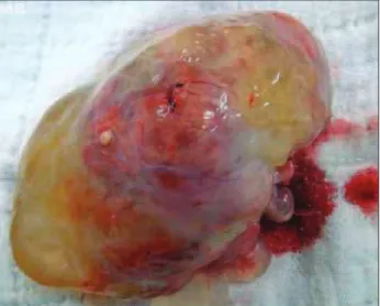
Figure 2 – Immature ovarian teratoma: macroscopic aspect.
Figure 3 – Photomicrographs of immature ovarian teratoma. On the left: an area of cartilage (C) and glandular tissue cys-tically dilated (G), 150x. On the right: portions of central nervous system tissue, 600x.
Normal preoperative tests were performed. An abdom-inal computed tomography revealed a complex mass in the pelvis, extending to mesogastrium; mild bilateral hydrone-phrosis; and presence of free luid in the pelvis (Figure 1).
he patient underwent laparotomy, with collection of ascitic luid for oncologic cytology. A complex cyst was found in the right ovary, weighing 680 g, multiseptated, with areas of capsular rupture. A right salpingo-oopho-rectomy was performed, and the material was sent for frozen section examination, diagnosed malignant. he let ovary showed increased volume with cystic areas (Figure 2).
Ater the family’s consent, a biopsy of the let ovary was performed, with frozen section examination, which was also positive for malignancy, followed by let salpin-go-oophorectomy, omentectomy, and periaortic and bi-lateral pelvic lymphadenectomy.
he patient had no complications in the postoperative period and was discharged in three days. She returned for removal of stitches, without incident.
he pathological report revealed a grade 3 immature teratoma; lymph nodes and omentum free of neoplastic involvement (Figure 3); ascitic luid positive for malig-nant neoplastic cells (FIGO stage 1C G3).
he patient was referred to the Clinical Oncology Department for adjuvant chemotherapy and received six cycles of vincristine/actinomycin/cyclophosphamide (VAC). Currently, she is being followed-up every six months without evidence of disease.
DISCUSSION
Bilateral immature teratoma is a rare condition, account-ing for 10% of cases3. Bilateral tumors are most oten
as-sociated with advanced staging, having a ive-year survival rate of 80.7%, compared with a survival rate of 93.6% for unilateral tumors13.
he degree of cell immaturity (grade 3) is another ad-verse prognostic factor, with high rate of reincidence14.
hese factors justify the radical approach taken, to the detriment of the patient’s reproductive future. Some au-thors advocate conservative treatment in germ cell tumors
grades 1 and 215. he tumor marker most commonly
relat-ed to immature teratoma is alpha-fetoprotein3. Diagnosis
of immature ovarian teratoma by tumor markers appears to be more sensitive when combined with detection of Ca125, Ca153, and alpha-fetoprotein16.
Imaging diagnosis of immature teratoma appear simi-lar to mature teratoma due to its cystic appearance with fat content. One way to distinguish them would be the pres-ence of contrast on computed tomography or magnetic resonance imaging17.
140
IMAGEINMEDICINE
Rev Assoc Med Bras 2012; 58(2):138-140 REFERENCES
1. Outwater EK, Siegelman ES, Hunt JL. Ovarian teratomas: tumor types and
im-aging characteristics. Radiographics. 2001;21(2):475-90.
2. Chabaud-Williamson M, Netchine I, Fasola S, Larroquet M, Lenoir M, Patte C,
et al. Ovarian-sparing surgery for ovarian teratoma in children. Pediatr Blood Cancer. 2011;57(3):429-34.
3. Saba L, Guerriero S, Sulcis R, Virgillio B, Melis G, Mallaraini G. Mature and
immature ovarian teratomas: CT, US and MR imaging characteristics. Eur J Radiol. 2009;72(3):454-63.
4. Ulbright TM. Germ cell tumors of the gonads: a selective review emphasizing
problems in diferential diagnosis, newly appreciated, and controversial issues. Mod Pathol. 2005;18(Suppl 2):S61-S79.
5. Abiko K, Mandai M, Hamanishi J, Matsumura N, Baba T, Horiuchi A, et al.
Oct4 expression in immature teratoma of the ovary: relevance to histologic grade and degree of diferentiation. Am J Surg Pathol. 2010;34(12):1842-8.
6. Devaja OM, Papadopoulos Andreas J. Current management of immature
tera-toma of the ovary. Arch Oncol. 2000;8(3):127-30.
7. Kurata A, Hirano K, Nagane M, Fujioka Y. Immature teratoma of the ovary
with distant metastases: favorable prognosis and insights into chemotherapeu-tic retroconversion. Int J Gynecol Pathol. 2010;29(5):438-44.
8. Mangili G, Scarfone G, Gadducci A, Sigismondi C, Ferrandina G, Scibilia G, et
al. Is adjuvant chemotherapy indicated in stage I pure immature ovarian tera-toma (IT)? A multicentre Italian trial in ovarian cancer (MITO-9). Gynecol Oncol. 2010;119(1):48-52.
9. Chen CH, Yang MJ, Cheng MH, Yen MS, Lai CR, Wang PH. Fertility
preser-vation with treatment of immature teratoma of the ovary. J Chin Med Assoc. 2007;70(5):218-21.
10. Tanaka T, Toujima S, Utsunomiya T, Yukawa K, Umesaki N. Experimental characterization of recurrent ovarian immature teratoma cells ater optimal surgery. Oncol Rep. 2008;20(1):13-23.
11. Barksdale EM, Obokhare I. Teratomas in infants and children. Curr Opin Pediatr. 2009; 21(3):344-9.
12. Weinberg LE; Lurain JR; Singh DK; Schink JC. Survival and reproductive outcomes in women treated for malignant ovarian germ cell tumors. Gynecol Oncol. 2011;121(2): 285-9.
13. Mahdi H, Kumar S, Seward S, Semaan A, Batchu R, Lockhart D, et al. Prog-nostic impact of laterality in malignant ovarian germ cell tumors. Int J Gynecol Cancer. 2011;21(2): 257-62.
14. Lai CH, Chang TC, Hsueh S, Wu TI, Chao A, Chou HH, et al. Outcome and prognostic factors in ovarian germ cell malignancies. Gynecol Oncol. 2005;96(3):784-91.
15. Ghaemmaghami F, Karimi Zarchi M, Naseri A, Mousavi AS, Gilani MM, Ramezanzadeh F, et al. Fertility sparing in young women with ovarian tumors. Clin Exp Obstet Gynecol. 2010;37(4):290-4.
16. Chen C, Li JD, Huang H, Feng YL, Wang LH, Chen L. Ai Zheng. Diagnostic value of multiple tumor marker detection for mature and immature teratoma of the ovary. Ai Zheng. 2008;27(1):92-5.
