Complex 121 Are Associated with Distinct Clinical
Entities
Kevin Kurt1, Jean-Philippe Rasigade2, Frederic Laurent2, Richard V. Goering3, Helena Zˇemlicˇkova´4, Ivana Machova4, Marc J. Struelens5, Andreas E. Zautner6, Silva Holtfreter7, Barbara Bro¨ker7,
Stephen Ritchie8, Sin Reaksmey9, Direk Limmathurotsakul10, Sharon J. Peacock10,11, Christiane Cuny1, Franziska Layer1, Wolfgang Witte1, Ulrich Nu¨bel1*
1Robert Koch Institute, Wernigerode, Germany,2National Reference Center for Staphylococci, Faculty of Medicine, University of Lyon, Lyon, France,3Creighton University School of Medicine, Omaha, Nebraska, United States of America,4National Institute of Public Health, Prague, Czech Republic,5European Centre for Disease Prevention and Control, Stockholm, Sweden,6Institut fu¨r Medizinische Mikrobiologie, Universita¨tsmedizin Go¨ttingen, Go¨ttingen, Germany,7Institute of Immunology and Transfusion Medicine, Ernst Moritz Arndt University, Greifswald, Germany,8Department of Molecular Medicine and Pathology, University of Auckland, Auckland, New Zealand,9Angkor Hospital for Children, Siem Reap, Cambodia,10Faculty of Tropical Medicine, Mahidol University, Bangkok, Thailand,11Department of Medicine, University of Cambridge, Cambridge, United Kingdom
Abstract
We investigated the population structure ofStaphylococcus aureusclonal complex CC121 by mutation discovery at 115 genetic housekeeping loci from each of 154 isolates, sampled on five continents between 1953 and 2009. In addition, we pyro-sequenced the genomes from ten representative isolates. The genome-wide SNPs that were ascertained revealed the evolutionary history of CC121, indicating at least six major clades (A to F) within the clonal complex and dating its most recent common ancestor to the pre-antibiotic era. The toxin gene complement of CC121 isolates was correlated with their SNP-based phylogeny. Moreover, we found a highly significant association of clinical phenotypes with phylogenetic affiliations, which is unusual forS. aureus. All isolates evidently sampled from superficial infections (including staphylococcal scalded skin syndrome, bullous impetigo, exfoliative dermatitis, conjunctivitis) clustered in clade F, which included the European epidemic fusidic-acid resistant impetigo clone (EEFIC). In comparison, isolates from deep-seated infections (abscess, furuncle, pyomyositis, necrotizing pneumonia) were disseminated in several clades, but not in clade F. Our results demonstrate that phylogenetic lineages with distinct clinical properties exist within anS. aureusclonal complex, and that SNPs serve as powerful discriminatory markers, able to identify these lineages. All CC121 genomes harboured a 41-kilobase prophage that was dissimilar toS. aureus phages sequenced previously. Community-associated MRSA and MSSA from Cambodia were extremely closely related, suggesting this MRSA arose in the region.
Citation:Kurt K, Rasigade J-P, Laurent F, Goering RV, Zˇemlicˇkova´ H, et al. (2013) Subpopulations ofStaphylococcus aureusClonal Complex 121 Are Associated with Distinct Clinical Entities. PLoS ONE 8(3): e58155. doi:10.1371/journal.pone.0058155
Editor:Malcolm James Horsburgh, University of Liverpool, United Kingdom
ReceivedJuly 30, 2012;AcceptedFebruary 4, 2013;PublishedMarch 7, 2013
Copyright:ß2013 Kurt et al. This is an open-access article distributed under the terms of the Creative Commons Attribution License, which permits unrestricted use, distribution, and reproduction in any medium, provided the original author and source are credited.
Funding:This work was partially funded by the European Commission, Framework Programme 7 (project TROCAR). No additional external funding was received for this study. The funders had no role in study design, data collection and analysis, decision to publish, or preparation of the manuscript.
Competing Interests:The authors have declared that no competing interests exist.
* E-mail: nuebelu@rki.de
Introduction
Staphylococcus aureus is one of the most common human pathogens and causes a wide range of infections, from skin and soft tissue infections (SSTIs) to life threatening diseases such as pneumonia, endocarditis, bacteremia and toxic-shock syndrome [1,2]. Among SSTI, two clinical entities predominate: (i) superficial infections that affect the upper skin layers exclusively (stratum corneum and stratum granulosum) and (ii) deep-seated infections associated with formation of abscesses such as furuncles, whitlows, and sweat-gland abscesses [3,4]. Superficial infections are associated with formation of exfoliative toxins, most commonly exfoliative toxins A (ETA) and B (ETB) [5–9]. Exfoliative toxins are structurally related to a family of serine proteases and associated with the binding and cleavage of desmosomal cadherins, in particular desmoglein-1 (Dsg-1). The degradation
The association of the two different clinical manifestations ofS. aureusinfections with either exfoliative toxins ETA and ETB or the PVL toxin, respectively, is particularly well documented for S. aureus clonal complex CC121 [4,14–16]. Complex CC121 is globally distributed, colonizing the anterior nares of asymptomatic subjects [14,17–21]; however, it is also a common cause of both superficial skin infections [9,14,15] and deep-seated infections [4,14–16]. Commonly, CC121 isolates are susceptible to methi-cillin. Recently, however, methicillin resistance had been found in CC121 among community-associated methicillin-resistantS. aureus
(caMRSA) from Cambodia [19].
In the present study, we examined the population structure of CC121 on the basis of polymorphisms discovered in 115 genetic loci scattered around the staphylococcal genome. We reconstruct-ed the evolutionary origin of caMRSA from PVL-producing methicillin-susceptible S. aureus in South-East Asia. We demon-strate that distinct phylogenetic lineages within CC121 are associated with specific courses of disease.
Results and Discussion
Global Population Structure
We tested for sequence polymorphisms at 115 housekeeping loci of about 400 basepairs each (Table S1), in total covering about 1.7% (46,811 basepairs) of the genome from each of 154S. aureus
CC121 isolates. Among these isolates, 138 displayed sequence type ST121, the presumed ancestral type of clonal complex CC121 (Table S2). Eleven isolates displayed various single-locus variants of ST121, and five isolates displayed ST123, a double-locus variant (Table S2). Onehundred-fiftythree isolates had been sampled between 1987 and 2009 in 27 countries on five continents (Table S2). In addition, we included the phage-propagating strain PS71 from approximately 1953 [22]. We identified 304 single-nucleotide polymorphisms, 5 insertions and 7 deletions ranging in size from 1 to 12 basepairs (Table S3). SNPs included 93 synonymous base substitutions and 211 non-synonymous substi-tutions (Table S3). Based on these polymorphisms, our 154 isolates were assigned to 121 haplotypes (Figure 1). Ninety-two SNPs were parsimony-informative (i. e., the respective derived alleles were found in more than one haplotype) and the level of homoplasy was very low (homoplasy index, 0.03). The nucleotide diversity = was 0.0002660.00001, which is nearly three times higher than the nucleotide diversity for S. aureus ST5 [23]. A SNP-based, maximum-likelihood phylogenetic analysis revealed at least six major clades, A to F, withinS. aureusCC121 (Figure 1).
We applied a Bayesian coalescent method of phylogenetic inference implemented in the BEAST program [24] to estimate evolutionary rates and divergence times for CC121. Based on our set of DNA sequences from isolates that had been sampled between 1953 and 2009 (Table S2), we estimated the nucleotide substitution rate at 261026
substitutions per nucleotide site per year (95% confidence intervals, 1.261026 to 2.861026). This short-term evolutionary rate is similar to rates previously reported forS. aureus[25,26]. Based on this rate and the sequence variation observed in our dataset, the most recent common ancestor of CC121 was dated to 1884 (95% confidence interval, 1827 to 1925). This age estimate should be interpreted with caution because it is based on a relatively long extrapolation from available sequence data (note the large confidence interval). However, it is several fold longer than estimates available for MRSA [25–27], which might reflect the widespread endemic nature of S. aureus
CC121 that precedes the antibiotic era.
The 154 isolates of our study displayed 38 differentspa types, with sixspatypes covering 67% of all isolates (Figure S1). Somespa
types were restricted to particular phylogenetic clades (t314 to clade B, t940 to clade C, t435 to clade D). However, the association of several otherspatypes (t284, t170, t272, t2155) with SNP-based phylogeny was imperfect, as each of them was found in several, unrelated haplotypes (Figure S2). The maximum mono-phyletic clade (MC) statistics calculated for thesespatypes by using BaTS software [28] confirmed the absence of significant associ-ations with phylogeny, because they were not significantly different from MC values for a randomized, null distribution (Table S4). These results suggestedspahomoplasies, similar to previous reports on otherS. aureussequence types [23,25,29].
Phylogeography
The isolates’ phylogenetic relationships reflect their geographic origin to a considerable extent, suggesting that specific subclones of CC121 are endemic in the regions sampled (Figure 1). Accordingly, for several geographic regions, Bayesian analysis with BaTS software indicated associations of geographic origins with phylogeny (Table S4). For example, clade A encompasses all 12 isolates from Oceania, including strains from New Zealand, Samoa, Tonga and Fiji. Similarly, clade C encompasses six out of seven isolates from South America, including Peru, Panama, Paraguay and the Bahamas. Further, all 20 isolates in clade E are from Southeast-Asia, including Thailand and Cambodia, clade B contains most of the isolates from Africa (Nigeria, Uganda, Togo, South Africa and Kenya), and clades D and F contain isolates mostly from Europe. Isolates from Europe are comparatively more diverse (Figure 1), possibly due to more extensive sampling coverage from this region, as 45% of the isolates originate from Europe.
The epidemic European fusidic acid-resistant impetigo clone (EEFIC), which is characterized by exfoliative toxin production, the fusB gene and, most commonly, spa type t171 [15], is a subclone of clade F (Figure 1). Evidently, EEFIC has spread clonally throughout several European countries [30,31], and here we report that EEFIC has been present also in Germany and the Czech Republic since at least 2007 and 2005, respectively (Table S2).
Our analysis included five isolates of ST121 community-associated MRSA from Cambodia, which had been reported only recently [19,32]. These MRSA isolates are very closely related to each other and to MSSA from the same area, displaying a maximum distance of only one point mutation in the 47 kb of DNA analysed (Figure 1). This result suggests that ST121 MRSA arose locally in Cambodia (and only once), which had been assumed previously based on the high prevalence of methicillin-susceptible S. aureus (MSSA) ST121 in the country and similar antibiotic resistance patterns among MRSA ST121 and MSSA ST121 from the same geographic region [19].
Comparative Genomics
more limited data (Figure 1). Genome sequences provided no evidence of recombination (homoplasy index, 0.05; no recombi-nation detected with Recombirecombi-nation Detection Program RDP 3.44). In addition, sequencing reads were assembled de-novo by applying Newbler software, revealing the gene content for each of the sequenced genomes (Table S6). All CC121 isolates harboured a 41-kb prophage that was dissimilar from S. aureus phages sequenced previously with respect to DNA sequence, gene content, integrase group, and capsid-gene based phage type (Table S5). The prophage encoded 55 open-reading frames, most of which could not be assigned any specific functions based on sequence similarity (Figure S3). CC121 genomes contained one to three additional prophages, including an integrase group Sa1
phage encoding theetagene or a groupSa2phage encoding the PVL gene (Table S6). Each genome had three pathogenicity islands (SaPI), with strain-specific variation (Table S6). MRSA carried a methicillin-resistance island that shared.99% sequence similarity with previously reported SCCmecV(sequence accession number GQ902038 [33]).
Because methicillin-resistant CC121 had not been found until recently, we investigated whether CC121 genomes may lack the DNA sequences necessary for integration of the SCCmecelement (the genetic determinant of methicillin resistance in staphylococci). SCCmecintegrates into a specific attachment site (attB) at the 3’-end of a gene namedorfXin the staphylococcal chromosome, and it has been proposed that the core of the attachment site itself and
the 100 basepairs left from it need to be highly conserved to enable efficient insertion or excision of the SCCmecelement [34]. In the ten genomes we have sequenced, this chromosomal region was identical to that from another ST121 isolate [34], except for a single point mutation in two ST121-MRSA and their close, methicillin-susceptible relative from Cambodia, indicating that the
attBsequence in CC121 does not prevent SCCmecintegration. It has also been proposed that a presumptive mobile genetic element encoding an enterotoxin gene (seh) interferes with SCCmecmotility, when inserted downstream of the SCCmecattachment site [35]. All of our CC121 genomes had a similar element at this position, encoding a different enterotoxin gene. However, this element was also present in ST121-MRSA, so it does not appear to hinder integration of SCCmecin CC121.
Correlation of Clinical Phenotype and Toxin Gene Complement with Phylogeny
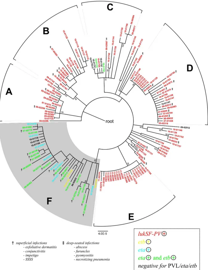
Bayesian analysis by using BaTS software indicated that the isolate toxin gene complement was strongly associated with their SNP-based phylogeny, because the MC values calculated for the presence of the toxin genes, the association index (AI) and the parsimony score (PS) all were significantly different from statistics estimated for a null distribution; p#0.01; Table S4). Clade F encompassed 35 isolates, 21 of which carried the exfoliative toxin genesetaandetb, ten carried either one of the two, and four tested Figure 1. Phylogenetic relationships of 154 CC121 isolates.Maximum-likelihood phylogenetic tree based on 304 SNPs ascertained in 115 genetic loci (47 kb total) and annotated with the country of origin and year of isolation. The continent of origin is indicated by the color of the isolate’s name: blue, Europe; orange, Asia; green, Oceania; red, North America; yellow, South America; black, Africa. Bootstrap values are shown where $85%. The scale bar represents substitutions per SNP site. Asterisks indicate isolates selected for whole-genome sequencing. The European epidemic fusidic acid-resistant impetigo clone (EEFIC) was identified on the basis of fusidic acid resistance, PCR detection of thefusBgene,spatyping (t171), and MLST (ST123).
doi:10.1371/journal.pone.0058155.g001
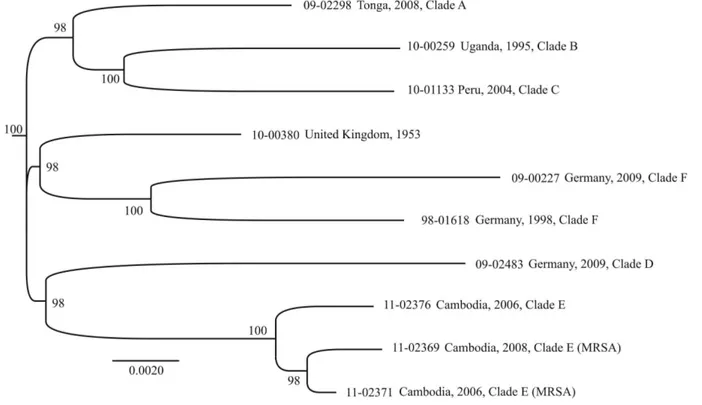
Figure 2. Maximum-likelihood phylogenetic tree based on an alignment of 1,828 SNPs ascertained in 2,209 kb from pyro-sequenced genomes.Bootstrap values.90% are indicated.
negative for both,etaandetb(Figure 3). We noted that three clade F isolates (97-01548, 97-01848-1, 00-01004-1) were etb-negative now, even though they had been testedetb-positive several years ago according to records in our strain collection database, presumably reflecting the instability of the etb carrying plasmid during long-term storage or sub-cultivation. None of the isolates in clade F tested positive for thelukS/Fgene encoding the PVL toxin. In contrast, isolates outside of clade F were preferentially linked with the presence of thelukS/Fgenes (Figure 3). Upon sequencing thelukS/Fgenes, we identified sequence variants R, H1, H2, and H3, all of which had been reported previously to be present in multiple other clonal complexes of S. aureus(24), plus six novel sequence variants (Table S7), indicating that CC121 has acquired thelukS/F-carrying phage multiple times.
It is not clear why lukS/F and eta/etb genes are mutually exclusive in CC121.LukS/Fandetareside on unrelated prophages that integrate at different positions into the genome, and etb is commonly encoded by a plasmid, whose mode of interaction with prophages is not known. We noted that one of the genomes from clade F (isolate 09-00227 with eta) lacked the attachment site for thelukS/F-carrying phage [36] due to a deletion event. However, by using a dedicated PCR (Table S8), we found that this deletion was not a common feature of clade F isolates. Rather, the attachment site was present in all clade F isolates except for those affiliated to the EEFIC clade (Figure 1) and one other isolate. It is possible that the observed association between gene complement and phylogeny is driven simply by the extensive international spread of clonal strains that carry one or other toxin genes, rather than any mechanistic hindrances to gene acquisition (Figure 1, Figure 3). For example, antibiotic usage promotes the spread of resistantS. aureusstrains, but in our sample of CC121 no specific resistance traits were associated with clade F (Table S2). Reportedly, Staphylococcus aureus strains with exfoliative toxins frequently also produce EDIN exotoxins [37]. Because the exact role of EDIN in staphylococcal infections has not yet been explored, this issue was not followed here.
We were able to ascertain the type of disease associated with 46 of the isolates in our international CC121 collection. Of these, 35 infections could be unequivocally classified as either superficial or deep-seated, whereas 11 were wound infections or unspecified ’skin and soft tissue infections’ (Table S2). Strikingly, all isolates associated with superficial infections (n = 14) (including SSSS, bullous impetigo, exfoliative dermatitis, and one case of conjunc-tivitis) clustered in clade F (Figure 3). In contrast, isolates from deep-seated infections (n = 21) (abscesses, furuncles, pyomyositis, necrotizing pneumonia) were disseminated in several clades, but not in clade F (Figure 3). Accordingly, the association of the clinical phenotype with the phylogenetic affiliation of the respective isolates was highly significant (i. e., MC, AI, and PS statistics were significantly different from those for a null distribution; p#0.01; Table S4).
Links between specific types of disease and phylogenetic groupings within the species S. aureus have been highly unusual findings in the past. For example, MLST-defined clonal lineages of
S. aureus generally do not correlate with virulence properties or with any other phenotypic traits that may be of interest to clinicians [38]. Our results demonstrate thatS. aureusstrains with specific clinical properties exist within clonal complexes, and that SNPs may be powerful discriminatory markers to identify them (Table S9). In the light of accelerated and abundant pathogen genome sequencing [25], we anticipate that additional S. aureus
strains with association to specific clinical symptoms may be recognized in the future, and that (SNP-based) typing could eventually be used to predict the pathogenic potential of a given
isolate. In many cases, however, the propensity to cause a specific clinical picture will depend on interactions between multiple proteins rather than a single toxin.
Methods
Bacterial Isolates
The sources and characteristics ofS. aureusisolates are provided in Table S2. Some of these isolates had been included in previous studies as indicated in Table S2 [16,19,22,32,39]. To represent global diversity, we included 154 isolates from 27 countries on five continents. Antimicrobial susceptibility was tested by using the microbroth dilution method according to DIN58940 and applying the EUCAST breakpoints.Spa-typing was performed according to the Ridom StaphType standard protocol (www.ridom.org) and
spa-types were assigned using the respective StaphType software (Ridom GmbH, Wu¨rzburg, Germany). MLST was performed as described previously [40]. Primers for the detection ofeta-,etb- and
lukSF-PV-genes are summarized in Table S8. The different lukSF-PVsequence variants were determined as specified by OHaraet al.
[41].
Mutation Discovery by dHPLC
Mutation discovery was performed as described previously [23,42]. Briefly, PCR-amplified gene fragments were analyzed by dHPLC (WaveR Nucleic Acid Fragment Analysis System, Transgenomic). The 115 genetic loci investigated, PCR primers, and dHPLC conditions are listed in Table S1. Subsequently, mutations were verified in all affected isolates by capillary sequencing. Discovered polymorphisms and their properties are summarized in Table S3.
Data Analysis
Sequences from 115 loci were concatenated for each of 154 isolates, resulting in a 46,811-bp sequence alignment. A maximum likelihood tree based on this alignment was constructed with PhyML 3.0.1. The ancestral node was determined by including the genome sequence from distantly relatedS. aureusN315 (ST5; GenBank accession number BA000018). DnaSP was used to calculate the mean pair-wise distance between alleles at synony-mous (Ks) and non-synonysynony-mous (Ka) sites and to estimate the nucleotide diversity (p) and nucleotide variation (hw) [43]. The homoplasy index was estimated with Paup 4.0. Short-term evolutionary rates and divergence dates were estimated by applying the BEAST software, version 1.6.2 (http://beast.bio.ed. ac.uk/) [24], dating sequences with the year of isolate sampling, and running 108iterations after a burn-in phase of 106iterations. BEAST results were virtually independent from applied clock models (strict, relaxed) and tree priors (constant population size, Bayesian skyline).
Figure 3. Toxin gene complement and clinical phenotype.Maximum likelihood phylogenetic tree as in Figure 1, indicating the presence of toxin genes lukSF-PV, eta, and etb (colours) and clinical phenotype where this was reported (symbols:{, superficial infection, i. e., impetigo, staphylococcal scalded skin syndrome, conjunctivitis, or exfoliative dermatitis);{, deep-seated infection (abscess, furuncle, pyomyositis, necrotizing pneumonia).
randomizations, and the null hypothesis was rejected if p,0.05 (Table S4).
Genome Sequencing
Staphylococcal genome sequences were generated commercially (GATC, Konstanz, Germany) on a 454 FLX machine, applying FLX+chemistry and resulting in 12- to 21-fold average coverage. De-novo assemblies with 454 Newbler software resulted in 68 to 156 contigs (.500 bp) per genome. Sequence annotation was performed by comparisons to previously published S. aureus
genomes, applying the annotation tool implemented in Kodon software (Applied Maths), and by BLAST searches in the GenBank database (http://blast.ncbi.nlm.nih.gov/Blast.cgi). In addition, prophage sequences were identified with PHAST (phage search tool, available at http://phast.wishartlab.com/[44]). Ge-nome-wide SNPs were ascertained by mapping the sequencing reads onto the N315 genome sequence (GenBank accession number BA000018), applying SSAHA2 software (available at http://www.sanger.ac.uk/resources/software/ssaha2/) and filter-ing the resultfilter-ing output for SNPs with a minimum consensus quality of 30, a minimum mapping quality of 30, and a minimum coverage of 5. SNPs in mobile genetic elements (SaPIs, prophages, transposons, IS elements), repetitive regions, homopolymeric regions, and within 100 bp from contig ends were removed manually, resulting in a dataset of 1,828 SNPs ascertained in the core genome of 2,208,736 basepairs. An alignment of core genome sequences from ten CC121 isolates and from the reference N315 was used to reconstruct a maximum likelihood phylogenetic tree with PhyML3.01. The Recombination Detection Program (RDP 3.44, [45]) was applied to screen the alignment for evidence of recombination based on the RDP, Geneconv, Chimaera, MaxChi, Bootscan, and SiScan methods (see RDP manual), applying default parameters and p,0.05.
Sequence data has been deposited at the NCBI Sequence Read Archive (http://www.ncbi.nlm.nih.gov/sra) under accession num-bers SRX209921 to SRX209927, SRX208966, and SRX209760.
Supporting Information
Figure S1 Distribution of the six major spa-types. Maximum likelihood phylogenetic tree based on 304 SNPs from a selection of housekeeping genes annotated with the respective
spa-types, indicated by the following colors: red, t159; light blue, t284; green, t314; yellow, t435; blue, t645; magenta, t940. (PDF)
Figure S2 Distribution of homoplasious spa-types. Maximum likelihood phylogenetic tree based on 304 SNPs from a selection of housekeeping genes annotated with the respective
spa-types. (PDF)
Figure S3 Prophage WSaCC121. Prophage modules are color coded: lysogeny, red; DNA replication, orange;
transcrip-tional regulation, yellow; DNA packaging and head, green; tail, blue; lysis, magenta; hypothetical proteins, black. Selected genes are indicated: int, integrase; rep, repressor; p.rep*, putative repressor HTH protein; ant, antirepressor; hel, helicase; pol, polymerase; pri, primase; terS/L, small and large subunit terminase; pro, portal; mhp, major head protein; tape, tape measure protein (tmp); hol, holin; ami, amidase.
(PNG)
Table S1 Genetic loci and PCR primers used for WAVE analysis.
(XLS)
Table S2 Bacterial isolates. (XLS)
Table S3 Polymorphisms discovered in the genome fragments from 154 isolates.
(XLS)
Table S4 Results of Bayesian tip-association signifi-cance testing.
(XLSX)
Table S5 Predicted ORFs in ST121 Phage. (XLSX)
Table S6 Genome content. (XLSX)
Table S7 PVL nucleotide variation. (XLS)
Table S8 PCR primers. (XLS)
Table S9 Mutations defining clades within CC121. (XLSX)
Acknowledgments
We thank Mike Henkel, Annette Weller, and the staff at our central sequencing lab for excellent technical assistance, and Elena Fuchs for PVL sequences. We thank Emma Nickerson, Premjit Amornchai and Vanaporn Wuthiekanun for their assistance in isolatingS. aureusin Cambodia and Thailand, and we are grateful to the staff of Angkor Hospital for Children, Siem Reap, Cambodia.
Author Contributions
Contributed to editing the manuscript: JR F. Laurent RVG HZ MJS AEZ SH BB S. Ritchie S. Reaksmey DL SJP. Provided the idea to study the lukS/F vs. eta/etb gene dichotomy in ST121: WW. Conceived and designed the experiments: KK WW UN. Performed the experiments: KK. Analyzed the data: KK F. Layer UN. Contributed reagents/materials/ analysis tools: JR F. Laurent RVG HZ MJS AEZ SH BB S. Ritchie S. Reaksmey DL SJP CC WW IM. Wrote the paper: KK CC F. Layer WW UN.
References
1. Iwatsuki K, Yamasaki O, Morizane S, Oono T (2006) Staphylococcal cutaneous infections: invasion, evasion and aggression. J Dermatol Sci 42: 203–214. 2. Lowy FD (1998)Staphylococcus aureusinfections. N Engl J Med 339: 520–532. 3. Durupt F, Mayor L, Bes M, Reverdy ME, Vandenesch F, et al. (2007)
Prevalence ofStaphylococcus aureustoxins and nasal carriage in furuncles and impetigo. Br J Dermatol 157: 1161–1167.
4. Masiuk H, Kopron K, Grumann D, Goerke C, Kolata J, et al. (2010) Association of recurrent furunculosis with Panton-Valentine leukocidin and the genetic background ofStaphylococcus aureus. J Clin Microbiol 48: 1527–1535. 5. Bukowski M, Wladyka B, Dubin G (2010) Exfoliative Toxins ofStaphylococcus
aureus. Toxins (Basel) 2: 1148–1165.
6. Lina G, Piemont Y, Godail-Gamot F, Bes M, Peter MO, et al. (1999) Involvement of Panton-Valentine leukocidin-producingStaphylococcus aureusin primary skin infections and pneumonia. Clin Infect Dis 29: 1128–1132. 7. Gravet A, Couppie P, Meunier O, Clyti E, Moreau B, et al. (2001)Staphylococcus
aureusisolated in cases of impetigo produces both epidermolysin A or B and LukE-LukD in 78% of 131 retrospective and prospective cases. J Clin Microbiol 39: 4349–4356.
9. Ruzickova V, Pantucek R, Petras P, Machova I, Kostylkova K, et al. (2012) Major clonal lineages in impetigoStaphylococcus aureusstrains isolated in Czech and Slovak maternity hospitals. Int J Med Microbiol.
10. Amagai M, Yamaguchi T, Hanakawa Y, Nishifuji K, Sugai M, et al. (2002) Staphylococcal exfoliative toxin B specifically cleaves desmoglein 1. J Invest Dermatol 118: 845–850.
11. Holmes A, Ganner M, McGuane S, Pitt TL, Cookson BD, et al. (2005) Staphylococcus aureus isolates carrying Panton-Valentine leucocidin genes in England and Wales: frequency, characterization, and association with clinical disease. J Clin Microbiol 43: 2384–2390.
12. Genestier AL, Michallet MC, Prevost G, Bellot G, Chalabreysse L, et al. (2005) Staphylococcus aureusPanton-Valentine leukocidin directly targets mitochondria and induces Bax-independent apoptosis of human neutrophils. J Clin Invest 115: 3117–3127.
13. Kaneko J, Kamio Y (2004) Bacterial two-component and hetero-heptameric pore-forming cytolytic toxins: structures, pore-forming mechanism, and organization of the genes. Biosci Biotechnol Biochem 68: 981–1003. 14. Larsen AR, Skov RL, Jarlier V, Henriksen AS (2008) Epidemiological
differences between the UK and Ireland versus France inStaphylococcus aureus isolates resistant to fusidic acid from community-acquired skin and soft tissue infections. J Antimicrob Chemother 61: 589–594.
15. O’Neill AJ, Larsen AR, Skov R, Henriksen AS, Chopra I (2007) Character-ization of the epidemic European fusidic acid-resistant impetigo clone of Staphylococcus aureus. J Clin Microbiol 45: 1505–1510.
16. Rasigade JP, Laurent F, Lina G, Meugnier H, Bes M, et al. (2010) Global distribution and evolution of Panton-Valentine leukocidin-positive methicillin-susceptible Staphylococcus aureus, 1981–2007. J Infect Dis 201: 1589–1597. 17. Aires de Sousa M, Conceicao T, Simas C, de Lencastre H (2005) Comparison of
genetic backgrounds of methicillin-resistant and -susceptibleStaphylococcus aureus isolates from Portuguese hospitals and the community. J Clin Microbiol 43: 5150–5157.
18. Aires-de-Sousa M, Conceicao T, de Lencastre H (2006) Unusually high prevalence of nosocomial Panton-Valentine leukocidin-positive Staphylococcus aureusisolates in Cape Verde Islands. J Clin Microbiol 44: 3790–3793. 19. Chheng K, Tarquinio S, Wuthiekanun V, Sin L, Thaipadungpanit J, et al.
(2009) Emergence of community-associated methicillin-resistantStaphylococcus aureusassociated with pediatric infection in Cambodia. PLoS One 4: e6630. 20. Melles DC, van Leeuwen WB, Boelens HA, Peeters JK, Verbrugh HA, et al.
(2006) Panton-Valentine leukocidin genes inStaphylococcus aureus. Emerg Infect Dis 12: 1174–1175.
21. Monecke S, Slickers P, Ellington MJ, Kearns AM, Ehricht R (2007) High diversity of Panton-Valentine leukocidin-positive, methicillin-susceptible isolates ofStaphylococcus aureusand implications for the evolution of community-associated methicillin-resistantS. aureus. Clin Microbiol Infect 13: 1157–1164.
22. Hood AM (1953) Phage typing ofStaphylococcus aureus.J Hygiene 51: 1–15. 23. Nu¨bel U, Roumagnac P, Feldkamp M, Song JH, Ko KS, et al. (2008) Frequent
emergence and limited geographic dispersal of methicillin-resistantStaphylococcus aureus. Proc Natl Acad Sci U S A 105: 14130–14135.
24. Drummond AJ, Rambaut A (2007) BEAST: Bayesian evolutionary analysis by sampling trees. BMC Evol Biol 7: 214.
25. Harris SR, Feil EJ, Holden MT, Quail MA, Nickerson EK, et al. (2010) Evolution of MRSA during hospital transmission and intercontinental spread. Science 327: 469–474.
26. Nu¨bel U, Dordel J, Kurt K, Strommenger B, Westh H, et al. (2010) A timescale for evolution, population expansion, and spatial spread of an emerging clone of methicillin-resistantStaphylococcus aureus. PLoS Pathog 6: e1000855.
27. McAdam PR, Templeton KE, Edwards GF, Holden MT, Feil EJ, et al. (2012) Molecular tracing of the emergence, adaptation, and transmission of
hospital-associated methicillin-resistantStaphylococcus aureus. Proc Natl Acad Sci U S A 109: 9107–9112.
28. Parker J, Rambaut A, Pybus OG (2008) Correlating viral phenotypes with phylogeny: accounting for phylogenetic uncertainty. Infect Genet Evol 8: 239– 246.
29. Basset P, Nu¨bel U, Witte W, Blanc DS (2012) Evaluation of Adding a Second Marker To Overcome Staphylococcus aureus spa Typing Homoplasies. J Clin Microbiol 50: 1475–1477.
30. Laurent F, Tristan A, Croze M, Bes M, Meugnier H, et al. (2009) Presence of the epidemic European fusidic acid-resistant impetigo clone (EEFIC) of Staphylococcus aureusin France. J Antimicrob Chemother 63: 420–421; author reply 421.
31. Rijnders MI, Wolffs PF, Hopstaken RM, den Heyer M, Bruggeman CA, et al. (2012) Spread of the epidemic European fusidic acid-resistant impetigo clone (EEFIC) in general practice patients in the south of The Netherlands. J Antimicrob Chemother 67: 1176–1180.
32. Nickerson EK, Wuthiekanun V, Kumar V, Amornchai P, Wongdeethai N, et al. (2011) Emergence of community-associated methicillin-resistant Staphylococcus aureuscarriage in children in Cambodia. Am J Trop Med Hyg 84: 313–317. 33. Chlebowicz MA, Nganou K, Kozytska S, Arends JP, Engelmann S, et al. (2010)
Recombination between ccrC genes in a type V (5C2&5) staphylococcal cassette chromosome mec (SCCmec) of Staphylococcus aureus ST398 leads to conversion from methicillin resistance to methicillin susceptibility in vivo. Antimicrob Agents Chemother 54: 783–791.
34. Noto MJ, Kreiswirth BN, Monk AB, Archer GL (2008) Gene acquisition at the insertion site for SCCmec, the genomic island conferring methicillin resistance in Staphylococcus aureus. J Bacteriol 190: 1276–1283.
35. Noto MJ, Archer GL (2006) A subset ofStaphylococcus aureusstrains harboring staphylococcal cassette chromosomemec(SCCmec) type IV is deficient in CcrAB-mediated SCCmecexcision. Antimicrob Agents Chemother 50: 2782–2788. 36. Wirtz C, Witte W, Wolz C, Goerke C (2010) Insertion of host DNA into
PVL-encoding phages of theStaphylococcus aureuslineage ST80 by intra-chromosomal recombination. Virology 406: 322–327.
37. Franke GC, Bockenholt A, Sugai M, Rohde H, Aepfelbacher M (2010) Epidemiology, variable genetic organization and regulation of the EDIN-B toxin inStaphylococcus aureusfrom bacteraemic patients. Microbiology 156: 860–872. 38. Turner KM, Feil EJ (2007) The secret life of the multilocus sequence type.
Int J Antimicrob Agents 29: 129–135.
39. Shittu AO, Okon K, Adesida S, Oyedara O, Witte W, et al. (2011) Antibiotic resistance and molecular epidemiology of Staphylococcus aureus in Nigeria. BMC Microbiol 11: 92.
40. Enright MC, Day NP, Davies CE, Peacock SJ, Spratt BG (2000) Multilocus sequence typing for characterization of resistant and methicillin-susceptible clones ofStaphylococcus aureus. J Clin Microbiol 38: 1008–1015. 41. O’Hara FP, Guex N, Word JM, Miller LA, Becker JA, et al. (2008) A geographic
variant of theStaphylococcus aureusPanton-Valentine leukocidin toxin and the origin of community-associated methicillin-resistantS. aureusUSA300. J Infect Dis 197: 187–194.
42. Roumagnac P, Weill FX, Dolecek C, Baker S, Brisse S, et al. (2006) Evolutionary history ofSalmonellaTyphi. Science 314: 1301–1304.
43. Rozas J, Sanchez-DelBarrio JC, Messeguer X, Rozas R (2003) DnaSP, DNA polymorphism analyses by the coalescent and other methods. Bioinformatics 19: 2496–2497.
44. Zhou Y, Liang Y, Lynch KH, Dennis JJ, Wishart DS (2011) PHAST: a fast phage search tool. Nucleic Acids Res 39: W347–352.