Preliminary communication
They know their trauma by heart: An assessment
of psychophysiological failure to recover in PTSD
Carlos Eduardo Norte
a, Gabriela Guerra L. Souza
b, Liliane Vilete
a,
Carla Marques-Portella
a, Evandro Silva F. Coutinho
c, Ivan Figueira
a, Eliane Volchan
d,naInstituto de Psiquiatria, Universidade Federal do Rio de Janeiro, Rio de Janeiro, Brazil bDepartamento de Ciˆencias Biolo´gicas, Universidade Federal de Ouro Preto, Ouro Preto, Brazil cEscola Nacional de Sau´de Pu´blica, Fundac
-ao Oswaldo Cruz, Rio de Janeiro, Brazil~
dInstituto de Biofisica Carlos Chagas Filho,Universidade Federal do Rio de Janeiro, Rio de Janeiro, Brazil
a r t i c l e
i n f o
Article history:
Received 9 October 2012 Accepted 20 November 2012 Available online 27 December 2012
Keywords:
Posttraumatic stress disorder Heart rate
Cardiac recovery Heart rate variability Re-experiencing Script-driven
a b s t r a c t
Background:Posttraumatic stress disorder (PTSD) develops following exposure to atraumatic event and is characterized by persistent intense reactivity to trauma related cues. Equally important, but less studied, is the failure to restore physiological homeostasis after these excessive reactions. This study investigates psychophysiological markers of sustained cardiac activity after exposure to reminders of traumatic event in PTSD patients.
Methods:Participants passively listened to neutral and personal traumatic event while electrocardio-gram was continuously recorded. Heart rate (HR) and heart rate variability (HRV) were analyzed in 19 PTSD patients and 16 trauma-exposed controls.
Results:Both PTSD patients and trauma exposed controls exhibited a significant increase in HR to the exposure of their personal trauma. PTSD patients sustained the increase of HR while controls recovered to basal levels. In PTSD patients, sustained HR was positively associated with re-experiencing symptoms. The PTSD group also showed a reduced HRV (a measure of parasympathetic influence on the heart) during personal trauma exposure and lack of recovery.
Limitations:The sample size was small and PTSD patients were under medication.
Conclusions: Our findings provide an experimental account of the failure of PTSD patients to exhibit physiological recovery after exposure to trauma-related stimuli. PTSD patients exhibited a sustained tachycardia with attenuation of HRV that persisted even after cessation of the stressor. Re-experiencing symptoms facilitated engagement in the trauma cues, suggesting that, in their daily-life, patients most likely present repeated episodes of sustained over-reactivity, which may underpin the emotional dysregulation characteristic of PTSD.
&2012 Elsevier B.V. All rights reserved.
1. Introduction
The diagnostic criteria for posttraumatic stress disorder (PTSD) require experiencing, witnessing, or being confronted with ‘‘an event or events that involved actual or threatened death or serious injury, or a threat to the physical integrity of self or others’’ (American Psychiatric Association, 1994). Diagnostic criteria also require that the individual experience intense fear, helplessness, or horror during the traumatic event. According to the DSM-IV, the definition of PTSD also includes three symptoms clusters: re-experiencing, avoidance and numbing, and
hyperarousal. These symptoms must continue for more than 1 month and must cause significant distress and impairment. PTSD is a chronic illness, and it leads to significant public health consequences; including increased rates of unemployment, mar-ital instability and suicide (Kessler, 2000); impaired role function-ing and health problems (Zayfert et al., 2002); cognitive impairments (Mueller-Pfeiffer et al., 2010); and secondary mental disorders, such as substance dependence (Breslau, 2002;Breslau et al., 2003).
PTSD pathology includes an excessive reaction to internal or external cues that resemble the traumatic event. Researchers have provided many relevant pathophysiological insights into PTSD symp-tomatology using laboratory assessments, which typically employ non-invasive biological recordings related to trauma memories. Autonomic hyperarousal during trauma-related cues has been demonstrated in many different experimental paradigms, including
Contents lists available atSciVerse ScienceDirect
journal homepage:www.elsevier.com/locate/jad
Journal of Affective Disorders
0165-0327/$ - see front matter&2012 Elsevier B.V. All rights reserved. http://dx.doi.org/10.1016/j.jad.2012.11.039
n
Corresponding author. Universidade Federal do Rio de Janeiro, Av. Carlos Chagas Filho 373, Rio de Janeiro 21941-902, Brazil. Tel.:þ55 21 38735545; fax:þ55 21 22808193.
hearing aversive unexpected sounds (Holstein et al., 2010), viewing pictures (Rabe et al., 2006), videos (Hauschildt et al., 2011) or words (Bremner et al., 2003) related to the trauma, or when the subject recounts and/or listens the traumatic event (Cohen et al., 1998; McTeague et al., 2010).
A meta-analysis of the psychophysiology of PTSD (Pole, 2007) showed that patients exhibit an elevated reactivity to external and internal trauma reminders. This striking excess in arousal and reactivity has led most investigators to emphasize biological systems involved in reacting to trauma-related cues. However, PTSD can also be conceptualized as a disorder related to failure of recovery mechanisms impeding the restitution of physiological homeostasis (Yehuda and LeDoux, 2007). Indeed, the meta-analysis byPole (2007)highlighted that, rather than the reactivity to trauma-related cues, the most robust effect to note in PTSD was the failure to show recovery.
The personal trauma script-driven imagery protocol is widely used to elicit PTSD symptoms. In this paradigm, the participant listens to an autobiographical script and is instructed to mentally relive the event as vividly as possible during and after audio playback (Lang et al., 1980; Pitman et al., 1987). Physiological reactivity during personal threat imagery is significant in PTSD patients as well as in controls (McTeague et al., 2010). A previous study (Volchan et al., 2011) employed passive listening to personal trauma script (without imagery instructions), an experi-mental condition with more resemblance to real contexts. Volchan et al. (2011)showed that even without imagery instruc-tions the trauma cue triggered a last-ditch defense response (tonic immobility) following the end of audio play in a larger proportion of PTSD patients than in trauma-exposed controls. The authors raised the possibility that re-experiencing symptoms was responsible for the different and sustained engagement of patients, relative to controls, following exposure to trauma cues without imagery instructions.
The aim of the present study is to investigate the psychophy-siological markers of sustained cardiac activity after exposure to reminders of the traumatic event in PTSD patients. For this purpose, we contrasted the cardiac activity of trauma-exposed victims with and without PTSD, both during and after presenta-tion of their personalized trauma script without imagery instruc-tions. We hypothesized that PTSD patients would show more cardiac reactivity than controls during exposure to their trauma script and, most importantly, we hypothesized that patients would sustain this high reactivity after exposure, while controls would recover. Furthermore, we speculated that re-experiencing symptoms would be critical for the sustained reactivity in PTSD patients.
2. Methods
2.1. Participants
Nineteen patients with primary diagnoses of PTSD (10 men, 9 women) aged 22–53 years (M¼39.8, SD¼7.53) were recruited
from an outpatient University clinic specialized in posttraumatic stress assessment and treatment. Psychiatric diagnoses were obtained using the Structured Clinical Interview for Diagnostic and Statistical Manual of Mental Disorders (DSM-IV) Axis I (First et al., 1997), translated and adapted into Portuguese byDel-Ben et al. (2001). Exclusion criteria were psychotic disorders, severe personality disorders, and significant cognitive impairment. Major depression was the most frequent comorbidity (84.2%).
Sixteen trauma-exposed controls (9 men, 7 women) aged 26–55 years (M¼41.7, SD¼8.75) without PTSD were also
recruited. This group met criteria A1 and A2 of DSM-IV.
Posttraumatic stress symptoms were assessed in both groups using the Posttraumatic Stress Disorder Checklist-Civilian Version (PCL-C) (Weathers et al., 1993), translated and adapted to Portuguese byBerger et al. (2004). The PCL-C is a 17-item self-report measure of the severity of symptoms in response to a traumatic experience. Using a 5-point Likert scale (1¼not at all, 5¼extremely), participants
rated the extent to which each symptom has disturbed them in the past month, providing a total symptom score and a score for each cluster (re-experiencing, avoidance and numbing, and hyperarousal). For the trauma-exposed control group, inclusion criterion required scoreso35 on the PCL-C, which is one of the most conservative
cutoffs for PTSD screening (McDonald and Calhoun, 2010).
The predominant index trauma for both PTSD patients and trauma-exposed controls was urban violence. PTSD patients were under pharmacological treatment with antidepressant drugs in adequate doses according to the recommended guidelines for PTSD (Friedman et al., 2008). Other data from some participants in this sample were described in a previous study (Volchan et al., 2011).
2.2. Autobiographical scripts
The protocol for personal traumatic scripts was based on previous studies (Lang et al., 1980; Lang, 1979; Pitman et al., 1987). Participants meticulously narrated their most traumatic event and described their physical reactions during the event. Details collected about traumatic events included the following: date, time, location, sensory stimuli, defense reactions, emotional reactions, and the presence of others. We summarized personal traumatic scripts so that they consisted of the most terrifying moment reported by the participants. The scripts comprised exactly 100 words, each chronologically portraying the traumatic experi-ence in the second person and the present tense. We also created neutral scripts composed of exactly 100 words that depicted shopping in a grocery store. Each neutral script was partially personalized in that we matched the number of people and the action verbs involved in the personal traumatic history. A male professional speaker recorded the 60-s scripts in a neutral voice for later playback and presentation to the participants.
2.3. Physiological measures
The scripts were presented and electrocardiogram data were acquired on two PCs running Presentation (Neurobehavioral Systems) and Acknowledge (BIOPAC Systems Inc.) software, respectively.
Electrocardiogram recordings were collected at a sampling frequency of 1000 Hz through an ECG100C module coupled to the MP150 system (BIOPAC Systems Inc.). An off-line peak detection algorithm (derivative plus threshold) was used to estimate R wave fiducial points, after which the series was screened by hand and corrected for artifacts. Heart rate was derived from the interval (in ms) between successive R waves and was later converted to beats per minute. A time-domain measure of heart rate variability was calculated as the root mean square of successive differences between adjacent R–R intervals (RMSSD).
Data processing followed the recommendations of the Task Force of the European Society of Cardiology and the North American Society of Pacing and Electrophysiology (1996). We employed KARDIA, a Matlab (MathWorks Inc., MA) software, for the analysis of cardiac parameters (Perakakis et al., 2010).
2.4. Procedure
The participants were informed that they could withdraw from the experiment at any time.
The experiment sessions occurred in a light- and sound-attenuated room. The participants were seated and had electro-cardiogram electrodes attached to their chest. Basal recordings were collected for 10 min. Scripts were played while participants remained in a standing position. The stimulus presentation followed a standard sequence: instructions (20 s), silent interval (60 s), neutral script (60 s), pre-exposure (60 s), traumatic script (60 s), and post-exposure (60 s) (Fig. 1). The scripts were presented without any explicit instructions to ‘‘pay attention’’ or to ‘‘vividly try to re-experience’’ the situation; the participants were simply instructed to passively listen to the scripts.
2.5. Statistical analysis
Data were analyzed using Statistica 7.0 (StatSoft, Inc.). Because the data did not follow a Gaussian distribution, only non-parametric tests were used.
Basal heart rate and heart rate variability were calculated for the last 5 min of the basal period, and comparisons between PTSD patients and trauma-exposed controls were performed using Mann–Whitney tests. Two patients were excluded from the ana-lysis of basal heart rate variability due to sporadic arrhythmias.
The modulation of heart rate along the conditions was calcu-lated for the 60-s duration of each condition (neutral script, pre-exposure, traumatic script and post-exposure). The modulation of heart rate variability along the conditions was calculated for a lesser duration (40 s) to include data from the two patients with sporadic arrhythmias. We used Friedman variance analyses for repeated measures, separately for PTSD patients and trauma-exposed controls. Where we found statistically significant results, we additionally used Wilcoxon signed rank post hoc tests to compare conditions of interest. To investigate cardiac activity, we compared corresponding conditions, that is, conditions with playback (traumatic script vs. neutral script) in one analysis and conditions without playback (pre-exposure vs. post-exposure) in another analysis. The former would represent cardiac reactivity to script audio play, whereas the latter would represent sustained cardiac reactivity after script audio play. Additionally, failure to recover was investigated by comparing cardiac parameters collected during playback of traumatic script with that during the post-exposure period.
We also investigated if failure to recover was correlated with PTSD cluster symptoms. One index of sustained cardiac reactivity consisted of the difference in heart parameters (heart rate, heart rate variability) between the period of post-exposure minus the period of pre-exposure divided by the period of pre-exposure (index 1). The other index of sustained cardiac reactivity con-sisted of the difference in heart parameters between the post-exposure period minus the traumatic script period (index 2). Spearman correlation tests were used to determine whether each cluster of posttraumatic symptoms correlated with either of the cardiac indexes.
In all analyses, we considered pvalueso0.05 as statistically
significant.
3. Results
Basal heart rate was significantly higher for PTSD patients than for trauma-exposed controls (z¼2.31, U¼69.0, p¼0.02). Basal
heart rate variability was significantly lower for PTSD patients than for trauma-exposed controls (z¼ 2.23,U¼74.0,p¼0.02).
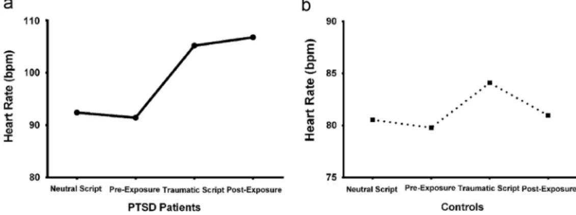
Analysis of the modulation of heart rate along the conditions (neutral script, pre-exposure, traumatic script and post-exposure) in PTSD patients revealed a significant main effect (
w
2¼31.86,p¼0.0001) (Fig. 2a). Post hoc Wilcoxon signed rank tests showed
an increase in heart rate during exposure to the traumatic script in comparison with the neutral script (z¼3.70,p¼0.0002) There
was also an increase in heart rate during the post-exposure period compared with the pre-exposure period (z¼3.42, p¼0.0006);
Finally, PTSD patients demonstrated no significant change in heart rate between the exposure to traumatic script and the post-exposure period (z¼0.20, p¼0.84), suggesting that they
sustained an accelerated heart rate even after cessation of the stressor stimulus.
The modulation of heart rate along the conditions in trauma-exposed controls also showed a significant main effect (
w
2¼14.02, p¼0.002) (Fig. 2b). Post hoc tests showed an increase in heart rate during exposure to the traumatic script in comparison with the neutral script (z¼2.27,p¼0.02). Comparison between thepost-exposure period and the pre-post-exposure period was not significant (z¼0.20, p¼0.8). Importantly, the control group showed a
sig-nificantly reduced heart rate during post-exposure relative to during traumatic script exposure (z¼2.63,p¼0.008).
Analysis of heart rate variability along the conditions in PTSD patients showed a significant main effect (
w
2¼14.31,p¼0.01) (Fig. 3).Post hoc tests showed a reduction in heart rate variability during the exposure to traumatic script in comparison with the neutral script (z¼2.69,p¼0.007), and a reduction in heart rate variability during
post-exposure period in comparison with pre-exposure period (z¼2.81, p¼0.004). Similarly to heart rate analysis, PTSD patients
Fig. 1.Sequential conditions of the experimental session. Neutral script: audio-play of a neutral history; pre-exposure silence: interval without audio-audio-play; traumatic script: audio-play of personal trauma; and post-exposure silence: interval without audio-play.
showed no significant change in heart rate variability between the post-exposure period and the exposure to traumatic script (z¼0.89,
p¼0.8), suggesting that the decrease in heart rate variability was
sustained even after cessation of the stressor.
In trauma-exposed controls, no significant modulation of heart rate variability along the conditions was found (
w
2¼2.28,p¼0.48).Investigation of the relationship between cardiac indexes and PTSD cluster symptoms were conducted only in PTSD patients, andTable 1summarizes the results.
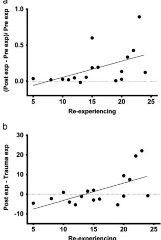
For heart rate, there was a significant correlation (
r
¼0.49, p¼0.03) between re-experiencing scores and index 1 (which contrasts heart rate post- with pre-exposure) indicating that patients with more re-experiencing symptoms showed the high-est enhance in heart rate (Fig. 4a). There was also a significant correlation (r
¼0.54, p¼0.01) between re-experiencing scoresand index 2 (which contrasts heart rate post-exposure with that during exposure) (Fig. 4b). The latter shows that tachycardia can rise beyond exposure levels in patients with high levels of re-experiencing symptoms.
There was a significant correlation (
r
¼0.51;p¼0.02) betweentotal symptoms’ scores and index 2. No other significant correla-tions for heart rate indexes and PTSD symptoms were found.
No significant correlations were found between heart rate variability indexes and PTSD symptoms.
4. Discussion
This study investigated failure of cardiac recovery following passive listening to an audiotaped personal trauma script and the
influence of the cluster of re-experiencing symptoms on sustained reactivity in PTSD patients.
4.1. Resting cardiac activity
PTSD patients showed a higher basal heart rate than trauma-exposed controls. This result is in line with other physiological studies of PTSD (Buckley and Kaloupek, 2001;Orr and Roth, 2000; Pole, 2007). It is interesting to note that a high basal heart rate in the aftermath of trauma was shown to be a significant predictor of the development of this disorder(Bryant et al., 2011).
Basal heart rate variability in PTSD patients was significantly lower than in trauma-exposed controls. This result is congruent with others studies that found lower basal heart rate variability in trauma-exposed participants with PTSD compared to those with-out PTSD (Cohen et al., 1997,1998,2000a,2000b;Blechert et al., 2007;Hughes et al., 2006,2007a, 2007b;Mitani et al., 2006;Tan et al., 2009).
4.2. Failure to recover: sustained cardiac reactivity in PTSD
The current findings demonstrate that both PTSD patients and trauma-exposed controls exhibit a significant heart rate reaction upon exposure to a traumatic script but that only PTSD patients present sustained cardiac reactivity following traumatic script presentation.
Previous script-driven protocols to evoke physiological reac-tivity in PTSD were based upon imagery instructions. These instructions required participants to vividly relive their trauma both during listening and during a post-exposure period (Lang et al.,
Fig. 3.Mean heart rate variability during each experimental condition for PTSD patients.
Table 1
Correlations between sustained heart rate indexes and posttraumatic symptomatology.
PTSD symptoms Spearmanr pvalue
Index 1
PCL-C Total 0.40 0.09
Re-experiencing 0.49 0.03
Avoidance/numbing 0.30 0.22
Hyperarousal 0.21 0.39
Index 2
PCL-C Total 0.51 0.02
Re-experiencing 0.54 0.01
Avoidance/numbing 0.42 0.08
Hyperarousal 0.43 0.08
Index 1, post-exposure minus pre-exposure, divided by pre-exposure; Index 2, post-exposure minus traumatic script; PCL-C, Posttraumatic Stress Disorder Checklist-Civilian Version
1980;Pitman et al., 1987). Studies of cardiac reactions in anxiety disorders that have employed this protocol have observed a significant acceleration in heart rate during script presentation and an even higher heart rate in the post-exposure period (e.g., Lang et al., 1998;Pitman et al., 1987). Furthermore, script-driven imagery studies in PTSD have found that both patients and controls show a significant reaction during imagery (albeit this reaction is less robust in controls) (Mcteague et al., 2010), revealing that imagery instructions are potent evokers of cardiac reactions even in the absence of psychopathology. In our study, in spite of not receiving any imagery instructions, PTSD patients still showed a robust increase in heart rate during traumatic script presentation and a sustained tachycardia in post-exposure (when no external stimuli were administered). However, trauma-exposed controls presented a significant (although feebler) heart rate response to traumatic script but recovered to basal levels when traumatic script presentation ended. Thus, compared to trauma-exposed controls, PTSD patients showed both an over-reaction during passive listen-ing to their traumatic script and a subsequent failure to recover. It has been shown (seeJovanovic et al., 2012for a review) that PTSD patients have impaired safety signal processing expressed as a difficulty in responding to safety cues physiologically, even when cognitively aware of it. Although they are safe in the laboratory setting, the present results showed that most PTSD patients respond to reminders of the trauma similarly to the real danger situation.
In the present study, heart rate variability in PTSD patients decreased significantly during traumatic script presentation and remained reduced thereafter. This reduction suggests a sustained decrease in parasympathetic outflow to the heart. Trauma-exposed controls did not show significant changes in heart rate variability at any point in the experimental session.
Taken together, the modulation of both cardiac parameters reported here seems to point to a deficient vagal control of the heart in PTSD patients.
4.3. Re-experiencing symptoms and over-engagement in external cues in PTSD
PTSD patients with more re-experiencing symptoms showed significantly higher heart rates following traumatic script expo-sure (seeFig. 4b). These results indicate that patients presenting more re-experiencing symptoms engaged more deeply in the script induction, as they showed robust heart rate acceleration to the traumatic script that was sustained or potentiated after exposure.
According toPitman (1989), high adrenergic activity during a traumatic event could over-consolidate memories. These mem-ories may become so deeply ingrained in the traumatized indivi-dual’s mind that memory extinction is inhibited and memories continually re-surface through re-experiencing symptoms. DSM-IV criteria for the re-experiencing cluster comprise spontaneous unwanted memories of the traumatic event. These include intru-sive recollections, nightmares and flashbacks as well as physical symptoms triggered by external cues. These criteria may be in line with our findings that among other symptoms only the re-experiencing cluster was significantly correlated with heart rate changes from pre-exposure to post-exposure, and with heart rate changes from trauma exposure to post-exposure.
4.4. Potential clinical implications
Our data suggest that the effects of a traumatic cue do not end with the removal of that stressor. Exposure to a personal trau-matic script without imagery instructions can trigger significant and sustained cardiac reactivity in PTSD patients.
Repeated events of strong and sustained tachycardia, accom-panied by sustained reduced heart rate variability (a measure of parasympathetic influence on the heart), may underpin the numerous findings of increased risk of cardiovascular disease in PTSD (Dedert et al., 2010). Several studies have shown that low heart rate variability is a strong predictor of cardiovascular morbidity and mortality (Thayer and Lane, 2007). Our study has significant clinical implications for the early detection and pre-vention of cardiovascular disease, and it suggests that integrated physical and mental health treatments could benefit long-term trajectories in PTSD.
Indeed, different PTSD treatments have in common the enhancement of heart rate variability in association with a reduction in PTSD severity. The studies include diverse treat-ments such as heart rate variability biofeedback (Zucker et al., 2009; Tan et al., 2011), eye movement desensitization and reprocessing (EMDR; Sack et al., 2008), fluoxetine treatment (Cohen et al., 2000b), and cognitive behavioral therapy (CBT; Nishith et al., 2003;Norte et al., 2011;Hinton et al., 2009).
Strong re-experiencing symptoms seems to prone PTSD patients to more deeply engage in traumatic script and trigger cardiac acceleration even in an experimental paradigm, which, as in our study, does not employ explicit instructions to relive the trauma. Moreover, given the present findings, it seems likely that patients repeatedly engage in strong and sustained stress reactiv-ity in their daily life. Psychotherapeutic interventions that focus directly on the management of re-experiencing symptoms should be emphasized in clinical settings, as they capture the emotional dysregulation characteristic of PTSD and its hyper-responsive expression.
4.5. Limitations
There are some limitations to the present study. First, our study is limited by its small sample size. Second, our PTSD patients were taking psychiatric medications and were known to have comorbid psychiatric illnesses.
5. Conclusions
The present work investigated cardiac activity in PTSD patients following passive exposure to their traumatic experience. This procedure is more closely related to potential real-life triggers than those more widely used in the psychophysiological assess-ment of PTSD. Here, there were no instruassess-mental instructions or cognitive/imagery tasks to be accomplished by the participant.
Our findings provided an experimental account for the failure of PTSD patients to exhibit physiological recovery after exposure to trauma-related stimuli. This failure to recover is associated with re-experiencing symptoms and with deficient vagal control. Replications of the current findings as well as the delineation of more qualitative and quantitative measures may help to clarify the emotional dysregulation characteristic of PTSD and its phy-siological expression.
Role of funding source
Conflict of interest
The other authors have no conflict of interest to disclose regarding this research.
Acknowledgments
We extend special thanks to Sonia Gleiser and Jose Magalhaes for technical~ assistance and to the following individuals for their assistance in data collection: Adriana Fiszman, Alessandra Lima, Ana Mendonc-a-de-Souza, Camila Franklin, Mariana Luz, Mauro Mendlowicz and William Berger.
References
American Psychiatric Association, 1994. Diagnostic and Statistical Manual of Mental Disorders. American Psychiatric Association, Washington, DC. Berger, W., Mendlowicz, M.V., Souza, W., Figueira, I., 2004. Semantic equivalence
of the Portuguese version of the Post-traumatic Stress Disorder Checklist-Civilian Version (PCL-C) for the screening of post-traumatic stress disorder. Revista de Psiquiatria do Rio Grande do Sul 26, 167–175.
Blechert, J., Michael, T., Grossman, P., Lajtman, M., Wilhelm, F.H., 2007. Autonomic and respiratory characteristics of posttraumatic stress disorder and panic disorder. Psychosomatic Medicine 69, 935–943.
Bremner, J.D., Vythilingam, M., Vermetten, E., Southwick, S.M., McGlashan, T., Staib, L.H., Soufer, R., Charney, D.S., 2003. Neural correlates of declarative memory for emotionally valenced words in women with posttraumatic stress disorder related to early childhood sexual abuse. Biological Psychiatry 53, 879–889.
Breslau, N., 2002. Epidemiologic studies of trauma, posttraumatic stress disorder, and other psychiatric disorders. Canadian Journal of Psychiatry 47, 923–929. Breslau, N., Davis, G.C., Schultz, L.R., 2003. Posttraumatic stress disorder and the
incidence of nicotine, alcohol, and other drug disorders in persons who have experienced trauma. Archives of General Psychiatry 60, 289–294.
Bryant, R.A., Creamer, M., O’Donnell, M., Silove, D., McFarlane, A.C., 2011. Heart rate after trauma and the specificity of fear circuitry disorders. Psychological Medicine 15, 1–8.
Buckley, T.C., Kaloupek, D.G., 2001. A meta-analytic examination of basal cardio-vascular activity in posttraumatic stress disorder. Psychosomatic Medicine 63, 585–594.
Cohen, H., Benjamin, J., Geva, A.B., Matar, M.A., Kaplan, Z., Kotler, M., 2000a. Autonomic dysregulation in panic disorder and in post-traumatic stress disorder: application of power spectrum analysis of heart rate variability at rest and in response to recollection of trauma or panic attacks. Psychiatry Research 96, 1–13.
Cohen, H., Kotler, M., Matar, M., Kaplan, Z., 2000b. Normalization of heart rate variability in post-traumatic stress disorder patients following fuoxetine treatment: preliminary results. The Israel Medical Association Journal 2, 296–301.
Cohen, H., Kotler, M., Matar, M.A., Kaplan, Z., Loewenthal, U., Miodownik, H., Cassuto, Y., 1998. Analysis of heart rate variability in posttraumatic stress disorder patients in response to a trauma-related reminder. Biological Psychiatry 44, 1054–1059.
Cohen, H., Kotler, M., Matar, M.A., Kaplan, Z., Miodownik, H., Cassuto, Y., 1997. Power Spectral analysis of heart rate posttraumatic stress disorder patients. Biological Psychiatry 41, 627–629.
Dedert, E.A., Calhoun, P.S., Watkins, L.L., Sherwood, A., Beckham, J.C., 2010. Posttraumatic stress disorder, cardiovascular, and metabolic disease: a review of the evidence. Annals of Behavioral Medicine 39, 61–78.
Del-Ben, C., Vilela, J., Hallak, J., Labate, C., Zuardi, A.W., 2001. Reliability of the structured clinical interview for DSM-IV – Clinical version translated into Portuguese. Revista Brasileira de Psiquiatria 23, 156–159.
First, M.B., Spitzer, R.L., Gibbon, M., Williams, J.B.W., 1997. Structured Clinical Interview for DSM-IV Axis I Disorders (SCID I), Clinical Version.
Friedman, M.J., Davidson, J.R., Stein, D.J., 2008. Effective treatments for PTSD: practice guidelines from the International Society for Traumatic Stress Studies. In: Foa, E.B., Keane, T.M., Friedman, M.J., Chen, J.A. (Eds.), Psychopharmacology for Adults, second ed. The Guilford Press, New York, pp. 245–268.
Hauschildt, M., Peters, M.J., Moritz, S., Jelinek, L., 2011. Heart rate variability in response to affective scenes in posttraumatic stress disorder. Biological Psychology 88, 215–222.
Hinton, D.E., Hofmann, S.G., Pollack, M.H., Otto, M.W., 2009. Mechanisms of efficacy of CBT for Cambodian refugees with PTSD: improvement in emotion regulation and orthostatic blood pressure response. CNS Neuroscience & Therapeutics 15, 255–263.
Holstein, D.H., Vollenweider, F.X., Jancke, L., Schopper, C., Csomor, P.A., 2010. P50 suppression, prepulse inhibition, and startle reactivity in the same patient cohort suffering from posttraumatic stress disorder. Journal of Affective Disorders 126, 188–197.
Hughes, J.W., Dennis, M.F., Beckham, J.C., 2007a. Baroreceptor sensitivity at rest and during stress in women with posttraumatic stress disorder or major depressive disorder. Journal of Traumatic Stress 20, 667–676.
Hughes, J.W., Feldman, M.E., Beckham, J.C., 2007b. Posttraumatic stress disorder is associated with attenuated baroreceptor sensitivity among female, but not male, smokers. Biological Psychology 71, 296–302.
Jovanovic, T, Kazama, A, Bachevalier, J, Davis, M., 2012. Impaired safety signal learning may be a biomarker of PTSD. Neuropharmacology 62, 695–704. Kessler, R.C., 2000. Posttraumatic stress disorder: the burden to the individual and
to society. Journal of Clinical Psychiatry 61, 4–12.
Lang, P.J., Kosak, M.J., Miller, G.A., Levin, D.N., McLean, A., 1980. Emotional imagery: conceptual structure and pattern of somato-visceral response. Psychophysiology 17, 179–192.
Lang, P.J., 1979. A bio-information theory of emotional imagery. Psychophysiology, 495–512.
Lang, P.J., Bradley, M.M., Cuthbert, B.N., 1998. Emotion and motivation: measuring affective perception. Journal of Clinical Neurophysiology 15, 397–408. McDonald, S.D., Calhoun, P.S., 2010. The diagnostic accuracy of the PTSD checklist:
a critical review. Clinical Psychology Review 30, 976–987.
McTeague, L.M., Lang, P.J., Laplante, M.C., Cuthbert, B.N., Shumen, J.R., Bradley, M.M., 2010. Aversive imagery in posttraumatic stress disorder: trauma recurrence, comorbidity, and physiological reactivity. Biological Psychiatry 67, 346–356.
Mitani, S., Fujita, M., Sakamoto, S., Shirakawa, T., 2006. Effect of autogenic training on cardiac autonomic nervous activity in high-risk fire service workers for posttraumatic stress disorder. Journal of Psychosomatic Research 60, 439–444. Mueller-Pfeiffer, C., Martin-Soelch, C., Blair, J.R., Carnier, A., Kaiser, N., Rufer, M., Schnyder, U., Hasler, G., 2010. Impact of emotion on cognition in trauma survivors: what is the role of posttraumatic stress disorder? Journal of Affective Disorders 126, 287–292.
Nishith, P., Duntley, S.P., Domitrovich, P.P., Uhles, M.L., Cook, B.J., Stein, P.K., 2003. Effect of cognitive behavioral therapy on heart rate variability during REM sleep in female rape victims with PTSD. Journal of Traumatic Stress 16, 247–250.
Norte, C.E., Souza, G.G.L., Pedrozo, A.L., Mendonc-a-de-Souza, A.C.F., Figueira, I., Volchan, E., Ventura, P.R., 2011. Impact of cognitive-behavior therapy on resilience-related neurobiological factors. Revista de Psiquiatria Clı´nica 38, 43–45.
Orr, S.P., Roth, W.T., 2000. Psychophysiological assessment: clinical applications for PTSD. Journal of Affective Disorders 61, 225–240.
Perakakis, P., Joffily, M., Taylor, M., Guerra, P., Vila, J., 2010. KARDIA: a Matlab software for the analysis of cardiac interbeat intervals. Computer Methods and Programs in Biomedicine 98, 83–89.
Pitman, R.K., 1989. Post-traumatic stress disorder, hormones, and memory. Biological Psychiatry 26, 221–223.
Pitman, R.K., Orr, S.P., Forgue, D.F., de Jong, J.B., Claiborn, J.M., 1987. Psychophy-siologic assessment of posttraumatic stress disorder imagery in Vietnam combat veterans. Archives of General Psychiatry 44, 970–975.
Pole, N., 2007. The psychophysiology of posttraumatic stress disorder: a meta-analysis. Psychological Bulletin 133, 725–746.
Rabe, S., Dorfel, D., Zollner, T., Maercker, A., Karl, A., 2006. Cardiovascular correlates of motor vehicle accident related posttraumatic stress disorder and its successful treatment. Applied Psychophysiology and Biofeedback 31, 315–330.
Sack, M., Lempa, W., Steinmetz, A., Lamprecht, F., Hofmann, A., 2008. Alterations in autonomic tone during trauma exposure using eye movement desensitization and reprocessing (EMDR)—results of a preliminary investigation. Journal of Anxiety Disorders 22, 1264–1271.
Tan, G., Dao, T.K., Farmer, L., Sutherland, R.J., Gevirtz, R., 2011. Heart rate variability (HRV) and posttraumatic stress disorder (PTSD): a pilot study. Applied Psychophysiology and Biofeedback 36, 27–35.
Tan, G., Fink, B., Dao, T.K., Hebert, R., Farmer, L.S., Sanders, A., Pastorek, N., Gevirtz, R., 2009. Associations among pain, PTSD, mTBI, and heart rate variability in veterans of Operation Enduring and Iraqi Freedom: a pilot study. Pain Medicine 10, 1237–1245.
Task Force of the European Society of Cardiology and the North American Society of Pacing and Electrophysiology, 1996. Heart rate variability: standards of measurement, physiological interpretation, and clinical use. Circulation 93, 1043–1065.
Thayer, J.F., Lane, R.D., 2007. The role of vagal function in the risk for cardiovas-cular disease and mortality. Biological Psychology 74, 224–242.
Volchan, E., Souza, G.G., Franklin, C.M., Norte, C.E., Rocha-Rego, V., Oliveira, J.M., David, I.A., Mendlowicz, M.V., Coutinho, E.S., Fiszman, A., Berger, W., Marques-Portella, C., Figueira, I., 2011. Is there tonic immobility in humans? Biological evidence from victims of traumatic stress. Biological Psychology 88, 13–19. Weathers, F.W., Litz, B.T., Herman, D.S., Huska, J.A., Keane, T.M., 1993. The PTSD
Checklist (PCL): reliability, validity, and diagnostic utility. In: Paper presented at the Annual Meeting of International Society for Traumatic Stress Studies, San Antonio, TX.
Yehuda, R., LeDoux, J., 2007. Response variation following trauma: a translational neuroscience approach to understanding PTSD. Neuron 56, 19–32.
Zayfert, C., Dums, A.R., Ferguson, R.J., Hegel, M.T., 2002. Health functioning impairments associated with posttraumatic stress disorder, anxiety disorders, and depression. Journal of Nervous and Mental Disease 190, 233–240. Zucker, T.L., Samuelson, K.W., Muench, F., Greenberg, M.A., Gevirtz, R.N., 2009. The