Effects of Decreased Occlusal Loading during
Growth on the Mandibular Bone
Characteristics
Natsuko Hichijo1, Eiji Tanaka2,3*, Nobuhiko Kawai2, Leo J. van Ruijven4, Geerling E. J. Langenbach4
1Department of Orthodontics and Dentofacial Orthopedics, Tokushima University Graduate School of Oral Sciences, Tokushima, Japan,2Department of Orthodontics and Dentofacial Orthopedics, Institute of Biomedical Sciences, Tokushima University Graduate School, Tokushima, Japan,3Department of Orthodontics, Faculty of Dentistry, King Abdulaziz University, Jeddah, Saudi Arabia,4Department of Oral Cell Biology and Functional Anatomy, Academic Centre for Dentistry Amsterdam (ACTA), Research Institute MOVE, University of Amsterdam and VU University Amsterdam, Amsterdam, Netherlands
Abstract
Background
Bone mass and mineralization are largely influenced by loading. The purpose of this study was to evaluate the reaction of the entire mandibular bone in response to decreased load during growth. It is hypothesized that decreased muscular loading will lead to bone changes as seen during disuse, i.e. loss of bone mass.
Methods and Findings
Ten 21-day-old Wistar strain male rats were divided into two groups (each n=5) and fed on either a hard- or soft-diet for 11 weeks. Micro-computed tomography was used for the inves-tigation of bone mineralization, bone volume, bone volume fraction (BV/TV) and morpholog-ical analysis. Mandibular mineralization patterns were very consistent, showing a lower degree of mineralization in the ramus than in the corpus. In the soft-diet group, mineraliza-tion below the molars was significantly increased (p<0.05) compared to the hard diet group. Also, bone volume and BV/TV of the condyle and the masseter attachment were decreased in the soft-diet group (p<0.05). Morphological analysis showed inhibited growth of the ramus in the soft-diet group (p<0.05).
Conclusion
Decreased loading by a soft diet causes significant changes in the mandible. However, these changes are very region-specific, probably depending on the alterations in the local loading regime. The results suggest that muscle activity during growth is very important for bone quality and morphology.
a11111
OPEN ACCESS
Citation:Hichijo N, Tanaka E, Kawai N, van Ruijven LJ, Langenbach GEJ (2015) Effects of Decreased Occlusal Loading during Growth on the Mandibular Bone Characteristics. PLoS ONE 10(6): e0129290. doi:10.1371/journal.pone.0129290
Academic Editor:Mohammed Elsalanty, Georgia Regents University, College of Dental Medicine, UNITED STATES
Received:November 20, 2014
Accepted:May 6, 2015
Published:June 10, 2015
Copyright:© 2015 Hichijo et al. This is an open access article distributed under the terms of the Creative Commons Attribution License, which permits unrestricted use, distribution, and reproduction in any medium, provided the original author and source are credited.
Data Availability Statement:All relevant data are within the paper.
Funding:This research was supported by Grants-in-Aid 26293436 (E.T.) and 26463095 (N.K.) for Science Research from the Ministry of Education, Culture, Sports, Science and Technology, Japan. The funders had no role in study design, data collection and analysis, decision to publish, or preparation of the manuscript.
Introduction
Bone characteristics are largely influenced by the loads imposed upon them. Increased natural
loads increase the bone mass [1,2]. Bone loss can be seen in individuals with decreased loading
pattern [3,4]. In vivo mechanical loading of bone tissue (eg three-point bending) has shown
that loading amplitude [5], cycle number [6] and frequency [7] are important factors in bone
adaptation. Indirect evidence shows that the majority of the natural loadings are generated by muscles, and that alterations in these natural muscles forces are important for bone adaptation
[8,9,10]. This adaptation can be local, as indicated by highly mineralized bones in the playing
arm of tennis players [11] or systemic impact physiological loads [12]. Paralysis has been
shown to lead to bone mass decrease [13,14]. In the craniofacial region, studies on the effect of
changed loading patterns show comparable but specific reactions. Myotonic dystrophy pa-tients, who have a lower masseter muscle activity, show some atypical mandibular forms, char-acterized by a large mandibular plane angle and changes of shape regarding the
temporomandibular joint [15,16,17]. A soft diet during development causes a reduction in jaw
bone development [18,19,20] and only a small change in the daily jaw muscle activity [18].
The purpose of this study was to investigate the regional reactions of the mandibular bone (including its form, and local bone volumes and mineralization degrees) in response to a, in time, limited decrease in daily load during growth. For this, the food consistency was decreased, assumingly resulting in a decreased masticatory muscle activity and occlusal load. We hypothe-sized that the decreased mandibular loading will result in a loss of bone mass, changes as seen during disuse.
Materials and Methods
Experimental animals
Ten Wistar strain male rats at the age of three-weeks were randomly divided into hard-diet and soft-diet groups (both n = 5). The use of a single gender was an attempt to eliminate any variation in bone characteristics due to sexual dimorphism. The hard-diet group was fed on an ordinary pellet (CE-2, CLEA Japan Inc., Tokyo, Japan), while a powder diet that contained the same constituents was used in the soft-diet group. Body weight was monitored once a week.
At 13-week-old, the animals were killed with an overdose of sodium pentobarbital (Nembu-tal; Dinabott, Osaka, Japan). The right mandibles were removed and examined by a micro-computed tomography system (micro-CT) for bone density, mineralization and morphometric analyses. These specimens were stored in 70% ethanol for maximally one month.
The protocol of the experiment was approved by the Animal Care and Use Committee at the Tokushima University.
Bones
Mineralization, bone volume and bone volume fraction (BV/TV). For a detailed study
of the bone characteristics, we used a micro-CT (μCT 40, Scanco Medical AG, Brüttisellen,
Switzerland). During scanning all mandibles were similarly oriented and submerged in water
to avoid dehydration. Scanning was performed at 10μm spatial resolution and 45 kV peak
volt-age (effective energy: 24 keV). An integration time of 1200 ms was applied to substantially re-duce noise and optimally discriminate between bone and background. An aluminum filter in
the micro-CT and a correction algorithm reduced the effects of beam hardening [21]. A
thresh-old of 642.8 mg hydroxyapatite/cm3 was used to distinguish bone from background. From the X-ray attenuation map, which contains the computed linear attenuation coefficients of each volume element (voxel) of the scan, mineralization of each voxel of the 3D reconstruction of
the mandible was determined. From the 3D reconstructions the outer two voxel layers were peeled off to avoid the partial volume effects. For each mandible a distribution map of the corti-cal mineralization was obtained by projecting the mineral densities of all voxels in a 0.288-mm thick layer below the removed voxels on the surface of the mandible. A more detailed
descrip-tion of the method can be found in de Jong et al. [22]. Visual comparison showed possible local
differences in surface mineralization degrees. Upon visual examination, various VOIs were de-termined selecting only the cortical bone at the attachment site of the masseter, the condylar
head and below the second molar (Fig 1). For these VOIs, the cortical bone parameters
(includ-ing bone volume, BV/TV and mineralization degree) were calculated tak(includ-ing into account the abovementioned threshold value. As mineralization was evaluated by including only "bone" voxels, the material density was calculated. Selection at the attachment site of the masseter and condyle is defined by the line connecting the two specific notches. For evaluation of alveolar cortical bone, the part of the second molar is picked up because of getting correct data from all mandibles.
Morphology. For evaluating morphology, a CT- scan of similarly oriented mandibles was
used (Latheta LCT-200, Hitachi Aloka Medical, Tokyo, Japan; 50kVp, 500μA, 48μm
resolu-tion). Three dimensional reconstructions of the mandibles were made for measurement of
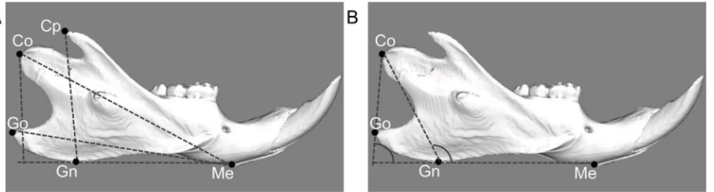
line-ar and angulline-ar dimensions (LEXI, Tokyo, Japan).Fig 2shows the specified landmarks and
measurements used in this study. Linear and angular dimensions were evaluated using five landmarks: menton (Me: most inferior point of mandibular symphysis), gnathion (Gn: most anterior point on bony contour of mandibular symphysis), gonion (Go: most outward and everted point on angle formed by junction of ramus and body of mandible), condylion (Co:
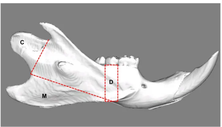
Fig 1. Lateral view of a reconstructed right hemimandible showing the volumes of interest.C: Condyle; Selection is defined by the line connecting the two notches superior and inferior of this area. M: Attachment site of the superficial masseter muscle; the border line connects the notches ventrocaudal and craniodorsal to the mandibular angle. D: Alveolar cortical bone; the selection is defined by two lines drawn perpendicular to the lower border of the mandible, mesial and distal of the second molar.
most posterior and superior point on mandibular condyle), coronoid point (Cp: most superior point on coronoid process of mandible). In between these landmarks four linear and two angu-lar measurements were assessed: total length of the mandible (Me-Co), base length of the man-dible (Me-Go), height of coronoid process (Cp-GnMe), mandibular ramus height (Co-GnMe), gonial angle (CoGo/GnMe), and ramus angle (CoGn/GnMe).
Statistics. Average and standard deviation values were obtained for each experimental group. All data were tested for normality of distribution (Kolmogorov-Smirnov test) and
uni-formity (Bartlett’s test). Statistical analyses in this study were tested for differences using
un-paired Student’st-test. Probabilities of less than 0.05 were considered to be significant.
Results
Animals in both groups showed an on average comparable increase of their body weight. Ani-mals in the soft-diet group did not show any noticeable change in their daily use of their masti-catory apparatus in response to the reduced food hardness.
Mineralization
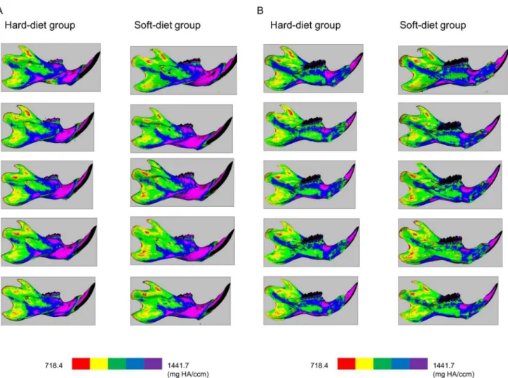
Mineralization maps of the mandibles (Fig3Aand3B) showed a remarkably similar
distribu-tion pattern of mineralizadistribu-tion. Also, both groups showed very similar values and patterns in
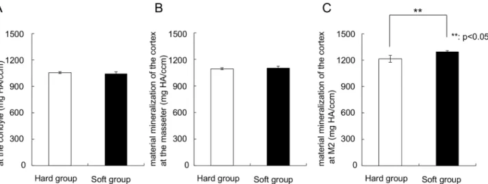
the regions of the condyle and the muscle attachments (Fig 4). However, below the dentition a
significantly higher (p = 0.003) regional mineralization (material density) was found in the
soft-diet group (1300 mg HA) compared to the hard-diet group (1200 mg HA) (Fig 4C).
Bone volume and BV/TV
The soft diet group showed a significantly (p = 0.01) lower bone volume in the condyle
(6.8 ± 1.3 mm3) than the hard diet group (9.0 ± 0.5 mm3). Moreover, bone volume of the
at-tachment of the masseter in the soft-diet group (10.9 ± 0.6 mm3) was also significantly lower
than in the hard-diet group (12.6 ± 0.9 mm3) (Fig 5, p = 0.01). In contrast, the region of
alveo-lar cortical bone did not show any difference in bone volume. In the same way, BV/TV of the
Fig 2. Landmarks and measurement items for linear analysis (A) and angular analysis (B) by 3D reconstructions.Me: Menton (most inferior point of mandibular symphysis). Gn: Gnathion (most anterior point on bony contour of mandibular symphysis). Go: Gonion (most outward and everted point on angle formed by junction of ramus and body of mandible). Co: Condylion (most superoposterior point on mandibular condyle). Cp: Coronoid process (most superior point on coronoid process of mandible). Me-Co: Total length of the mandible (distance measured between menton and condylion). Me-Go: Base length of the mandible (distance measured between menton and gonion). CpH: Height of coronoid process (a perpendicular line from coronoid process to the line connected to gnathion and menton). CoH: Height of mandibular ramus (a perpendicular line from condylion to the line connected to gnathion and menton). CoGo/GnMe: Gonial angle (angle made from the line connected to condylion and gonion and the line connected to gnathion and menton). CoGn/GnMe: Ramus angle (angle made from condylion, gnathion and menton).
doi:10.1371/journal.pone.0129290.g002
condyle (0.789 ± 0.018 mm3/mm3, p = 0.004) and the attachment of the masseter (0.863 ±
0.011 mm3/mm3, p = 0.042) in the soft-diet group showed significantly lower values than those
in the hard diet group (0.834 ± 0.012 mm3/mm3and 0.878 ± 0.005 mm3/mm3, respectively)
(Fig 6).
Morphometric findings
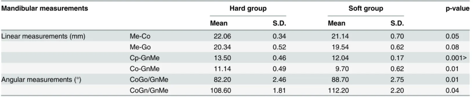
Table 1shows means and standard deviations for linear and angular measurements in both groups. The soft-diet group (9.70 ± 0.62 mm) demonstrated significantly (p = 0.007) shorter mandibular ramus height than the hard-diet group (11.14 ± 0.49 mm). Me-Co for soft-diet
group also showed shorter length than for hard-diet group. Moreover,Fig 7presents the 3D
re-constructions for each group, and shows the overall smaller mandible of the soft-diet group compared to the hard-diet group. The mandibular gonial and ramus angles in the soft-diet group (mean ± s.d.) were 88.7 ± 2.8°and 112.2 ± 2.20°, respectively. These values were signifi-cantly larger (p = 0.008, 0.038), compared to the hard-diet group (82.2 ± 2.5°and 108.6 ± 1.8°).
Fig 3. Lateral (3-A) and medial (3-B) views of the cortical mineralization.Left column: bones of the hard-diet group, Right column: bones of the soft-diet group. The colours red, yellow, green, blue, and purple indicate mineralization ranges 718.4–863.0, 863.0–1007.7, 1007.7–1152.4, 1152.4–1297.0, and 1297.0–1441.7 mg HA/ccm, respectively.
Discussion
The aim of this study was to evaluate the changes in mandibular bone due to decreased muscu-lar loading caused by a soft diet during growth. The experimental results indicate that these changes are different for various mandibular regions. Compared to the hard-diet group, the mineralization degree was increased only at the occlusion region, while the bone volume and BV/TV was lowered at the condylar region and the attachment areas of the masseter. Appar-ently, a limited decrease in daily loading results in significant changes in bone characteristics.
Fig 4. Graphs depicting the material mineralization of the cortex at the (A) Condyle, (B) Attachment of the masseter, (C) The part below the second tooth.**Significant difference between both groups (p<0.05).
doi:10.1371/journal.pone.0129290.g004
Fig 5. Graphs depicting the bone volume.(A) Condyle, (B) Attachment of the masseter, (C) The part below the second tooth.**Significant difference between both groups (p<0.05).
doi:10.1371/journal.pone.0129290.g005
However, the changes in bone characteristics varied with location, including bone mass loss and increase in mineralization degree.
We already showed by using a telemetric system that a decrease in food consistency resulted
in weaker jaw closer activity [23]. Simultaneously, the expression of insulin-like growth
factor-1 (IGF-factor-1), which is often used for evaluating the effect of mechanical load, decreased and the condylar growth was inhibited. In the present research, we can assume that the soft diet again resulted in lower muscle activity. The deficiency of masticatory demand during growth led to minor mandibular development. This inhibited growth resulted in less bone volume in the
soft-diet group, although it didn’t show the correlation with mineralization.
In this study, the cancellous bone was not included because the masseter site did not contain any cancellous bone. At the other sites, the volume of the cancellous bone was also too little to get reliable means. As only the cortical bone was analyzed, bone mineralization was calculated
as material density including only voxels exceeding the threshold value. As far asFig 3showed,
the pattern of mineralization was very consistent and a clearly lower mineralization degree was found at the ramus compared to the corpus in both groups. This is quite similar to the pattern
of mineralization found in rabbits by de Jong et al. [22]. They also found a very consistent
Fig 6. Graphs depicting the BV/TV.(A) Condyle, (B) Attachment of the masseter, (C) The part below the second tooth.**Significant difference between both groups (p<0.05).
doi:10.1371/journal.pone.0129290.g006
Table 1. Comparison of mandibular measurements between the hard- and soft-diet groups.
Mandibular measurements Hard group Soft group p-value
Mean S.D. Mean S.D.
Linear measurements (mm) Me-Co 22.06 0.34 21.14 0.70 0.05
Me-Go 20.34 0.52 19.54 0.62 0.08
Cp-GnMe 13.50 0.46 12.04 0.17 0.001>
Co-GnMe 11.14 0.49 9.70 0.62 0.01
Angular measurements (°) CoGo/GnMe 82.20 2.46 88.70 2.75 0.01
CoGn/GnMe 108.60 1.81 112.20 2.20 0.04
mineralization pattern among used rabbits, and a lower mineralization degree at the ramus. As in mice, a major part of the ramus serves as an attachment site of the powerful jaw muscles. We can expect that this direct loading activates remodeling and creates lower mineralization com-pared to the corpus of the mandible, where much smaller and weaker muscles attach. Here, the occlusal force is the principle loading.
At the ramus, as mentioned above, many muscle forces are working, generated during vari-ous behaviors: grooming, licking, posture, yawning, chewing. As chewing is just one of the many loading patterns during daily life, a change in the muscle activity is probably not deter-mining for the bone, as all other behaviors remain unaffected, generating equal amounts of loading. For the region below the molars, the only loading is when the teeth make contact (chewing). So, the loading pattern at the corpus is seriously affected by the soft diet. This may explain why, in the soft diet animals, the value of mineralization is unaffected at the ramus re-gion, but increased at the region below the molars. Moreover, another possible factor for these results is the clearly defined VOI. Some previous studies showed remarkable regional
differ-ences in mineral density using a very small VOI [20,24]. However, even more important than
the size of the VOI is its location. As can be seen inFig 3, the mineralization can vary
enor-mously, even over very short distances. Choosing a VOI at a slightly different location in differ-ent animals can create large differences in the degree of mineralization. Therefore, the VOIs were standardized by clear reference points. A clearly defined location of measurement, as per-formed in this study, will minimize such errors.
The increased mineral content below the dentition seems to contrast the increased mineral content found in the seriously used tennis arm. However, there is a difference in time path. The increased mineral content in the occlusal area is probably a temporary state, caused by an acute decrease in remodeling rate due to the locally decreased loading regime. Eventually, the area will remodel towards a state that less bone volume will be present. The increased mineral con-tent in the tennis arm is the result of years of training, and can be seen as an adaptation to the continuously increased use of the arm muscles.
The increased mineralization below the molars is in line with previous studies. The periodon-tal ligament is the connective tissue localized between the root of the tooth and the alveolar
bone, and is known to react to mechanical loading such as occlusal force [25–27]. Several papers
show the relation with loading and alveolar bone. In rats with reduced masticatory function by a
soft diet, the alveolar bone of the mandible, was thinner [28,29] and taller [28] and its bone
den-sity [28–30] was lower. Balazs et al. also indicated differences in the alveolar process and
evi-dence that a reduction of occlusal loading induces a simultaneous response in both tissues [31].
This is in line with the increased mineralization below the teeth found in the current study.
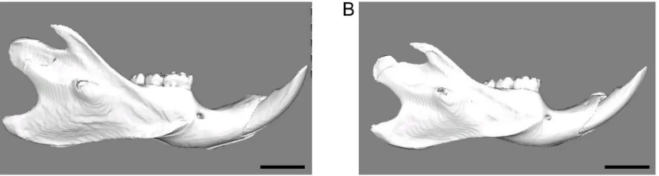
Fig 7. A representative 3D reconstructions of the hard-diet group (A) and the soft-diet group (B).Scale bar = 5 mm. Note the much smaller size of the soft-diet mandible.
doi:10.1371/journal.pone.0129290.g007
For these experiments, ten rats were used. Although the sample size for each group looks
small, we checked the minimum number of animals by the software named“GPower 3.1.9.2”
[32]. It followed that 5 rats per group is sufficient.
The results of the present study suggest that muscular loading is an important factor for bone characteristics and morphology during growth. Normal bone characteristics cannot be maintained after a limited decrease in daily loading. Also, the changes in bone characteristics are very region-specific, conceivably depending on the locally specific changes in the loading regime.
Acknowledgments
This research was supported in part by Grants-in-Aid 26293436 (E.T.) and 2643095 (N.K.) for Science Research from the Ministry of Education, Culture, Sports, Science and Technology, Japan. The funders had no role in study design, data collection and analysis, decision to pub-lish, or preparation of the manuscript.
Author Contributions
Conceived and designed the experiments: GEJL NK ET. Performed the experiments: NH LJvR. Analyzed the data: NH LJvR GEJL. Contributed reagents/materials/analysis tools: NH NK ET. Wrote the paper: NH GEJL.
References
1. Nurmi-Lawton JA, Baxter-Jones AD, Mirwald RL, Bishop JA, Taylor P, Cooper C, et al. (2004) Evidence of sustained skeletal benefits from impact-loading exercise in young females: a 3-year longitudinal study. J Bone Miner Res 19:314–322. PMID:14969402
2. Daly RM, Bass SL (2006) Lifetime sport and leisure activity participation is associated with greater bone size, quality and strength in older men. Osteoporos Int 17:1258–1267. PMID:16680498
3. Lang T, LeBlanc A, Evans H, Lu Y, Genant H, Yu A (2004) Cortical and trabecular bone mineral loss from the spine and hip in long duration spaceflight. J Bone Miner Res 19:1006–1012. PMID:15125798
4. Watanabe Y, Ohshima H, Mizuno K, Sekiguchi C, Fukunaga M, Kohri K, et al. (2004) Intravenous pami-dronate prevents femoral bone loss and renal stone formation during 90-day bed rest. J Bone Miner Res 19:1771–1778. PMID:15476576
5. Rubin CT, Lanyon LE (1985) Regulation of bone mass by mechanical strain magnitude. Calcif Tissue Int 37:411–417. PMID:3930039
6. Cullen D M, Smith R T, Akhter M P (2001) Bone-loading response varies with strain magnitude and cycle number. J Appl Physiol 91:1971–1976. PMID:11641332
7. Qin Y-X, Lam H, Ferreri S, Rubin C. Dynamic Skeletal Muscle Stimulation and its Potential in Bone Ad-aptation (2010) J Musculoskelet Neuronal Interact 10:12–24. PMID:20190376
8. Burr DB (1997) Muscle strength, bone mass, and age-related bone loss. J Bone Miner Res 12: 1547–1551. PMID:9333114
9. Turner CH (2000) Muscle-bone interactions, revisited. Bone 27:339–340. PMID:10962342
10. Frost HM (2001) From Wolff's law to the Utah paradigm: insights about bone physiology and its clinical applications. Anat Rec 262:398–419. PMID:11275971
11. Kannus P, Haapasalo H, Sievänen H, Oja P, Vuori I (1994) The site-specific effects of long-term unilat-eral activity on bone minunilat-eral density and content. Bone 15:279–284. PMID:8068448
12. Daly RM, Saxon L, Turner CH, Robling AG, Bass SL (2004) The relationship between muscle size and bone geometry during growth and in response to exercise. Bone 34:281–287. PMID:14962806
13. Warner SE, Sanford DA, Becker BA, Bain SD, Srinivasan S, Gross TS (2006) Botox induced muscle paralysis rapidly degrades bone. Bone 38:257–264. PMID:16185943
15. Kiliaridis S, Mejersjo C, Thilander B (1989) Muscle function and craniofacial morphology: a clinical study in patients with myotonic dystrophy. Eur J Orthod 11:131–138. PMID:2767145
16. Odman C, Kiliaridis S (1996) Masticatory muscle activity in myotonic dystrophy patients. J Oral Rehabil 23:5–10. PMID:8850154
17. Zanoteli E, Yamashita HK, Suzuki H, Oliveira AS, Gabbai AA (2002) Temporomandibular joint and masticatory muscle involvement in myotonic dystrophy: a study by magnetic resonance imaging. Oral Surg Oral Med Oral Pathol Oral Radiol Endodont 94:262–271. PMID:12221397
18. Kawai N, Sano R, Korfage JAM, Nakamura S, Kinouchi N, Kawakami E, et al. (2010) Adaptation of rat jaw muscle fibers in postnatal development with a different food consistency: an immunohistochemical and electromyographic study. J Anat 216:717–23. doi:10.1111/j.1469-7580.2010.01235.xPMID: 20579175
19. Yonemitsu I, Muramoto T, Soma K (2007) The influence of masseter activity on rat mandibular growth. Arch Oral Biol 52:487–493. PMID:17126288
20. Tanaka E, Sano R, Kawai N, Langenbach GE, Brugman P, Tanne K, et al. (2007) Effect of food consis-tency on the degree of mineralization in the rat mandible. Ann Biomed Eng 35:1617–1621. PMID: 17522978
21. Mulder L, Koolstra JH, van Eijden TMGJ (2004) Accuracy of microCT in the quantitative determination of the degree and distribution of mineralization in developing bone. Acta Radiol 45:769–777. PMID: 15624521
22. de Jong WC, van Ruijven LJ, Brugman P, Langenbach GE (2013) Variation of the mineral density in cortical bone may serve to keep strain amplitudes within a physiological range. Bone 55:391–399. doi: 10.1016/j.bone.2013.04.026PMID:23659830
23. Hichijo N, Kawai N, Mori H, Sano R, Ohnuki Y, Okumura S, et al. (2014) Effects of the masticatory de-mand on the rat de-mandibular development. J Oral Rehabil 41:581–587. doi:10.1111/joor.12171PMID: 24702545
24. Mavropoulos A, Kiliaridis S, Bresin A, Ammann P. (2004) Effect of different masticatory functional and mechanical demands on the structural adaptation of the mandibular alveolar bone in young growing rats. Bone 35:191–197. PMID:15207756
25. Kusters ST, Kuijpers-Jagtman AM, Maltha JC (1991) An experimental study in dogs of transseptal fiber arrangement between teeth which have emerged in rotated or non-rotated positions. J Dent Res 70:192–197. PMID:1999558
26. Shuttleworth CA, Smalley JW (1983) Periodontal ligament. Int Rev Connect Tissue Res 10:211–247. PMID:6358097
27. Enlow DH (1990) Physiologic tooth movements and alveolar remodeling. In: Enlow DH, ed. Facial growth. Philadelphia: Saunders, pp130–148.
28. Mavropoulos A, Odman A, Ammann P, Kiliaridis S (2010) Rehabilitation of masticatory function im-proves the alveolar bone architecture of the mandible in adult rats. Bone 47:687–692. doi:10.1016/j. bone.2010.06.025PMID:20601301
29. Bresin A, Kiliaridis S, Strid KG (1999) Effect of masticatory function on the internal bone structure in the mandible of the growing rat. Eur J Oral Sci 107:35–44. PMID:10102749
30. Sato H, Kawamura A, Yamaguchi M, Kasai K (2005) Relationship between masticatory function and in-ternal structure of the mandible based on computed tomography findings. Am J Orthod Dentofacial Orthop 128:766–773. PMID:16360919
31. Denes BJ, Mavropoulos A, Bresin A, Kiliaridis S (2013) Influence of masticatory hypofunction on the al-veolar bone and the molar periodontal ligament space in the rat maxilla. Eur J Oral Sci 121:532–537. doi:10.1111/eos.12092PMID:24206071
32. Faul F, Erdfelder E, Lang AG, Buchner A (2007) G*Power 3: a flexible statistical power analysis pro-gram for the social, behavioral, and biomedical sciences. Behav Res Methods 39:175–191. PMID: 17695343