Autonomic Regulation of Mechanical Properties in Porcine Mitral
Valve Cusps
Xiang Hu, Qiang Zhao, Xiaofeng Ye
Department of Cardiac Surgery, Rui Jin Hospital, Shanghai Jiao Tong University School of Medicine, Shanghai, 200025, PRC
Mailing address: Qiang Zhao •
Department of Cardiac Surgery, Rui Jin Hospital, Shanghai Jiao Tong University School of Medicine, Shanghai, 200025, PRC
E-mail: [email protected]
Manuscript received September 01, 2010; revised manuscript received November 01, 2010; accepted November 23, 2010.
Abstract
Background: The presence of nerves in heart valves was first depicted decades ago and identified into subpopulations: sympathetic, parasympathetic. The autonomic nerve is expected to greatly affect heart valves. However, regarding the aspect of mechanical properties regulated by nerves, very little focus has been placed on that.
Objective: We sought to identify the role of the autonomic nervous system in the regulation of the mechanical properties of porcine mitral valve tissues.
Methods: Mechanical properties of porcine mitral valve leaflets were evaluated in response to norepinephrine (NE) and acetylcholine (ACH), the main neurotransmitters. At the same time, phentolamine (Phent), metoprolol (Metop), atropine (Atrop) and endothelial denudation were added to the reactive system.
Results: Under physiological conditions, the stiffness was not affected by endothelial denudation (p > 0.05). NE elevated the valve stiffness significantly per 10-fold increase in concentration (10-6vs 10-7, p < 0.05; 10-5vs 10-6, p < 0.05). The
stiffness elevated by NE was mitigated by Phent and Metop addition or endothelial denudation (p < 0.05). However, the stiffness remained significant when compared to Control (p < 0.05). ACH caused a decrease in stiffness accompanied by an increase in its concentration (significant change in stiffness per 10-fold increase in ACH concentration, 10-6vs
Control, p < 0.05; 10-5vs 10-6, p < 0.05), which were reversed by endothelial denudation and Atrop (p > 0.05 vs Control).
Conclusion: These findings highlight the role of the autonomic nervous system in the regulation of the mechanical
properties of porcine mitral valve cusps, which underline the importance of autonomic nervous status for optimal valve function. (Arq Bras Cardiol 2012;98(4):321-328)
Keywords: Mitral valve; autonomic pathways; bioprosthesis; autonomic nervous system.
Introduction
Heart valve function is quite complicated, as the passive movements of valve do not account for its ability to adapt to the hemodynamic and humoral milieu. This is exemplified by the presence of nerves in heart valves, which was first depicted decades ago and identified into subpopulations: sympathetic, parasympathetic, sensory and cholinergic nerve markers1,2. Cardiac arrhythmia is frequently detected
in patients with heart diseases, including the coronary-derived and valve-coronary-derived ones. Improper autonomic tone, the supposed etiology, is considered prevalent in majority of heart diseases, so valves are expected to be greatly affected by the autonomic nerves. However, very little focus has been given to the regulation of heart valves by the autonomic nervous system. Recent studies have
indicated that the addition of norepinephrine (NE) and acetylcholine (ACH) produced dose-dependent increases in mitral valve contractility and aortic valve tissue relaxation in an ex vivo model, respectively3,4. The functional role of
neurotransmitters in the mechanical properties of cusp tissue remains undetermined. This could be vital, as the valve is subjected to high mechanical forces at each cardiac cycle. Understanding valvular mechanical properties is crucial to address an increasingly prevalent heart valve disease entity, even more so considering the increase in life expectancy of the general population and the improvement in long-term survival of patients with heart valve disease undergoing allogenic valve replacement surgery. This study will focus mainly on mitral valves, considering that, of all four heart valves, the mitral valve is the most commonly affected in disease processes requiring surgical intervention.
Methods
Tissue isolation
Porcine hearts (20 to 24 months old) were harvested from a slaughterhouse (FuXing Meats, Shanghai, China). Mitral cusps (anterior or posterior leaflet, randomly), with no visible evidence of lesion, were immersed immediately in ice-cold Krebs buffer and used within 24 hours of the animal’s sacrifice. Specimens were prepared by first trimming away all chordae and were divided into identical 15 x 10 mm rectangular strips from the “belly” of each cusp in radial and circumferential directions. The circumferential direction was defined as parallel to the free edge and the radial direction went from the free edge to the basement of mitral valves. Radial and circumferential strips were analyzed separately. Sections were assigned to different groups, according to the neurotransmitters used at random: norepinephrine (NE) (10-7 - 10-4 M; n = 10; SIGMA), acetylcholine (ACH) (10-6 - 10-4
M; n = 10; SIGMA) and normal Controls (n = 10). In order to identify the location and mechanism of the neuroagents involved in stiffness, endothelial-denuded strips, which were mechanically accomplished with a cell scraper, were also employed (denuded group, n = 10). Each tissue section was only exposed to a single neurotransmitter to avoid interactions between various agents and the serial solution was replaced by a new one every time, ensuring the concentration. Procedures involving animals and their care were approved by Animal Care and Use Committee of the Shanghai Jiao Tong University School of Medicine.
Role of the neurotransmitter receptors
To evaluate the contribution of the neurotransmitter receptors to changes in tissue mechanical properties, strips were preincubated for 1 hour with phentolamine (Phent, a nonselective α adrenoceptor inhibitor, SIGMA), prazosin (Praz,
a selective α1 adrenoceptor inhibitor, SIGMA), metoprolol
(Metop, a selective β1 adrenoceptor inhibitor, SIGMA),
propranolol (Propr, a nonselective β-adrenoceptor inhibitor, SIGMA), and atropine (Atrop, a M cholinergic receptor inhibitor, SIGMA), before testing.
Experimental protocol
Gripped at both ends with 2-piece stainless clamps lined with sandpaper, the specimens were mounted in a modified mechanical testing device (model 8511; Instron, Canton, Mass) in a Krebs bath heated at 37° C and
continuously gassed with 95% O2 and 5% CO2. The pH
of solutions was stabilized between 7.35 and 7.45. Strain gauge force transducers were attached along long axes to real-time monitor the changes of tension in the radial and circumferential orientations respectively.
Tissue stiffness (modulus) measurements
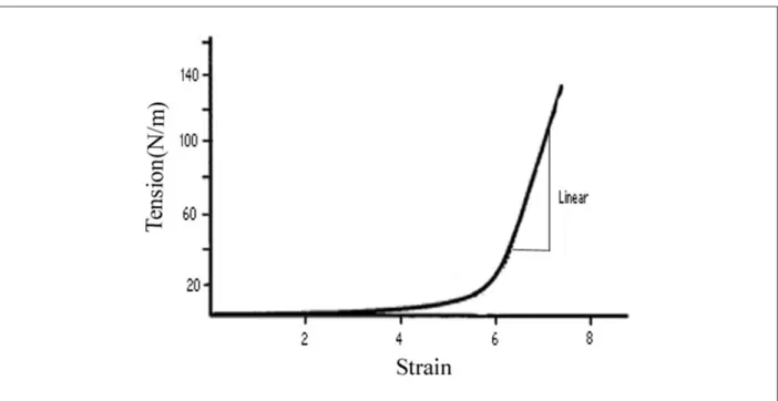
The grips were first placed 1 to 2 mm closer together to slightly wrinkle the tissue and to ensure no preload. The specimens were then preconditioned by continuously loading and unloading at 4 mm/s to a predetermined load (between 200 and 400 g) for 20 cycles, to allow the tension-strain response of the valve to become repeatable. Another three cycles of tension-strain measurements were then recorded for radial and circumferential axes (baseline group). The tissue was then challenged with increasing concentrations of neurotransmitters and some respective receptor blockers. Three more cycles of tension versus strain were recorded (treatment group). Each sample served as its own control and was used only once. The stiffness of the specimens was calculated from the gradient of the linear portion of this tension-strain curve (Figure 1). In this manuscript, the terms stiffness, modulus, and elastic modulus are used interchangeably.
Data analysis
Data are expressed as mean ± SD. Comparisons of mechanical properties between normal and treated tissues were performed by means of a 2-sided Student t test for two independent samples or One-way ANOVA test for multiple groups. All values of p < 0.05 were considered statistically significant. The statistical analyses were performed using SPSS version 13.0 (SPSS Institute).
Results
Baseline of stiffness in both axes
Baseline stiffness was assessed for the initial groups: normal Control and endothelial-denuded leaflets. Endothelial denudation did not affect valve stiffness (Control vs.
Denudation; Circumferential: 5.08 ± 0.41 vs 4.71 ± 0.64,
p > 0.05; Radial: 2.40 ± 0.10 vs 2.18 ± 0.41, p > 0.05). In addition, there was a significant difference between radial and circumferential axes within group in compliance with with the anisotropy of this tissue (Circumferential vs Radial; Control: 5.08 ± 0.41 vs 2.40 ± 0.10, p < 0.05; Denudation: 4.71 ± 0.64 vs 2.18 ± 0.41, p < 0.05). See Table 1.
Sympathetic regulation of change in tissue stiffness
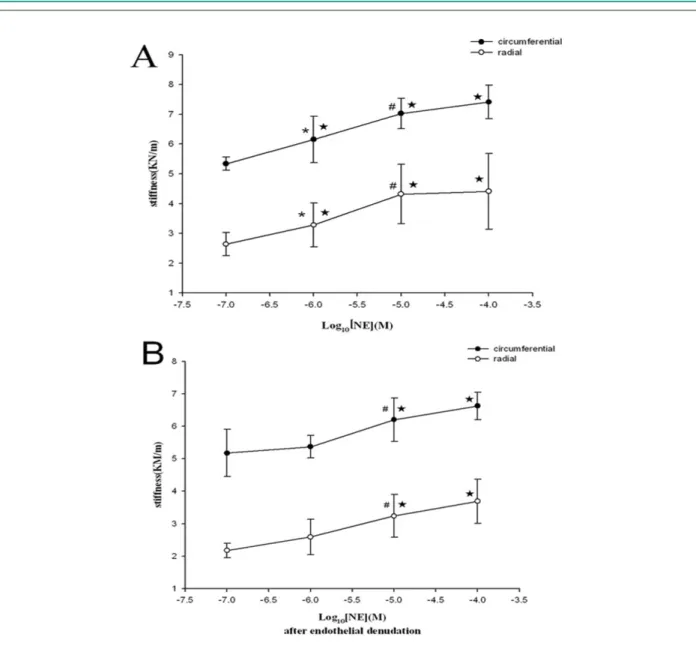
When challenged with NE in gradient concentration, the valves presented increasing stiffness in a concentration-dependent manner (Fig 2A). NE elevated the valve stiffness statistically per 10-fold increase in concentration (10-6vs 10-7:
Circumferential: 6.15 ± 0.78 vs 5.34 ± 0.23, Radial: 3.28 ± 0.74 vs 2.64 ± 0.39, p < 0.05; 10-5vs 10-6: Circumferential:
7.03 ± 0.51 vs 6.15 ± 0.78, Radial: 4.32 ± 0.99 vs 3.28 ± 0.74, p < 0.05), reaching the concentration-effect plateau in the end (10-4vs 10-5, p>0.05).
However, endothelium-denudation blunted the ability of NE in increasing stiffness (Fig 2B), only maintaining the
high-dose NE (10-5) significant compared to lower-dose
groups (10-5vs 10-6: Circumferential: 6.20 ± 0.67 vs 5.37
± 0.35, Radial: 3.24 ± 0.66 vs 2.59 ± 0.55, p < 0.05) with the plateau effect unchangeable.
Similarly, when Phent was present in the organ bath, there was a diminishing trend in cusp stiffness after addition of maximal responsive NE (Phent vs 10-5; Circumferential:
5.96 ± 1.06 vs 7.03 ± 0.51, Radial: 3.14 ± 0.64 vs 4.32
± 0.99, p < 0.05), still significant compared with Control (Phent vs control; Circumferential: 5.96 ± 1.06 vs 5.08 ± 0.41, Radial: 3.14 ± 0.64 vs 2.40 ± 0.10, p < 0.05), see Fig 3A. However, this response did not occur in the presence of Praz or Phent alone.
At last, after incubation of cusps in Metop, addition of NE resulted in a significant difference in cusp stiffness compared
to NE alone (Metop vs NE; Circumferential: 5.67 ± 0.73 vs
7.03 ± 0.51, Radial: 3.09 ± 0.68 vs 4.32 ± 0.99, p < 0.05). However, direct comparison of changes after Metop and NE versus control was still statistically significant (p < 0.05). See Fig 3B. The stiffness was not affected by addition of Propr or Metop alone.
Vagal contribution to tissue stiffness
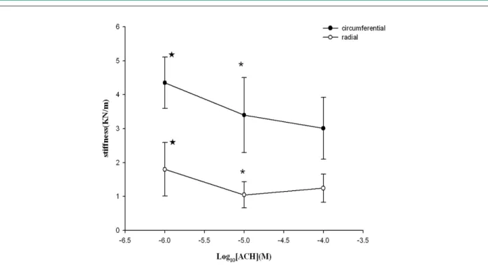
The stiffness of mitral cusps was significantly decreased in response to increasing concentrations of ACH (significant change in stiffness per 10-fold increase in ACH concentration, 10-5vs 10-6; Circumferential: 3.40 ± 1.10 vs 4.35 ± 0.76,
Radial: 1.05 ± 0.38 vs 1.80 ± 0.79, p < 0.05). The lowest scale group (10-6 mol/l) still exhibited significant effects
in diminishing modulus when compared with Controls
(Circumferential: 4.35 ± 0.76 vs 5.08 ± 0.41, Radial: 1.80
± 0.79 vs 2.40 ± 0.10, p < 0.05 vs control), with the plateau effect occurring at the concentration of 10-5 mol/l (10-5vs 10-4,
p > 0.05). See Fig 4.
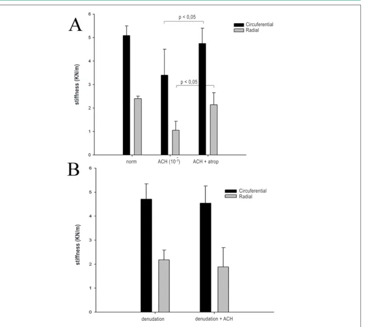
When valve specimens were preincubated in Atrop or denuded with mechanical means, the response to ACH was abrogated completely (p > 0.05 vs control). See Fig 5.
Discussion
We strove to create a near-natural internal environment, in which the mitral valve lies and is mediated by the autonomic nervous system. The main findings from this article are, under physiological conditions, the autonomic nerve (sympathetic and vagus) significantly modulates the mechanical properties of porcine mitral cusps through the neurotransmitters released by nervous ends. Stiffness of mitral valves was significantly decreased in response to increasing concentrations of ACH in a concentration-dependent fashion, and diversely, elevated by NE in a dose-effect manner. At the same time, we
observed that valvular endothelium, α/β-adrenoceptor and
cholinergic receptor played a fundamental role in regulating mechanical properties.
It has been proposed that autonomic nervous system dysfunction prevails in the vast majority of patients with heart diseases. This proposal is based mostly on the results of diverse heart rate variability studies and some dysautonomic symptoms, such as reflex syncope, anxiety, psychological distress, palpitation and so on. Mitral valves, which are far more than passive leaflets by transvalvular pressure, have been considered to be also regulated by the autonomic nervous system. However, little is known about the details of such process, so this study was performed aiming to identify the role of autonomic nerve in the regulation of porcine mitral valve mechanical properties.
The autonomic nervous end releases a number of
bioactive substances in vivo, notably NE and ACH, exerting
very complicated reactions on the target tissue and organ. Moreover, ACH is a smooth muscle cell relaxant specific in valve tissue, and muscarinic 2 acetylcholine receptors (M2) have been found to be functional, whereas the other subtypes
Table 1 - Baseline of stiffness in both axes
Circumferential Radial
Normal control (n = 10) 5.08 ± 0.41* 2.40 ± 0.10
Endothelial denuded (n = 10) 4.71 ± 0.64# 2.18 ± 0.41
* value denotes signiicance regarding Radial in normal control, p < 0.05; # value
Figure 2 -Concentration-Response Curves to NE. Concentration-dependent changes in stiffness (mean ± SD) of porcine mitral valve specimens in response to (A)
norepinephrine (NE) alone, and (B) NE + endothelial denudation in the radial and circumferential direction respectively. p < 0.05 vs Control, * p < 0.05 vs 10-7, # p <
0.05 vs 10-6 (mol/l).
have no apparent physiological effect5. ACH was shown to
induce aortic valve tissue relaxation in an ex vivo model4. In
our study, the use of exogenous ACH demonstrated the effect of this peptide by evaluating its effect on the mitral valvular stiffness. It turned out that under the present experimental conditions, ACH decreased the stiffness in a concentration-dependent manner, as expected. In addition, it appears that ACH may induce mitral valve tissue relaxation resulting in stiffness diminished through M2, as evidenced after the addition of Atrop, when responses to ACH were abrogated thoroughly. In order to specifically understand the effect
of ACH stimulation on valves, Ohashi et al6 showed that
ACH produced transient and then sustained membrane hyperpolarizations on rabbit aortic valvular endothelium; and ACH withdrawal evoked a transient depolarization. These
mechanisms contribute to release of endothelium-dependent vasoactive factors such as NO7,8 in canine and porcine mitral
valves. This may be one of the ACH mechanisms that mediate stiffness. NE can cause a potent contraction of smooth muscle cells through the stimulation of specific receptor subtypes in equine aortic valve9,10. There are more known adrenoceptor
subtypes, a number of which have been found in human heart valve tissue: α1, α2, β1 and β2 adrenoceptors10,11. Williams
TH et al3 reported that rat mitral valves had been shown to
display spontaneous contraction-like movements localized to the leaflet itself, immediately after being dissected from the heart, which was enhanced by addition of norepinephrine (NE) in a dose-dependent fashion. Otherwisely, in an effort to examine valve cusps’ responses to neuromodulators ex vivo
these effects, porcine aortic valve cusps, mounted uniaxially in an organ chamber, contracted significantly in response
to increasing concentrations of α adrenoreceptor agonists,
including norepinephrine. These contractions were specifically mediated by α2-adrenoreceptors as indicated by the absence of any response following the addition of yohimbine, an α 2-adrenoreceptor antagonist, but not after addition of prazosin, an inhibitor of α1-adrenoreceptors in equine aortic valves9.
In our experiment, we observed the valvular response to NE in stiffness, which could be mitigated by the nonselective α adrenoceptor inhibitor Phent, not prazosin. These results all confirmed the reports before and suggested β1 adrenoceptor might take part in the modulation of stiffness. It seemed that changes in elastic modulus consistently correlated with relaxation or contraction responses of the porcine tissue to the
mediators. Therefore, obviously the status of the autonomic nervous system is more likely to influence the long-term function and pliability of the valves due to the rapidity of valve opening and closing, and dysautonomia, particularly in concurrent heart valve disease, appears non-negligible.
Recent studies have indicated that porcine aortic valve endothelial cells have specific cellular and molecular characteristics that play a fundamental role in regulating heart valve tone12,13. It was reported that the valve endothelium
exerted its effects by modulating human aortic valve cusp stiffness to different mediators: ON and ET-114. Nevertheless,
the physiological role of the autonomic nervous system in heart valve endothelium has not been adequately defined. Based on the outcomes of this study, it was apparently observed that the stiffness altered by ACH was completely
Figure 3 -Role of adrenoceptors in the regulation of stiffness. Changes in stiffness maximal response to NE in the presence of (A) phentolamine (Phent) and (B) metoprolol (Metop).* p < 0.05 vs control (radial), # p < 0.05 vs NE (10-5 M, radial).
p < 0.05 p < 0.05
Circuferential Radial
S
tiffness (K
N
/m)
norm NE (10–5m) NE + fent
Circuferential Radial p < 0.05
p < 0.05
stiffness (K
N
/m)
Figure 4 -Concentration-Response Curves to ACH. Concentration-dependent changes in stiffness (mean ± SD) of porcine mitral valve specimens in response to
acetylcholine (ACH) in the radial direction and circumferential direction, respectively. p < 0.05 vs Control, * p < 0.05 vs 10-6.
dependent on the valvular endothelium, whereas the stiffness altered by NE was partially dependent on the valvular endothelium and other mechanisms might be involved. These responses may be elicited by the distribution of the
respective receptors. The findings by Pompilio et al4 on
porcine aortic and pulmonary valves may help appreciate this reaction. The effect of ACH, inducing aortic valve tissue relaxation, was absent when the endothelium was mechanically removed, underlining the essential role of the endothelium and M2 receptors (above mentioned). The changes in the mechanical properties of valve tissue in response to different mediators can be explained by the presence of myofibroblasts and smooth muscle cells in the
human valves15. The porcine valvular endothelium alone
cannot anatomically facilitate the interstitial cell populations in modulus, as evidenced by the absence of change in valve stiffness after mechanical denudation of the endothelium. However, the cross-talk between endothelial and interstitial myofibroblasts and smooth muscle cells was suggested by this study, seen from the observation that endothelium-denudation blunted the ability of NE in increasing modulus, only preserving the high-dose NE (10-5) significant compared
to lower-dose groups. This may be the means by which the mitral valve endothelium regulates the mechanical properties of porcine valve cusps. Therefore, the unique structure and function of valve endothelial cells should be highlighted.
This is an ex vivo study, and actual values of the overall environment: endocrine, paracrine and neurocrine signals may differ from those in vivo. The acute experimental setup does not account for the long-term trophic effects of these neurotransmitters or their ability to modulate the extracellular
matrix in human beings16. Further studies are required to
evaluate any other biological parameters involved in response to autonomic nervous stimuli, as well as the responsive differences between endothelial cells from interstitial cell populations.
Clinical implication
Changes in heart valves mechanical responses with autonomic nervous-dependent pathways are likely to be significant in the pathophysiology of valve disease. It has been shown that parasympathetic and sympathetic reflex arcs differed with aging, including increased circulating norepinephrine levels at baseline and in response to stress, decreased catecholamine concentration in senescent rat heart, increased synthesizing and degrading enzymes for
catecholamines17. Heart valve dysfunctions show increased
Conclusions
In conclusion, this study has shown that the mechanical properties of mitral valve cusps are actively regulated through autonomic nervous-dependent pathways. The autonomic nerve (sympathetic and vagus) exerts its effects by modulating the stiffness of the cusps through α2, β1 and M2 receptors in the valve endothelium, in addition to interstitial cells. We are grateful that these findings will help to understand valve pathophysiology and establish a blueprint of adaptive responses of valve mechanical properties for future tissue-engineered heart valves.
Potential Conflict of Interest
No potential conflict of interest relevant to this article was reported.
Sources of Funding
There were no external funding sources for this study.
Study Association
This study is not associated with any post-graduation program.
Figure 5 -Role of cholinergic receptor and valve endothelium in regulating stiffness. Change in stiffness maximal response to ACH in the presence of (A) atropine (Atrop) and (B) endothelium denuded.
p < 0,05
p < 0,05
Circuferential Radial
stiffness (K
N
/m)
norm ACH (10–5) ACH + atrop
Circuferential Radial
stiffness (K
N
/m)
References
1. Voloshchenko AA. Afferent innervation of atrioventricular valve. Fed Proc Transl Suppl. 1965;24(4):571-4.
2. Williams TH. Mitral and tricuspid valve innervation. Br Heart J. 1964;26:105-15.
3. Williams TH, Jew JY. Is the mitral valve passive flap theory overstated? An active valve is hypothesized. Med Hypotheses. 2004;62(4):605-11.
4. Pompilio G, Rossoni G, Sala A, Polvani GL, Berti F, Dainese L, et al. Endothelial-dependent dynamic and antithrombotic properties of porcine aortic and pulmonary valves. Ann Thorac Surg. 1998;65(4):986-92.
5. van Zwieten PA, Doods HN. Muscarinic receptors and drugs in cardiovascular medicine. Cardiovasc Drugs Ther. 1995;9(1):159-67.
6. Ohashi M, Satoh K, Itoh T. Acetylcholine-induced membrane potential changes in endothelial cells of rabbit aortic valve. Br J Pharmacol. 1999;126(1):19-26.
7. Ku DD, Nelson JM, Caulfield JB, Winn MJ. Release of endothelium-derived relaxing factors from canine cardiac valves. J Cardiovasc Pharmacol. 1990;16(2):212-8.
8. Siney L, Lewis MJ. Nitric oxide release from porcine mitral valves. Cardiovasc Res. 1993;27(9):1657-61.
9. Bowen IM, Marr CM, Chester AH, Wheeler-Jones CP, Elliott J. In vitro contraction of the equine aortic valve. J Heart Valve Dis. 2004;13(4):593-9.
10. Chester AH, Misfeld M, Yacoub MH. Receptor-mediated contraction of aortic valve leaflets. J Heart Valve Dis. 2000;9(2):250-4.
11. Osman L, Chester AH, Sarathchandra P, Latif N, Meng W, Taylor PM, et al. A novel role of the sympatho-adrenergic system in regulating valve calcification. Circulation. 2007;116(11 Suppl):I282-7.
12. Butcher JT, Penrod AM, Garcia AJ, Nerem RM. Unique morphology and focal adhesion development of valvular endothelial cells in static and fluid flow environments. Arterioscler Thromb Vasc Biol. 2004;24(8):1429-34.
13. Butcher JT, Tressel S, Johnson T, Turner D, Sorescu G, Jo H, et al. Transcriptional profiles of valvular and vascular endothelial cells reveal phenotypic differences: influence of shear stress. Arterioscler Thromb Vasc Biol. 2006;26(1):69-77.
14. El-Hamamsy I, Balachandran K, Yacoub MH, Stevens LM, Sarathchandra P, Taylor PM, et al. Endothelium-dependent regulation of the mechanical properties of aortic valve cusps. J Am Coll Cardiol. 2009;53(16):1448-55.
15. Roy A, Brand NJ, Yacoub MH. Molecular characterization of interstitial cells isolated from human heart valves. J Heart Valve Dis. 2000;9(3):459-64.
16. Hafizi S, Taylor PM, Chester AH, Allen SP, Yacoub MH. Mitogenic and secretory responses of human valve interstitial cells to vasoactive agents. J Heart Valve Dis. 2000;9(3):454-8.