Left Ventricular Diastolic Function in Morbidly Obese Patients in the
Preoperative for Bariatric Surgery
Irlaneide da Silva Tavares
1,4, Antonio Carlos Sobral Sousa
1,2, Raimundo Sotero Menezes Filho
3, Manuel H.
Aguiar-Oliveira
1, José Augusto Barreto-Filho
1,2, Amanda Ferreira de Brito
1, Joselina Luzia Menezes Oliveira
1,2Departamento de Medicina - Universidade Federal de Sergipe1, São Cristóvão, SE; Centro de Ensino e Pesquisa do Hospital e Fundação São Lucas2; Dimese - Centro de Diabetes de Sergipe3; Hospital do Coração de Aracaju4, Aracaju, SE
Abstract
Background: Obesity is a chronic and multifactorial disease, associated with increased cardiovascular risk, especially diastolic heart failure.
Objective: To evaluate left ventricular diastolic function in morbidly obese patients in the pre-operative for bariatric surgery, correlating it with cardiovascular risk factors and heart structure.
Methods: This is a cross-sectional study with 132 patients eligible for bariatric surgery submitted to transthoracic echocardiography assessment and of cardiovascular risk factors, as follows: 97 women (73.5%), mean age 38.5 ± 10.5 years and BMI of 43.7 ± 7.2 kg / m². Patients were divided into three groups: 61 with normal diastolic function, 24 with mild diastolic dysfunction and 47 with moderate/severe diastolic dysfunction, of which 41 with moderate diastolic dysfunction (pseudonormal pattern) and six with severe diastolic dysfunction (restrictive pattern).
Results: Hypertension, age and gender were different in the groups with diastolic dysfunction. Groups with dysfunction had higher left atrial diameter, left ventricular diameter, left atrial volume in four and two chambers, left atrial volume index and left ventricular mass index corrected for body surface area and height.
Conclusion: The high frequency of left ventricular diastolic dysfunction in the preclinical phase in morbidly obese patients justifies the need for careful echocardiographic assessment, aiming at identifying individuals at higher risk, so that early intervention measures can be carried out. (Arq Bras Cardiol 2012;98(4):300-306)
Keywords: Obesity morbid; heart failure; preoperative care; bariatric surgery.
Mailing address: Irlaneide Da Silva Tavares •
Rua Gerson dos Santos, 08 - Residencial Parque dos Coqueiros – Inácio Barbosa – 49040-293 – Aracaju, SE – Brazil
E-mail: [email protected]
Manuscript received August 29, 2011; revised mansucript received August 29, 2011; accepted December 27, 2011.
comorbidities7. The presence of diastolic dysfunction of the left ventricle (LV), in the general population, is associated with the development of HF and shorter survival8.
The high prevalence of obesity, increased risk of Diastolic Heart Failure (DHF), the frequent association with comorbidities that increase cardiovascular risk and the fact that it is an independent and modifiable risk factor for the development of several diseases, require a thorough echocardiography investigation in obese individuals, respecting their peculiarities, in order to identify patients at higher risk, so that intervention measures can be carried out early.
To date, we are unaware of publications that investigated morbidly obese patients according to the criteria of the recommendations for quantifying cardiac chambers9 and diastolic dysfunction10, so the aim of this study is to assess left ventricular diastolic function in morbidly obese patients that are candidates to bariatric surgery, according to these guidelines, associating cardiovascular risk factors and cardiac structure.
Methods
The study was carried out in the Cardiology Clinic of Hospital Universitário da Universidade Federal de Sergipe and in a private institution in Aracaju, Hospital do Coração, both
Introduction
Obesity is a chronic and multifactorial disease, which represents a high risk for health1. It is a public health problem that has reached epidemic proportions in adults and children2. It is often associated with conditions that increase cardiovascular risk3, including: dyslipidemia, systemic arterial hypertension (SAH), glucose intolerance, left ventricular hypertrophy (LVH), hyperuricemia, elevated fibrinogen, metabolic syndrome (MS) and sleep apnea (SA). Obese individuals have a 104% increase in the risk of developing heart failure (HF) compared with nonobese ones4. The centripetal distribution of body fat is associated with higher concentrations of plasma lipoprotein levels and blood pressure in both sexes, regardless of the amount of body fat5.
in the state of Sergipe, Brazil, from August 2009 to October 2010. This is a cross-sectional and analytical study, with a convenience sample of morbidly obese patients concomitantly referred for preoperative evaluation for bariatric surgery. Patients with inadequate echocardiographic windows and the following diseases: established atherosclerosis, higher-than-mild degree of valvular disease, presence of prosthetic valve, pericardial disease, atrial fibrillation, frequent extrasystoles, ventricular systolic dysfunction, hyperthyroidism and hypothyroidism were excluded.
To identify the profile of the studied population a semistructured interview was developed at the clinical assessment. An individual was considered to be a smoker if he/she had smoked at least one cigarette over the past thirty days and those who performed less than 30 minutes of physical activity, three times a week were considered sedentary. Abdominal circumference measurement was performed with the patient standing at the end of expiration, in the midpoint between the lower margin of the last palpable rib and the top of the anterosuperior iliac crest using an inelastic tape in the horizontal position11. BMI12 (BMI: weight/height²) and body surface area13 [BS: w 0.425 x h 0.725 x 71.84 x 104] were also calculated.
Blood pressure measurement followed the recommendations of the Brazilian Society of Hypertension14, which defines
hypertension as systolic blood pressure ≥ 140 mmHg and / or diastolic blood pressure ≥ 90 mmHg, or use of
antihypertensive medication; heart rate was obtained with the patient in the sitting position, after five minutes of rest, by counting the heartbeats in 60 seconds using the radial pulse. To detect diabetes, we followed the recommendations of the American Diabetes Society15; for dyslipidemia, we followed the recommendations of the IV Brazilian Guidelines on Dyslipidemia16 and for the characterization of MS, we followed the criteria of the First Brazilian Guideline for the Diagnosis and Treatment of Metabolic Syndrome11.
All echocardiograms were performed by an experienced professional, using a TOSHIBA echocardiography device, model Istyle or GE device model Vivid 3, with a 2.5 MHz sectorial transducer, obtaining second harmonic images with 100 mm/s scanning, using one-dimensional, two-dimensional and Doppler (pulsed, color and tissue) echocardiography in the parasternal (longitudinal and transverse) and apical (two, four and five chambers) windows. The average of three measurements was used for each variable. The assessment of cardiac chamber dimensions and LV systolic and diastolic functions was based on the recommendations of the American Society of Echocardiography9,10.
The following echocardiographic variables were analyzed: cardiac dimensions (aorta, left atrium, left ventricle during systole and diastole, interventricular septum and posterior wall), percentage of systolic shortening (%), ejection fraction (LVEF), left ventricular mass index indexed to body surface area in g/ m2 (LVMIbs) and for height in g/m (LVMIh), left ventricular geometric pattern, two and four-chamber left atrial volume (LAV2c and LAV4c) and indexed for body surface area (LAVi - ml/m²), velocities (cm/s) of E and A mitral flow waves, velocity of E and A waves of the septal and lateral annulus of the mitral valve, E/A and E/e ratios, isovolumetric relaxation time (IVRT)
(ms), deceleration time (DT) (ms), duration (ms) of A wave (Adur), velocity (cm/s) and duration (ms) of the pulmonary atrial reversal flow (Ar and Ardur), relation (ms) of the duration of the atrial reversal flow subtracted from the duration of A wave of the mitral flow (Ardur - Adur), velocity (cm/s) of the systolic (S) and diastolic (D) wave of the pulmonary flow and S / D ratio. The size of the aorta and left atrium were evaluated using one-dimensional mode (Mode M) in the longitudinal parasternal view. Measurements of systolic and end-diastolic diameters, septal and LV posterior wall thickness were performed in the parasternal transversal view. Atrial volume was measured by Simpson’s method, left ventricular ejection fraction by Teichholz formula17, using M-mode and LV mass
was calculated using the modified Deveraux formula18: 0·80
(1·04 [(IVST+PWT+LVED)3- LVED3]+0.6 g, and the relative
wall thickness (RWT) was calculated by the ratio of twice the posterior wall divided by the left ventricular diastolic diameter9
(RWT = 2PP/DD).
The characterization of the left ventricular geometric pattern was considered as mass related to height9, and a
normal pattern was considered when mass and RWT were normal, concentric remodeling when there was normal mass and high RWT, concentric hypertrophy when both mass and RWT were high and eccentric hypertrophy when mass was high and RWT was normal.
Diastolic dysfunction was considered when the velocities of septal e < 8 cm / and lateral < 10 cm/s. It was classified as
mild when E / A <0.8, DT> 200 ms, E/e ≤ 8 and Ardur - Adur
< 0 ms; as moderate when E/A = 0.8 to 1.5, DT = 160-200 ms, E/e > 13 and Ardur – Adur > 30 ms; and as severe when E/A> 2, DT < 160, E/e > 13 and Ardur - Adur < 30 ms10.
The 132 patients were divided into three groups according to LV diastolic function: normal (N), mild diastolic dysfunction (mild DD) and moderate or severe diastolic dysfunction (M/S DD). The inclusion of patients with a restrictive pattern in the third group was due to their small number (six patients).
For statistical calculations, we used the SPSS (Statistical Package for Social Sciences) release 18.0. Numerical variables were described as mean and standard deviation and categorical variables as simple and relative frequencies. The Shapiro-Wilk test was used to evaluate the assumption of normality. To test hypotheses related to categorical variables the Chi-square test or Fisher’s exact test was used when appropriate.
The comparison between the groups (N vs. mild DD vs.
M/S DD) was carried out using the general linear model with one factor (ventricular dysfunction), adjusting for the variables. Confidence level was 0.05 for the α error, with a power of 0.80, and the tests were assumed to be two-tailed. For the analysis of factors associated with the outcome variable (presence of diastolic dysfunction) the forward stepwise logistic regression method was used, considering p = 0.25 for entering the model and p = 0.05 for remaining in the same model; simple and adjusted odds ratio were also calculated.
Results
The sample consisted of 132 patients, of which 97 (73.5%) were females, resulting in a ratio of 2.7 women for each man. The mean age was 38.5 ± 10.5 years and ranged from 16 to 62 years. The mean BMI was 43.7 ± 7.2 kg/m² and ranged from 35.2 to 71.2 kg/m². Forty-eight (36.4%) patients had class II obesity, and 84 patients (63.6%) had grade III obesity. The frequency of LVDD was 53.8% (71 patients), and M/S DD was more frequent than the mild one. The comparative analysis of the clinical and anthropometric variables that showed significant differences between groups were: age, gender and waist circumference (Table 1).
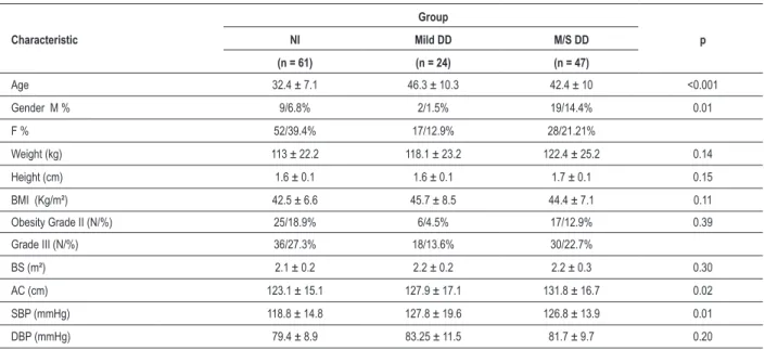
Patients with mild DD and M/S DD were older and had significantly higher mean SBP (p <0.001) than group N. Regarding the waist circumference, the M/S DD group showed higher mean values (p = 0.02) than group N. There was a positive association between abdominal obesity and degree of LVDD (Figure 1).
With regard to cardiovascular risk factors (CVRF), it was observed that mild DD and M/S DD groups had significantly more diabetes (p <0.0001), hypertension (p <0.0001) and MS (p <0.001) when compared to N, without, however, showing any difference between them (Table 2). Among the cardiovascular risk factors, only age, gender and hypertension were independently correlated with the presence of DD at the multivariate analysis.
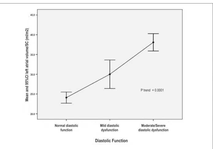
The echocardiographic variables (LAD, LVDD, LAV4c, LAV2c, MLAV, LAVi, and LVMIbs, LVMIh) showed significantly higher values in groups with DD when compared to N (Table 3). There is also a linear trend of increasing severity between these variables and diastolic
dysfunction, as shown in Figure 2. As for the LA dimensions, it was also possible to discriminate the two groups with DD. The LV mass index indexed for height was also higher in patients with DD; however, only the group M/S DD showed to be statistically different (p <0.001) from group N (Table 3).
Left ventricular hypertrophy was diagnosed in 20% of the sample when the mass was indexed for body surface, and 55.3% when indexed for height. The geometric pattern showed the following distribution when the LV mass was indexed for body surface area and height, respectively: normal in 102 (77.3%) and 58 (43.9%), concentric remodeling in 1 (0.8%) and concentric hypertrophy in 7 (5.3%) for both and eccentric hypertrophy in 22 (16.7%) and 66 (50%) patients. There was a significant association between the eccentric geometric pattern (p = 0.03) with the presence of diastolic dysfunction and a higher prevalence of that in the mild (62.5%) and M/S (50.9%) DD groups when compared to group N.
Discussion
The findings of the present study show a predominance of female patients, which seems show a greater demand for treatment among women. Regarding age, the data are similar to that from other studies19-22, such as a study by Matos et al23 of 50 patients, 10 men and 40 women that reported a BMI of 40 to 81.7 kg/m2 (mean 52.2 ± 9.2 kg / m²) and age between 18 and 56 years (mean 38, 5 ± 9.7). As for the BMI, there was a similar result in a study by Okawa24, with 104 morbidly obese patients at the pre-operative period of bariatric surgery, which showed an average BMI of 42.8% ± 5.45 kg / m².
Table 1 – Characterization of the clinical and anthropometric variables according to LV diastolic function
Characteristic
Group
p
NI Mild DD M/S DD
(n = 61) (n = 24) (n = 47)
Age 32.4 ± 7.1 46.3 ± 10.3 42.4 ± 10 <0.001
Gender M % 9/6.8% 2/1.5% 19/14.4% 0.01
F % 52/39.4% 17/12.9% 28/21.21%
Weight (kg) 113 ± 22.2 118.1 ± 23.2 122.4 ± 25.2 0.14
Height (cm) 1.6 ± 0.1 1.6 ± 0.1 1.7 ± 0.1 0.15
BMI (Kg/m²) 42.5 ± 6.6 45.7 ± 8.5 44.4 ± 7.1 0.11
Obesity Grade II (N/%) 25/18.9% 6/4.5% 17/12.9% 0.39
Grade III (N/%) 36/27.3% 18/13.6% 30/22.7%
BS (m²) 2.1 ± 0.2 2.2 ± 0.2 2.2 ± 0.3 0.30
AC (cm) 123.1 ± 15.1 127.9 ± 17.1 131.8 ± 16.7 0.02
SBP (mmHg) 118.8 ± 14.8 127.8 ± 19.6 126.8 ± 13.9 0.01
DBP (mmHg) 79.4 ± 8.9 83.25 ± 11.5 81.7 ± 9.7 0.20
When left ventricular diastolic function was assessed, there was a reduction in diastolic function in 53.8% of patients, a fact which is not common in the young age group as in this sample, aged 38.5 years (± 10.5). Okawa’s study24 with 104 morbidly obese patients, found 62.4% of diastolic dysfunction, while another study by Rocha et al25 found in 30 patients in the preoperative period for bariatric surgery, 54.6% of diastolic dysfunction. Studies on the prevalence of diastolic dysfunction in obese diabetic patients showed a prevalence of 47% when using mitral flow26; Boyer et al27, when associated flow propagation and color M-mode tissue Doppler to mitral flow analysis, found a prevalence of 75% of LV diastolic dysfunction, and tissue Doppler detected diastolic dysfunction in 63% of patients.
Regarding dysfunction severity, there was a predominance of moderate diastolic dysfunction, probably to the detriment of the restrictive pattern due to the diagnostic criteria used, characterizing this pattern when all variables were altered together (E/A> 2, DT < 160 ms, E/e ‘ > 13 and Ar - A < 30 ms).
In relation to left ventricular systolic function, which was an exclusion criterion, none of the patients referred for evaluation had systolic dysfunction. Several studies in obese subjects28-32 have shown preserved systolic function, and systolic dysfunction was found only in obese patients who had been so for a long time. The LV ejection fraction is a reliable index, but can be within normal limits even
Table 2 – Distribution of cardiovascular risk factors according to LV diastolic function
Groups
Variables N Mild DD M/S DD Total p
(n = 61) (n = 24) (n = 47)
Diabetes Yes 6 (10.2%) 6 (27.3%) 20 (43.5%) 32 (25.2%) <0.0001
Dyslipidemia Yes 42 (76.4%) 17 (85%) 35 (79.5%) 94 (79%) 0.71
SAH Yes 22 (36.1%) 18 (75%) 39 (83%) 79 (59.8%) <0.0001
Metabolic Syndrome Yes 25 (44.6%) 18 (90%) 38 (86.4%) 81 (67.5%) <0.001
Smoking Yes 2 (3.3%) 0 (0%) 0 (0%) 2 (1.5%) 0.37
Sedentary life style Yes 51 (83.6%) 20 (83.3%) 36 (76.6%) 107 (81.1%) 0.62
Alcohol consumption Yes 3 (4.9%) 0 (0%) 2 (4.3%) 5 (3.8%) 0.55
N - group with normal diastolic function; DD - diastolic dysfunction; M/S - moderate/severe; SAH - systemic arterial hypertension.
Figure 1 – Abdominal circumference according to LV diastolic function.
Diastolic Function
P trend = 0.006
Normal diastolic function
Mild diastolic dysfunction
Moderate/Severe diastolic dysfunction
M
édia e IC
95% da cir
cunfer
ência abdominal (cm)
135.0
130.0
125.0
Table 3 – Echocardiographic variables according to LV diastolic function
Echocardiographic Variables
Group
p
N Mild DD M/S DD
(n = 61) (n = 24) (n = 47)
LAD (cm) 4.08 ± 0.04 4.22 ± 0.06 4.47 ± 0.05 <0.001
LVDD (cm) 5.24 ± 0.05 5.35 ± 0.08 5.52 ± 0.05 <0.005
LAV4c (mL) 53.3 ± 2.3 62.9 ± 3.4 81.8 ± 2.5 <0.001
LAV2c (mL) 54.7 ± 2.3 61.8 ± 3.4 82.9 ± 2.4 <0.001
MLAV (mL) 54.0 ± 2.7 62.4 ± 3.3 82.3 ± 2.4 <0.001
LAVi (mL/m2) 25.2 ± 1.0 28.5 ± 1.5 37.4 ± 1.1 <0.001
LVMIbs (g/m²) 78.1 ± 2.5 90.2 ± 3.6 94.5 ± 2.6 <0.001
LVMIh (g/m) 104.3 ± 3.4 121.4 ± 4.9 125.1 ± 3.5 <0.001
Linear general model adjusted for age, gender, abdominal circumference and systolic blood pressure, com p < 0.05. N - Group with normal diastolic function; mild
DD - Group with mild diastolic dysfunction and M/S DD - Group with moderate or severe diastolic dysfunction. LAD - left atrial diameter; LVDD - left ventricular diastolic diameter; LAV4c - Left atrial volume in 4 chambers; LAV2c - Left atrial volume in 2 chambers; MLAV - mean left atrial volume; LAVi - Left atrial volume index; LVMIbs - left ventricular mass index for body surface; LVMIh - left ventricular mass index for height.
Figure 2 – Left Atrial Volume Index (LAVi) chart according to LV diastolic function.
Diastolic Function
P trend = 0.0001
Normal diastolic function
Mild diastolic dysfunction
Moderate/Severe diastolic dysfunction
M
ean and 9
5%C
I left atr
ial volume/S
C
(ml/m2)
45.0
40.0
35.0
30.0
25.0
though there are significant compensatory changes in contractile state.
SAH is the most prevalent comorbidity when measured alone. Several authors have described the association between BMI and higher prevalence of MS, indicating that obesity is related to an unfavorable risk profile for cardiovascular disease. Similar data were found in other studies that showed hypertension as the most frequent isolated comorbidity20,21. A similar result was observed by Costa et al32, with a prevalence of SAH of 63.4% among 252 morbidly obese patients. The presence of DM occurred in 25.2% of patients, similar to the 26% rate found by Rocha e Silva et al25.
It was demonstrated that the greater left atrial volume, the worse the diastolic function, proving the theory that this is a sensitive index that expresses the severity of LV diastolic dysfunction33. Left ventricular hypertrophy was more often diagnosed when the LVM criterion was indexed for height (73 patients = 55.3%) than for body surface area (29 patients = 22%), even when following the recent guidelines from the American Society of Echocardiography in that the cutoff for left ventricular mass is greater when indexed for height9. The LV geometric patterns found in this study were in agreement with other authors7,28,34,35, with eccentric LVH being the most common geometric abnormality, regardless of whether the mass index is related to the height or body surface. The concentric remodeling, associated with decreased cardiac index, elevated peripheral vascular resistance and reduced circulating plasma volume36 was observed in only one (0.7%) case, confirming the pathophysiological model proposed for severe obesity, in which the circulating volume is high and peripheral resistance is normal or low.
Study limitations
To prevent measurement bias, patients were evaluated by a single observer and three consecutive echocardiographic measurements were recorded for each variable; selection bias was minimized by the fact that it was not random, with the evaluation of consecutive patients; as for sampling bias, it is believed that the sample is representative of the morbidly obese population in the preoperative period for bariatric surgery (patient from the public health system and from a private practice).
Conclusion
Obesity is associated with high frequency of left ventricular diastolic dysfunction and changes in cardiac structure, including increased left atrial volume in the pre-clinical phase in morbidly obese patients. These data justify the need for careful echocardiographic assessment using the combined analysis of all available echocardiographic techniques, aiming at identifying individuals at greater risk, so that early intervention measures can be adopted.
Potential Conflict of Interest
No potential conflict of interest relevant to this article was reported.
Sources of Funding
There were no external funding sources for this study.
Study Association
This article is part of the thesis of master submitted by Irlaneide da Silva Tavares, from Universidade Federal de Sergipe.
References
1. Chopra M, Galbraith S, Darnton-Hill I. A global response to a global problem: the epidemic of overnutricion. Bull World Health Organ. 2002; 80(12): 952-8.
2. Poirier P, Giles TD, Bray GA, Hong Y, Stern JS, Pi-Sunyer FX, et al. Obesity and cardiovascular disease: pathophysiology, evaluation, and effect of weight loss: an update of the 1997 American Heart Association scientific statement on obesity and heart disease from the obesity committee of the council on nutrition, physical activity, and metabolism. Circulation. 2006; 113(6): 898-918.
3. Kannel WB, Agostinho RBD, Cobb JL. Effect of weight on cardiovascular disease. Am J Clin Nutr. 1996; 63(3 Suppl): 419s-22s.
4. Kenchaiah S, Evans JC, Levy D, Wilson PWF, Benjamin EJ, Larson MG, at al. Obesity and the risk of heart failure. N Engl J Med.2002;347(5):305-13.
5. Guedes DP, Guedes JERP. Distribuição de gordura corporal, pressão arterial e níveis de lipídios-lipoproteínas plasmáticas. Arq Bras Cardiol. 1988;70(2):93-8.
6. Alpert MA. Obesity cardiomyopathy: pathophysiology and evolution of the clinical syndrome. Am J Med Sci. 2001; 321(4): 225–36.
7. Iacobellis G, Ribaudo MC, Zappaterreno A, Iannucci CV, Di Mario U, Leonetti F. Adapted changes in left ventricular structure and function in severe uncomplicated obesity. Obes Res. 2004; 12(10): 1616-21.
8. Gaasch WH, Little WC. Assessment of left ventricular diastolic function and recognition of diastolic heart failure. Circulation. 2007; 116(6):591-3.
9. Lang RM, Bierig M, Devereux RB, Flachskampf FA, Foster E, Pellikka PA, et al. Recommendations for Chamber Quantification: A Report from the American Society of Echocardiography’s Guidelines and Standards Committee and the Chamber Quantification Writing Group, Developed in Conjunction with the European Association of Echocardiography, a Branch of the European Society of Cardiology. J Am Soc Echocardiogr. 2005; 18(12): 1440-63.
10. Nagueh SF, Appleton CP, Gillebert TC, Marino PN, Oh JK, Smiseth OA, et al. Recommendations for the Evaluation of left ventricular diastolic function by echocardiography. J Am Soc Echocardiogr. 2009; 22 (2):107-33.
11. Sociedade Brasileira de Hipertensão/Sociedade Brasileira de Cardiologia/ Sociedade Brasileira de Nefrologia. I Diretriz brasileira de diagnóstico e tratamento da síndrome metabólica. Arq Bras Cardiol. 2005; 84 (Supl. 1): s3-s28.
12. Onis M de, Habicht JP. Anthropometrics reference data for international use: recommendations from a World Health Organization Expert Committee. Am J Clin Nutr. 1996; 54(4):650-8.
14. Sociedade Brasileira de Cardiologia/ Sociedade Brasileira de Hipertensão/ Sociedade Brasileira de Nefrologia. V Diretrizes brasileiras de hipertensão arterial. Arq Bras Cardiol. 2007; 89 (3): e24-e79.
15. American Diabetes Association. Standards of Medical Care in Diabetes. Diabetes Care. 2010; 33( Suppl 1): 11-61.
16. Sposito AC, Caramelli B, Fonseca FA, Bertolami MC, Afiune Neto A, Souza AD, et al;Sociedade Brasileira de Cardiologia. IV Diretriz brasileira sobre dislipidemias e prevenção da aterosclerose. Arq Bras Cardiol. 2007; 88(1 supl): S2-19.
17. Te i c h h o l z L E , K r e u l e n T, H e r m a n M V, G o r l i n R . P r o b l e m s i n echocardiographic volume determinations: echocardiographic-angiographic correlations in the presence of absenceof asynergy. Am J Cardiol.1976;37(1):7-11.
18. Ganau A, Devereux RB, Roman MJ, de Simone G, Pickering TG, Saba PS, et al. Patterns of left ventricular hypertrophy and geometric remodeling in essential hypertension. J Am Coll Cardiol. 1992; 19(7): 1550-8.
19. Porto MC, Brito IC, Calfa AD, Amoras M, Villela N, Araújo LM. Perfil do obeso classe III do ambulatório de obesidade de um hospital universitário de Salvador, Bahia. Arq Bras Endocrinol Metab. 2002; 46 (6): 668-73.
20. Anderi Júnior E, Araújo LGC, Fuhro FE, Godinho CA, Henriques AC. Experiência inicial do serviço de cirurgia bariátrica da Faculdade de Medicina do ABC. Arq Med ABC. 2007; 32 (1): 25-9.
21. Faria OP, Pereira VA, Gangoni CM, Lins RD, Leite S, Rassi V, et al. Obesos mórbidos tratados com gastroplastia redutora com Bypass gástrico em Y de Roux: análise de 160 pacientes. Brasília Méd. 2002; 39 (1/4): 26-34.
22. Souza LJ, Gicovate Neto C, Chalita FEB, Reis AFF, Bastos DA, Souto Filho JTD, et al. Prevalência de obesidade e fatores de risco cardiovascular em Campos, Rio de Janeiro. Arq Bras Endocrinol Metab. 2003; 47 (6): 669-76.
23. Matos MIR, Aranha LS, Faria AN, Ferreira SRG, Bacaltchuck J, Zanella MT. Binge eating disorder, anxiety, depression and body image in grade III obesity patients. Rev Bras Psiquiatr. 2002; 24 (4): 165-9.
24. Okawa RTP. Avaliação ecoDopplercardiográfica da função diastólica do ventrículo esquerdo em indivíduos obesos pré e pós cirurgia bariátrica[tese]. São Paulo: Faculdade de Medicina . Universidade de São Paulo;2006.
25. Rocha IEGM, Victor EG, Braga MC, Silva OB, Becker MMC. Avaliação ecocardiográfica em obesos graves assintomáticos. Arq Bras Cardiol. 2007; 88 (1): 52-8.
26. Zabalgoitia M, Ismaeil MF, Anderson L, Maklady FA. Prevalence of diastolic dysfunction in normotensive, asymptomatic patients with well controlled type 2 diabetes mellitus. Am J Cardiol. 2001; 87(3): 320-3.
27. Boyer JK, Thanigaraj S, Schechtman KB, Perez JE.. Prevalence of ventricular diastolic dysfunction in asymptomatic, normotensive patients with diabetes mellitus. Am J Cardiol. 2004; 93(7): 870-5.
28. Pascual M, Pascual DA, Soria F, Vicente T, Hernandez AM, Tebar FJ, et al. Effects of isolated obesity on systolic and diastolic left ventricular function. Heart . 2003; 89(10): 1152-6.
29. Peterson LR, Waggoner AD, Schechtman KB, Meyer T, Gropler RJ, Barzilai B, et al. Alterations in left ventricular structure and function in young healthy obese women: assessment by echocardiography and tissue Doppler imaging. J Am Coll Cardiol. 2004; 43(8): 1399-404.
30. De Divitis O, Fazio S, Petitto M, Madalena G, Contaldo F, Mancini M. Obesity and cardiac function. Circulation. 1981; 64(3): 477-82.
31. Messerli FH, Sundgard-Riise K, Reisin E, Dreslinski G, Dunn FG, Frohlinch E. Disparate cardiovascular effects of obesity and arterial hipertension. Am J Med. 1983; 74(5): 808-12.
32. Costa AC, Ivo ML, Cantero WB, Tognini JR. Obesidade em pacientes candidatos a cirurgia bariátrica. Acta Paul Enferm. 2009; 22 (1): 55-9.
33. Sousa ACS. Volume atrial esquerdo como indice de função diastólica. Arq Bras Cardiol. 2006; 87 (3): e27-e33.
34. De Simone G, Devereux RB, Roman MJ, Alderman MH, Laragh JH. Relation of obesity and gender to left ventricular hypertrophy in normotensive and hypertensive adults. Hypertension. 1994; 23(5): 600-6.
35. Alexander JK. The cardiomyopathy of obesity. Prog Cardiovasc Dis. 1985; 27(5): 325-34.