Sildenafil vs. Sodium Nitroprusside for the Pulmonary Hypertension
Reversibility Test Before Cardiac Transplantation
Aguinaldo Figueiredo Freitas Jr, Fernando Bacal, José de Lima Oliveira Júnior, Alfredo Inácio Fiorelli, Ronaldo
Honorato Santos, Luiz Felipe Pinho Moreira, Christiano Pereira Silva, Sandrigo Mangini, Jeane Mike Tsutsui,
Edimar Alcides Bocchi
Instituto do Coração (InCor) - Hospital das Clínicas da Faculdade de Medicina da Universidade de São Paulo, São Paulo, SP - Brazil
Mailing Address: Aguinaldo Figueiredo Freitas Jr. •
Rua T - 27, nº 300, Apto 902, Bueno. Postal Code 74210-030, Goiânia, GO - Brazil
E-mail: [email protected], [email protected]
Manuscript received January 8, 2012; manuscript revised January 20, 2012; accepted April 10, 2012.
Abstract
Background: Pulmonary hypertension is associated with a worse prognosis after cardiac transplantation. The pulmonary hypertension reversibility test with sodium nitroprusside (SNP) is associated with a high rate of systemic arterial hypotension, ventricular dysfunction of the transplanted graft and high rates of disqualification from transplantation.
Objective: This study was aimed at comparing the effects of sildenafil (SIL) and SNP on hemodynamic, neurohormonal and echocardiographic variables during the pulmonary reversibility test.
Methods: The patients underwent simultaneously right cardiac catheterization, echocardiography, BNP measurement, and venous blood gas analysis before and after receiving either SNP (1 – 2 µg/kg/min) or SIL (100 mg, single dose).
Results: Both drugs reduced pulmonary hypertension, but SNP caused a significant systemic hypotension (mean blood pressure - MBP: 85.2 vs. 69.8 mm Hg; p < 0.001). Both drugs reduced cardiac dimensions and improved left cardiac function (SNP: 23.5 vs. 24.8%, p = 0.02; SIL: 23.8 vs. 26%, p < 0.001) and right cardiac function (SIL: 6.57 ± 2.08 vs. 8.11 ± 1.81 cm/s, p = 0.002; SNP: 6.64 ± 1.51 vs. 7.72 ± 1.44 cm/s, p = 0.003), measured through left ventricular ejection fraction and tissue Doppler, respectively. Sildenafil, contrary to SNP, improved venous oxygen saturation, measured on venous blood gas analysis.
Conclusion: Sildenafil and SNP are vasodilators that significantly reduce pulmonary hypertension and cardiac geometry, in addition to improving biventricular function. Sodium nitroprusside, contrary to SIL, was associated with systemic arterial hypotension and worsening of venous oxygen saturation. (Arq Bras Cardiol 2012;99(3):848-856)
Keywords: Pulmonary Hypertension; Sildenafil; Sodium Nitroprusside; Tecidual Doppler; Cardiac Transplant.
Introduction
Pulmonary hypertension (PH) in cardiac transplantation candidates with chronic heart failure is a well-established risk factor for early death due to right ventricular or biventricular dysfunction of the transplanted graft1. To minimize the failure of
donated organs, those patients are routinely submitted to the PH reversibility test and preoperative right cardiac catheterization, aiming at measuring the responsiveness of pulmonary pressure, and its hemodynamic variables, to vasodilators 2-4.
Sodium nitroprusside (SNP) is the vasodilator routinely used, but systemic arterial hypotension is a common and limiting side effect. In patients with chronic heart disease, the borderline blood pressure and ventricular dysfunction reduce the compensatory capacity of the cardiovascular system to maintain systemic
arterial blood pressure stable, and hemodynamic instabilities are associated with high indices of heart decompensation and patient’s disqualification from transplantation.
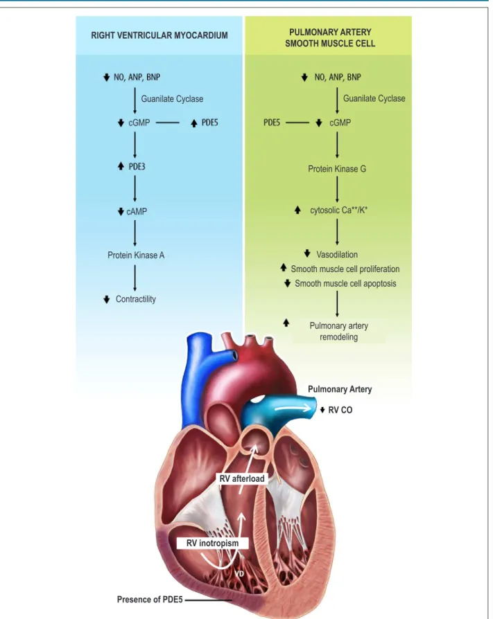
Sildenafil (SIL) is a potent and selective inhibitor of phosphodiesterase type 5 (PDE-5), the enzyme responsible for cGMP degradation. That inhibition determines an increase in the cytosolic cGMP concentration and consequent vascular smooth muscle relaxation, resulting in vasodilation. As a PDE-5 inhibitor, SIL has direct effects on pulmonary circulation and right ventricular myocardium (Figure 1), promoting, in addition to a reduction in pulmonary vascular resistance (PVR), a direct and indirect increase in cardiac output5.
The effects of SIL on the cardiovascular system of patients with heart failure and PH have been demonstrated with both acute and chronic uses. Gómez-Sánchezet al.6 and Freitas Jr. et al.7 have shown that a single dose of 100 mg of sublingual
Figure 1 – Pathophysiological changes in the pulmonary circulation and right ventricular myocardium of patients with pulmonary hypertension. The inhibition of
phosphodiesterase type 5 (PDE-5) by sildenail has a positive inotropic effect on the right ventricle and delays the pulmonary artery remodeling process.
* RV: right ventricle; CO: cardiac output; BNP: brain natriuretic peptide; NO: nitric oxide; ANP: atrial natriuretic peptide; cGMP: cyclic guanosine monophosphate; PDE3: phosphodiesterase type 3; cAMP: cyclic adenosine monophosphate. (Adapted from Archer SL et al., NEJM. 2009).
Guanilate Cyclase Guanilate Cyclase
RIGHT VENTRICULAR MYOCARDIUM
cGMP cGMP
cAMP
Protein Kinase A
Contractility
PULMONARY ARTERY SMOOTH MUSCLE CELL
Protein Kinase G
cytosolic Ca**/K*
Vasodilation
Smooth muscle cell proliferation Smooth muscle cell apoptosis
Pulmonary artery remodeling
Pulmonary Artery
RV CO
RV afterload
RV inotropism
vasodilation and improves physical capacity, measured by use of the six-minute walk test.
Thus, an ideal vasodilator would serve not only to test vascular responsiveness of pulmonary pressure, but would not interfere negatively with systemic circulation, would enhance right cardiac output, and would maintain a sustained reduction in PH.
The major objective of this study was to compare the acute hemodynamic effects of SIL and SNP on PH of heart transplantation candidates with advanced heart failure. Its second objective was to assess the effects of two vasodilators on cardiac geometry, microcirculation, and neurohormonal variables.
Methods
This cross-sectional, prospective and randomized study was performed at the Heart Failure and Transplantation Clinical Unit of the Instituto do Coração of the Hospital das Clínicas of the Medical School of the Universidade de São Paulo (InCor – HCFMUSP), from June 2006 to June 2009. Patients meeting the inclusion criteria were included in the study randomly. The study protocol was approved by the Scientific Committee of the Instituto do Coração – InCor (SDC 2698/05/118) and by the Ethics Committee on Scientific Research of the Hospital das Clínicas of the Medical School of the Universidade de São Paulo (935/05). The patients or their guardians (for children and adolescents) provided written informed consent.
Patients
The study population consisted of patients from the Heart Failure Outpatient Clinic of the InCor-HCFMUSP, who had moderate or severe left ventricular dysfunction [ejection fraction (EF) < 45%], NYHA functional class III or IV heart failure and formal indication for cardiac transplantation, according to the International Society for Heart and Lung
Transplantation guidelines (ISHLT)9 and the Brazilian Society
of Cardiology for Heart Transplantation guidelines10.
Patients with the following characteristics were excluded from the study: arteriovenous shunts; associated pulmonary disease; use of vasoactive drugs; and hemodynamic instability.
Study design
After undergoing right heart catheterization and two-dimension Doppler echocardiography simultaneously, as part of the routine preoperative assessment for heart transplantation, the patients were selected by this study’s author, according to the inclusion and exclusion criteria already described.
Then, general clinical data of all patients were recorded, and venous blood samples were also collected from the pulmonary artery for the analysis of biochemical variables.
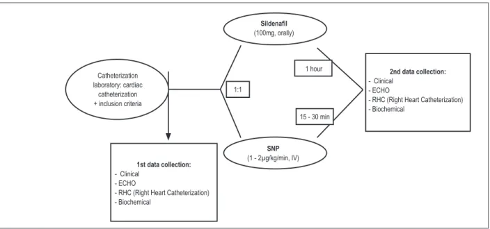
Once the baseline tests were performed, the patients were randomized to receive either SNP or SIL, at standardized doses. A second collection of clinical, hemodynamic, echocardiographic and biochemical data was performed after a predetermined period of time for each group (Figure 2).
Sildenafil was orally administered during fasting at the single dose of 100 mg (two tablets of 50 mg), and, after 60 minutes, the time interval considered ideal to obtain the highest blood concentration of that drug, a second round of tests was performed8,9.
The SNP solution, consisting of 50 mg of SNP into 250 mL of 5% glucose solution, was administered by use of a continuous intravenous infusion pump, at the velocity of 1 µg/ kg/min. After 15 minutes, the patients underwent non-invasive measurement of their systemic arterial blood pressure and of their systolic pressure of the pulmonary artery and its variables through right heart catheterization. The criteria for interrupting the administration of SNP were as follows: systemic arterial hypotension (systolic blood pressure (SBP) ≤ 85 mm Hg) and/or
Figure 2 – Study design.
Sildenail
(100mg, orally)
1st data collection: - Clinical
- ECHO
- RHC (Right Heart Catheterization) - Biochemical
SNP (1 - 2µg/kg/min, IV)
2nd data collection: - Clinical
- ECHO
- RHC (Right Heart Catheterization) - Biochemical
1:1 Catheterization
laboratory: cardiac catheterization + inclusion criteria
1 hour
appropriate reduction in PH (pulmonary artery systolic pressure – PASP < 50 mm Hg; PVR < 3 Wood units; and transpulmonary gradient - TPG < 15 mm Hg). When the interruption criteria were not reached, the infusion velocity was increased to 2 µg/kg/min, and new measurements of the hemodynamic parameters were taken every 15 minutes, with a gradual and proportional increase in the drug administration velocity.
Hemodynamic variables
Pressure readings were taken by use of the Swan-Ganz catheter in the following sites: right atrium (mean pressure); right ventricle (systolic, diastolic and mean pressure); and pulmonary artery (systolic, diastolic and mean pressure). The pulmonary artery occlusion pressure or mean pulmonary capillary wedge pressure(PCWP) was also recorded.
Pulmonary and systemic cardiac output, the later assumed as equal to the first, was measured by using the thermodilution method.
Based on those measures, PVR and systemic vascular resistance (SVR) were determined, in addition to TPG.
Echocardiographic variables
Echocardiographic assessment was performed by using the HDI 5000 device (Philips Medical System, Andover, MA, USA), equipped with a broadband 4-2-MHz transducer.
Echocardiography was performed by the same skilled examiner at the heart catheterization laboratory, simultaneously with the hemodynamic study.
Assessment of the left and right cavities allowed the determination of the atrial and ventricular diameters, in addition to left ventricular EF (LVEF). The right atrial and right ventricular volumes were measured in the apical four-chamber view, using the area-length method, during systole and diastole. Right ventricular function was estimated by the tricuspid annular plane systolic excursion (TAPSE, cm/s) using tecidual Doppler.
Biochemical variables
Venous blood samples were collected from the pulmonary artery during right cardiac catheterization, and the following were measured: brain natriuretic peptide and venous blood gas analysis.
Statistical Analysis
Initially, the variables assessed were tested regarding their adherence to normal distribution (Gaussian) for determining the adequate statistical test to be used. Only the p values < 0.05 were considered statistically significant, with a 95% confidence interval.
Comparison between groups
For comparing the baseline characteristics of both groups, the qualitative variables (gender, etiology of the underlying disease, and heart failure functional class) were assessed by using the chi-square test, and presented in a contingency table containing absolute (n) and relative (%) frequencies.
The quantitative variables were assessed according to the normality of distribution. Those with a non-normal distribution (mean dose of digoxin, spironolactone, losartan and hydrochlorothiazide) were assessed based on the Mann-Whitney U test, and those with normal distribution (all other variables) were assessed by using the independent Student t test.
Analysis of each group before and after the administration of vasodilators
The hemodynamic, echocardiographic and biochemical quantitative variables had Gaussian distribution, and their means were compared by use of Student t test for paired data.
Results
Of the 30 patients selected according to the established criteria, one was excluded due to atrial fibrillation during echocardiography. The 29 patients effectively participating in the study had a mean age of 49.1 ± 14.9 years, and most were males (62%). Tables 1 and 2 synthesize the baseline characteristics of the population studied.
Hemodynamic variables
The group receiving SIL showed a significant reduction in PVR (4.26 ± 2.65 vs. 1.94 ± 1.39 Wood units; p < 0.001)
and an increase in cardiac output (3.64 ± 0.81 vs. 4.31 ±
0.81 L/min; p = 0.003). Those effects were not accompanied by negative systemic repercussions, as shown in Table 3.
The group receiving SNP showed a different hemodynamic response. A moderate reduction in PH was observed (PVR: 4.52 ± 3.15 vs. 4.03 ± 3.13 Wood units; p = 0.2), but no
interference with cardiac output (3.64 ± 1.06 vs. 3.86 ±
1.19 L/min; p = 0.2). Unlike the group on SIL, patients on SNP showed important systemic repercussions, such as systemic hypotension SBP: 85.29 ± 13.1 vs. 69.86 ± 13.48 mmHg;
p < 0.001) and increased heart rate (66.79 ± 8.22 vs. 73.79
± 12.42 bpm; p = 0.001).
Echocardiographic variables
The acute administration of SIL was associated with significant reductions in the mean area of the right atrium (24.4 ± 6.33 vs. 21.6 ± 5.46 mm2; p = 0.008) and left ventricle
(29.4 ± 5.63 vs. 23.8 ± 5.57 mm2; p < 0.001), as shown in
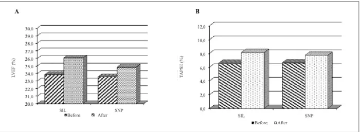
Figure 3. The reduction in cardiac geometry was accompanied by an increase in the left and right cardiac functions, measured by LVEF (23.87 ± 8 vs. 26.07 ± 7.46%; p = 0.001) and TAPSE:
6.57 ± 2.08 vs. 8.11 ± 1.81 cm/s; p = 0.002), respectively,
shown in Figure 4.
The group receiving SNP as vasodilator, however, also showed a significant reduction in the right ventricular area (29.21 ± 5.24 vs. 25.79 ± 4.9 mm2; p = 0.007), but only a
tendency towards reduction in the right atrial area (29.71 ± 6.52 vs. 27.86 ± 6.18 mm2; p = 0.08). Nevertheless, SNP
was associated with an improvement in LVEF (23.57 ± 4.69
vs. 24.86 ± 5.64 %; p = 0.02) and TAPSE (6.64 ± 1.51 vs.
Table 1 – Baseline demographic characteristics of the patients studied
All Sildenail Sodium nitroprusside p
Patients 29 15 14 NS
Age, years (mean ± sd) 49.10 ± 14.9 49.73 ± 11.17 48.42 ± 17.45 0.08
Sex, male (%) 18 (62%) 11 (73.3%) 7 (50%) 0.19
NYHA functional class n (%) III
IV
11 (37.9%) 18 (62.1%)
05 (33.3%) 10 (66.4%)
06 (42.8%)
08 (57.2%) 0.01
Etiology n (%) Ischemic Idiopathic Chagas’ disease Hypertensive Other
10 (34.5%) 06 (20.7%) 08 (27.5%) 02 (7%) 03 (10.3%)
05 (33.3%) 02 (13.3%) 05 (33.3%) 02 (13.3%) 01 (6.8%)
05 (35.7%) 04 (28.5%) 03 (21.5%)
0 02 (14.3%)
0.29
Drug (mean dose ± sd) Carvedilol (mg/day) Losartan (mg/day) Spironolactone (mg/day) Digoxin (mg/day) Furosemide (mg/day) Hydrochlorothiazide (mg/day)
35.9 ± 24.5 73.2 ± 33.3 22.4 ± 7.7 0.18 ± 0.06 80 ± 40.3 13.2 ± 17
41.6 ± 27.8 71.6 ± 32.5 21.6 ± 8.7 0.15 ± 0.07 86.6 ± 45.1 13.3 ± 18.6
29.9 ± 19.7 75 ± 35.3 23.2 ± 6.6 0.1 ± 0.04 72.8 ± 34.7 13.1 ± 15.9
0.20 0.79 0.60 0.03 0.36 0.97
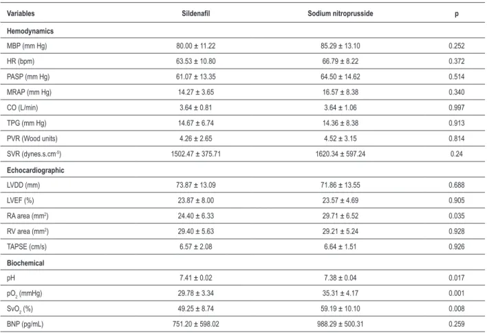
Table 2 – Baseline hemodynamic, echocardiographic and biochemical variables in both groups
Variables Sildenail Sodium nitroprusside p
Hemodynamics
MBP (mm Hg) 80.00 ± 11.22 85.29 ± 13.10 0.252
HR (bpm) 63.53 ± 10.80 66.79 ± 8.22 0.372
PASP (mm Hg) 61.07 ± 13.35 64.50 ± 14.62 0.514
MRAP (mm Hg) 14.27 ± 3.65 16.57 ± 8.38 0.340
CO (L/min) 3.64 ± 0.81 3.64 ± 1.06 0.997
TPG (mm Hg) 14.67 ± 6.74 14.36 ± 8.38 0.913
PVR (Wood units) 4.26 ± 2.65 4.52 ± 3.15 0.814
SVR (dynes.s.cm-5) 1502.47 ± 375.71 1620.34 ± 597.24 0.24
Echocardiographic
LVDD (mm) 73.87 ± 13.09 71.86 ± 13.55 0.688
LVEF (%) 23.87 ± 8.00 23.57 ± 4.69 0.905
RA area (mm2) 24.40 ± 6.33 29.71 ± 6.52 0.035
RV area (mm2) 29.40 ± 5.63 29.21 ± 5.24 0.928
TAPSE (cm/s) 6.57 ± 2.08 6.64 ± 1.51 0.926
Biochemical
pH 7.41 ± 0.02 7.38 ± 0.04 0.017
pO2 (mmHg) 29.78 ± 3.34 35.31 ± 4.17 0.001
SvO2 (%) 49.25 ± 8.74 59.19 ± 10.10 0.008
BNP (pg/mL) 751.20 ± 598.02 988.29 ± 500.31 0.259
Table 3 – Pulmonary and systemic hemodynamic effects of both vasodilators
Variables Before SIL After SIL p Before SNP After SNP p
MBP (mm Hg) 80 ± 11.2 76.4 ± 9 0.054 85.2 ± 13.1 69.8 ± 13.4 < 0.001
HR (bpm) 63.5 ± 10.8 63.6 ± 10.5 0.902 66.7 ± 8.2 73.7 ± 12.4 0.001
PVR (Wood units) 4.26 ± 2.65 1.94 ± 1.39 < 0.001 4.52 ± 3.15 4.03 ± 3.13 0.2
CO (L/min) 3.64 ± 0.81 4.31 ± 0.81 0.003 3.64 ± 1.06 3.86 ± 1.19 0.2
PCWP (mm Hg) 23.6 ± 6.4 23.5 ± 7.3 0.964 28.5 ± 7.5 23.6 ± 9.3 0.001
MRAP (mm Hg) 14.2 ± 3.6 11.6 ± 3.9 0.06 16.5 ± 8.3 15.5 ± 8.7 0.404
SVR (dynes.s.cm-5) 1502.4 ± 375.7 1241.6 ± 298.9 0.006 1620.3 ± 597.2 1204.5 ± 545.2 0.004
SIL: sildenail; SNP: sodium nitroprusside; MBP: mean systemic blood pressure; HR: heart rate; PVR: pulmonary vascular resistance; CO: cardiac output; PCWP:
pulmonary capillary wedge pressure; MRAP: mean right atrial pressure; SVR: systemic vascular resistance.
Figure 3 – Signiicant reductions (p < 0.05) in the right atrial and ventricular areas after administration of sildenail and sodium nitroprusside.
* RA: right atrium; RV: right ventricle; SIL: sildenail; SNP: sodium nitroprusside.
SIL (RA area) SNP (RA area) SIL (RV area) SNP (RV area)
Before After
Figure 4 – Increase in left ventricular ejection fraction (A) and S’ (B) after administration of sildenail and sodium nitroprusside.
*LVEF: left ventricular ejection fraction; TAPSE: tricuspid annular plane systolic excursion; SIL: sildenail; SNP: sodium nitroprusside.
L
V
E
F
(%)
T
A
P
S
E
(%)
SNP
SNP SIL
SIL
Before After
Biochemical variables
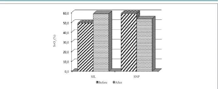
SIL associated with an improvement in pO2 (29.78 ± 3.34 vs. 34.36 ± 2.94 mm Hg; p < 0.001) and in venous
oxygen saturation (SvO2: 49.25 ± 8.74 vs. 58.92 ± 6.84 %; p < 0.001), as shown in Figure 5. This group also showed a significant reduction in mean pCO2 (44.25 ± 6.61 vs. 41 ±
6.11 mm Hg; p < 0.001) and a decrease in the serum levels of BNP (751.2 ± 598.01 vs. 622.27 ± 497.08 pg/mL; p = 0.15),
but with no statistical significance.
The SNP group showed different results as follows: worsening of pO2 (35.31 ± 4.17 vs. 31.34 ± 3.94 mm Hg;
p < 0.001) and SvO2 (59.19 ± 10.1 vs. 53.58 ± 9.36%;
p < 0.001); and no interference in pCO2 (45.18 ± 3.69
vs. 44.91 ± 4.29 mm Hg; p = 0.49). A tendency towards a
reduction in the serum levels of BNP was observed (988.29 ± 500.31 vs. 836 ± 365.31 pg/mL; p = 0.07).
Discussion
Pulmonary hypertension in patients with heart failure, although considered an important comorbidity with potential risk for death after heart transplantation, has not been widely explored, and studies on its acute perioperative management are scarce.
Some studies have shown that the PH reversibility test with SNP is associated with high indices of resistant PH and systemic hypotension, situations that contraindicate heart transplantation11. That group of patients had a lower survival
rate (~29%) in the first 30 days following transplantation as compared with patients who had no arterial hypotension
11-13 (~67%).
In that context, SIL has emerged as a feasible option, because it promotes selective and sustained pulmonary vasodilation, with minimum systemic effects14-16. However,
the use of SIL in patients with left ventricular dysfunction is recent and uncertain, which emphasizes the importance of this study.
Figure 5 – Improvement and worsening of venous oxygen saturation (SvO2) after using sildenail (p < 0.001) and sodium nitroprusside (p < 0.001), respectively.
S
vO
2
(%)
SNP SIL
Before After
The first limitation of this study is its open design, because two drugs with different administration routes were compared, resulting in loss of the double-blind study characteristic of avoiding prejudgment.
The epidemiological profile of the population sample studied is in accordance with that of the literature, most patients being males (62%) and with heart failure of ischemic etiology (34.5%).
Our study showed that SIL and SNP were effective in reducing PH of candidates for heart transplantation, but they had different effects on systemic circulation. Such findings had already been shown before, when we evidenced that the administration of a single dose of SIL reduced PH, increased cardiac output, and did not interfere with systemic blood pressure7.
The effects of vasodilators on cardiac geometry and function were extremely important. Both drugs reduced the mean diameters of the right cardiac chambers and concomitantly improved biventricular function. The change in cardiac dimension and function might be secondary to the reduction in afterload, but the direct effects of SIL on right ventricular inotropism do not exclude the hypothesis of an additional action on myocardial performance. Such findings support the hypothesis that SIL and SNP act on the acute and reverse cardiac remodeling process. That expression has been widely used, even without molecular and histopathological data, as demonstrated in some studies. Bacal et al.17 have
assessed the normalization of right ventricular function and cardiac remodeling after heart transplantation by using only cardiac magnetic resonance imaging. Similarly, Yu et al.18
and Ferrazzi et al.19 have already used the term ‘reverse
remodeling’ to designate reductions in the mean ventricular diameters and increase in systolic function, obtained from resynchronization therapy and/or ventriculoplasty.
1. Zakliczynski M, Maruszewski M, Pyka L, Trybunia D, Nadziakiewicz P, Przybylski R, et al. Effectiveness and safety of treatment with sildenafil for secondary pulmonary hypertension in heart transplant candidates. Transplant Proc. 2007;39(9):2856-8.
2. Costard-Jackle A, Fowler MB. Influence of preoperative pulmonary artery pressure on mortality after heart transplantation: testing of potential reversibility of pulmonary hypertension with nitroprusside is useful in defining a high risk group. J Am Coll Cardiol. 1992;19(1):48-54.
3. Chen JM, Levin HR, Michler RE, Prusmack CJ, Rose EA, Aaronson KD. Reevaluating the significance of pulmonary hypertension before cardiac transplantation: determination of optimal thresholds and quantification of the effect of reversibility on perioperative mortality. J Thorac Cardiovasc Surg. 1997;114(4):627-34.
4. Klotz S, Wenzelburger F, Stypmann J, Welp H, Drees G, Schmid C, et al. Reversible pulmonary hypertension in heart transplant candidates: to transplant or not to transplant. Ann Thorac Surg. 2006;82(5):1770-3. 5. Archer SL, Michelakis ED. Phosphodiesterase type 5 inhibitors for pulmonary
arterial hypertension. N Engl J Med. 2009;361(19):1864-71.
6. Angel Gómez-Sánchez M, Saenz De La Calzada C, Escribano Subías P, Francisco Delgado Jiménez J, Lázaro Salvador M, Albarrán González A, et al. Pilot assessment of the response of several pulmonary hemodynamic variables to sublingual sildenafil in candidates for heart transplantation. Eur J Heart Fail. 2004;6:615-7.
7. Freitas Jr AF, Bacal F, Oliveira Jr J de L, Santos RH, Moreira LF, Silva CP, et al. Impact of sublingual sildenafil on pulmonary hypertension in patients with heart failure. Arq Bras Cardiol. 2009;92(2):116-26.
8. Katz SD, Balidemaj K, Homma S, Wu H, Wang J, Maybaum S. Acute type 5 phosphodiesterase inhibition with sildenafil enhances flow-mediated vasodilation in patients with chronic heart failure. J Am Coll Cardiol. 2000;36(3):845-51.
9. Mehra MR, Kobashigawa J, Starling R, Russell S, Uber PA, Parameshwar J, et al. Listing criteria for heart transplantation: International Society for Heart and Lung Transplantation guidelines for the care of cardiac transplant candidates --2006. J Heart Lung Transplant. 2006;25(9):1024-42. 10. Bacal F, Souza-Neto JD, Fiorelli AI, Mejia J, Marcondes-Braga FG, Mangini S,
et al. / Sociedade Brasileira de Cardiologia. II Diretriz brasileira de transplante cardíaco. Arq Bras Cardiol. 2009;94(1 supl.1):e16-e73.
11. Zakliczynski M, Zebik T, Maruszewski M, Swierad M, Zembala M. Usefulness of pulmonary hypertension reversibility test with sodium nitroprusside in stratification on early death risk after orthotopic heart transplantation. Transplant Proc. 2005;37(2):1346-8.
12. Chen JM, Levin HR, Michler RE, Prusmack CJ, Rose EA, Aaronson KD. Reevaluating the significance of pulmonary hypertension before cardiac transplantation: determination of optimal thresholds and quantification of the effect of reversibility on perioperative mortality. J Thorac Cardiovasc Surg. 1997;114(4):627-34.
13. Klotz S, Wenzelburger F, Stypmann J, Welp H, Drees G, Schmid C, et al. Reversible pulmonary hypertension in heart transplant candidates: to transplant or not to transplant. Ann Thorac Surg. 2006;82(5):1770-3. 14. Lee AJ, Chiao TB, Tsang MP. Sildenafil for pulmonary hypertension. Ann
Pharmacoter. 2005;39(5):869-84.
15. Watanabe H. Inhibition of type-5 phosphodiesterase: promising therapy for pulmonary hypertension. Intern Med. 2004;43(10):891-3.
16. Humbert M, Sitbon O, Simonneau G. Treatment of pulmonary arterial hypertension. N Engl J Med. 2004;351(14):1425-36.
17. Bacal F, Pires PH, Moreira LF, Silva CP, Parga Filho JR, Costa UM, et al. Normalization of right ventricular performance and remodeling evaluated by magnetic resonance imaging at late follow-up of heart transplantation: relationship between function, exercise capacity and pulmonary vascular resistance. J Heart Lung Transplant. 2005;24(12):2031-6.
References
and the lack of standardized methods for its evaluation; however, on clinical practice, they are usually estimated qualitatively. The assessment of systolic function on tissue Doppler (TAPSE) was the most accurate and available way20,21
to satisfy the initial objectives of this study.
When assessing the biochemical variables, the initial objective was to correlate the central cardiovascular effects with neurohormonal and microcirculatory markers, such as BNP and venous blood gas analysis, respectively. The improvement in the venous oxygenation indices after the administration of SIL, contrary to that of SNP, was due to the balanced reduction in SVR and PVR. The BNP levels showed no significant changes, maybe because of the short time interval between the two collections.
Conclusion
Both SIL and SNP significantly reduce PH in patients with chronic left ventricular dysfunction, being associated
with a reduction in cardiac geometry and improvement in biventricular function. Only SIL improved the rates of peripheral oxygenation, with no interference with systemic blood pressure.
Potential Conflict of Interest
No potential conflict of interest relevant to this article was reported.
Sources of Funding
There were no external funding sources for this study.
Study Association
This article is part of the thesis of doctoral submitted by Aguinaldo Figueiredo de Freitas Jr. , from Faculdade de Medicina da USP.
Erratum
18. Yu CM, Chau E, Sanderson JE, Fan K, Tang MO, Fung WH, et al. Tissue Doppler echocardiographic evidence of reverse remodeling and improved synchronicity by simultaneously delaying regional contraction after biventricular pacing therapy in heart failure. Circulation. 2002;105(4):438-45. 19. Ferrazzi P, Matteucci MLS, Merlo M, Iacovoni A, Rescigno G, Bottai M,
et al. Surgical ventricular reverse remodeling in severe ischemic dilated cardiomyopathy: the relevance of the left ventricular equator as a prognostic factor. J Thorac Cardiovasc Surg. 2006;131(2):357-63.
20. Quiñones MA, Otto CM, Stoddard M, Waggoner A, Zoghbi W. Recommendations for quantification of Doppler echocardiography: a report from the Doppler Quantification Task Force of the Nomenclature and Standards Committee of the American Society of Echocardiography. J Am Soc Echocardiogr. 2002;15(2):167-84.