DOI: 10.5935/2359-4802.20180033
Mailing Address: Rafael Alessandro Ferreira Gomes
Rua dos Palmares, s/n, Santo Amaro. Postal Code: 50100-060, Recife, PE - Brazil. E-mail: rafaelgomes@cardiol.br
Carotid Atherosclerosis in Pre- and Post-Menopausal Women with a History of
Pregnancy-Induced Hypertension: Case-Control Study
Rafael Alessandro Ferreira Gomes, Isly Maria Lucena de Barros, Moacir de Novaes Lima Ferreira, Laura Olinda Bregieiro Fernandes Costa
Universidade de Pernambuco (UPE), Recife, PE - Brazil
Manuscript received on July 10, 2017, revised manuscript received on November 18, 2017, accepted on December 20, 2017.
Abstract
Background: Cardiovascular disease mortality among women remains high. Observational studies are controversial
about the participation of a history of gestational hypertensive disorder in cardiovascular risk.
Objective: To verify the association between carotid atherosclerosis in menopausal women who had
pregnancy-induced hypertension.
Methods: Case-control study, with cases consisting of women with carotid atherosclerosis, defined as carotid
intima-media thickness > 1 mm and/or presence of carotid plaques; the controls did not have these alterations. The significance level was set at 95%.
Results: A total of 504 women without previous cardiovascular disease were assessed, 126 cases and 378 controls.
Of the total, 67% were hypertensive; 76% were dyslipidemic; and 16% were diabetic. Approximately 10% reported a history of hypertension during pregnancy. Women with carotid atherosclerosis had higher values of systolic blood pressure (134.18 mmHg vs. 128.59 mmHg, p = 0.008) and LDL-cholesterol (156.52 mg% vs. 139.97 mg%; p = 0.0005). No statistical difference was found regarding the presence of carotid atherosclerosis and history of hypertension during pregnancy (OR 1.672, 95% CI: 0.883-3.131).
Conclusion: The history of hypertension during pregnancy was not associated with subclinical carotid
atherosclerosis in menopausal women. However, an association was observed between carotid atherosclerosis and classic risk factors, such as elevated systolic blood pressure and LDL-cholesterol levels. (Int J Cardiovasc Sci. 2018;31(4)359-366)
Keywords: Carotid Artery Diseases/physiopathology; Hypertension, Pregnancy-Induced; Women,
Premenopause; Postmenopause; Case-Control Studies.
Introduction
Cardiovascular diseases (CVD) are the leading cause of death among women worldwide.1,2 In the United States, they account for almost a third of all causes of death in the female gender,3,4 and similar data are observed in Europe5 and in Brazil.6 Advances in CVD treatment in the last three decades have allowed a sustained decrease in mortality. However, socioeconomic and behavioral aspects have interrupted this process in recent years.7 In 2014, there were 340,284 CVD deaths among Brazilian women, representing an increase of almost 20% in relation
to those occurring 10 years earlier.6 The cardiovascular risk stratification in the female population has failed to detect and prevent the disease. The exploration of new risk factors thus becomes essential to reduce such indices.
the number of complicated pregnancies,12 even after the normalization of blood pressure levels after childbirth.
Nonetheless, Romundstad et al.13 questioned whether such an association would be a factor of ambiguity, because pre-gestational characteristics – especially obesity, hypertension and dyslipidemia – would attenuate the effect that the gestational hypertensive disorder has on the late cardiovascular outcome. Unfortunately, most studies have insufficient evidence, such as limited sample size and clinical follow-up.
Considering that atherosclerosis is a gradual process that starts in childhood, the aim of this study was to verify the association between carotid atherosclerosis in menopausal women who had pregnancy-induced hypertension.
Methods
A case-control study was carried out, with a population of women aged between 45 and 65 years, who had had menstrual irregularities or interruption in the last year. Women receiving hormone replacement therapy, those with chronic inflammatory conditions or any previously diagnosed conditions with high cardiovascular risk or heart disease were excluded from the analysis.
The sample was calculated based on the systematic review performed by Brown et al.,14 using as reference a hypertension exposure during pregnancy of around 8% and Odds Ratio (OR) to increase the risk of atherosclerosis of 2.28. For a paired study with a one-tailed hypothesis test, we calculated at least 116 cases and 348 controls in order to obtain a 95% level of significance and 80% of test power with a ratio of one case for three controls. Controls were obtained from the same database, and were paired by age group.
All women underwent carotid ultrasound with the same examiner; the carotid intima-media thickness (CIMT) was quantified and the presence of carotid plaques was assessed. For image acquisition, a high-resolution device (EnVisor, Philips) was used with a 12.3 MHz linear transducer. The data were recorded for subsequent analysis using the QLAB-Intima Media Thickness (QLAB-IMT, Philips) software.
The presence of carotid atherosclerosis was defined when the CIMT was greater than 1 mm (mean values obtained in the analyzed segments of the right and left carotid arteries) and / or the presence of atheroma plaque. Atheroma plaque was defined as: (1) localized parietal structure with a thickness greater than 1.5 mm;
(2) protrusion into vessel lumen > 0.5 mm or; (3) thickness > 1.5-fold the adjacent CIMT, according to the Mannheim Carotid Intima-Media Thickness and Plaque Consensus.15
The cases consisted of women who had carotid atherosclerosis and the controls, of women who did not have this alteration at the ultrasonographic assessment.
The independent variable was pregnancy-induced hypertension, considered as the self-reported information of blood pressure increase during pregnancy. According to Diehl et al.,16 this information shows good accuracy (specificity of 96% and sensitivity of 79.6%) for the antecedents of pregnancy-induced hypertension, even 24.5 years after the pregnancy. Other variables were considered, namely: blood pressure, income, smoking, type 2 diabetes mellitus, family history of coronary artery disease (CAD), body mass index (BMI), number of pregnancies, preterm birth, low birth-weight offspring, fasting glycemia, total cholesterol (CT), high-density lipoprotein cholesterol (HDL-cholesterol), low-density lipoprotein cholesterol (LDL-cholesterol), triglycerides and ultrasensitive C-reactive protein (us-CRP).
The study was approved by the Research Ethics Committee of Complexo Hospitalar Hospital Universitário Oswaldo Cruz/ Pronto-Socorro Cardiológico de Pernambuco
under CAAE number 55361416.0.0000.5192 and Opinion number 1,593,189 of June 16, 2016.
Statistical analysis
Results
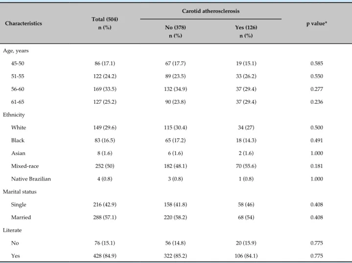
A total of 504 women were studied, of which 126 had carotid atherosclerosis and 378 did not. The groups did not differ regarding age, ethnicity, marital status and literacy (Table 1). There was also no difference regarding the number of pregnancies, preterm birth and low birth-weight offspring (Table 2).
Carotid atherosclerosis showed a higher association with systemic arterial hypertension (OR 1.837, 95% CI 1.154-2.925, p = 0.01) and dyslipidemia (OR 1.971, 95% CI, 1.149-3.380, p = 0.01). There was a tendency to a higher prevalence of carotid atherosclerosis in women with metabolic syndrome (OR 1.442, 95% CI, 0.957-2.172, p = 0.08). Carotid atherosclerosis was also directly associated with higher systolic blood pressure (134.18 mmHg vs. 128.59 mmHg, p < 0.01), LDL-cholesterol (156.52 mg% vs. 139.97 mg%, p < 0.01) and TC levels (229.68 mg% vs.
214.31 mg%, p < 0.01). There was no difference in relation to diastolic blood pressure, BMI, waist circumference, hip circumference, glycemia, HDL-cholesterol, triglycerides or CRP levels (Tables 3 and 4).
Approximately 10% of the sample had a history of pregnancy-induced hypertension. No statistically significant difference was observed between carotid atherosclerosis in the menopausal period and history of pregnancy-induced hypertension (OR 1.631, 95% CI: 0.874-3.042, p = 0.12). When analyzing only the women with a history of pregnancy-induced hypertension and those with systemic arterial hypertension in the menopausal period, no statistical difference was observed either (OR 1.862, 95% CI: 0.955-3.628, p = 0.07).
When the mean CIMT was evaluated, no statistical association was observed with the history of pregnancy-induced hypertension (0.8516 ± 0.1491 vs. 0.8101 ± 0.1441,
Table 1 - Comparison of sociodemographic characteristics with carotid atherosclerosis in menopausal women
Characteristics Total (504)
n (%)
Carotid atherosclerosis
p value* No (378)
n (%)
Yes (126) n (%)
Age, years
45-50 86 (17.1) 67 (17.7) 19 (15.1) 0.585
51-55 122 (24.2) 89 (23.5) 33 (26.2) 0.550
56-60 169 (33.5) 132 (34.9) 37 (29.4) 0.277
61-65 127 (25.2) 90 (23.8) 37 (29.4) 0.236
Ethnicity
White 149 (29.6) 115 (30.4) 34 (27) 0.500
Black 83 (16.5) 65 (17.2) 18 (14.3) 0.491
Asian 8 (1.6) 6 (1.6) 2 (1.6) 1.000
Mixed-race 252 (50) 182 (48.1) 70 (55.6) 0.181
Native Brazilian 4 (0.8) 3 (0.8) 1 (0.8) 1.000
Marital status
Single 216 (42.9) 158 (41.8) 58 (46) 0.408
Married 288 (57.1) 220 (58.2) 68 (54) 0.408
Literate
No 76 (15.1) 56 (14.8) 20 (15.9) 0.775
Yes 428 (84.9) 322 (85.2) 106 (84.1) 0.775
Table 2 - Comparison of pregnancy-related characteristics with carotid atherosclerosis in menopausal women
Characteristics Total (504)
n (%)
Carotid atherosclerosis
p value* No (378)
n (%)
Yes (126) n (%)
Number of pregnancies
None 38 (7.5) 27 (7.1) 11 (8.7) 0.561
One 42 (9) 36 (10.2) 6 (5.2) 0.132
Two 112 (24) 82 (23.3) 30 (26.1) 0.530
Three 114 (24.4) 86 (24.4) 28 (24.3) 1.000
Four 67 (14.3) 50 (14.2) 17 (14.8) 0.879
Five 41 (8.8) 29 (8.2) 12 (10.4) 0.453
Six 32 (6.9) 22 (6.3) 10 (8.7) 0.396
Pregnancy-induced hypertension
No 454 (90.1) 345 (91.3) 109 (86.5) 0.124
Yes 50 (9.9) 33 (8.7) 17 (13.5) 0.124
Low birth-weight newborn
No 475 (94.2) 358 (94.7) 117 (92.9) 0.507
Yes 29 (5.8) 20 (5.3) 9 (7.1) 0.507
Preterm birth
No 454 (90.1) 340 (89.9) 114 (90.5) 1.000
Yes 50 (9.9) 38 (10.1) 12 (9.5) 1.000
* Teste do qui quadrado.
p = 0.06). Also, no statistical difference was observed when only the presence of carotid plaques was compared with a history of pregnancy-induced hypertension (OR 1,332, 95% CI: 0.668-2.655, p = 0.41).
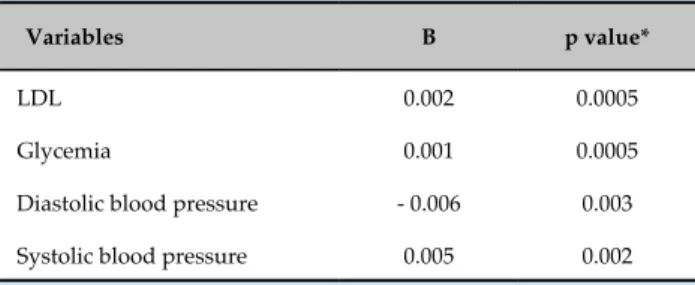
In the logistic regression model, only systemic arterial hypertension (B = 0.108, p = 0.01) and dyslipidemia (B = 0.122, p = 0.01) showed statistical significance with carotid atherosclerosis in the menopausal period (Table 5).
Discussion
In our study, carotid atherosclerosis was associated with systemic arterial hypertension and dyslipidemia, but not with a history of pregnancy-induced hypertension, although the CIMT and the presence of carotid plaques were analyzed separately. These results indicate that pregnancy-induced hypertension is not associated with subclinical atherosclerosis.
Increased CIMT and the presence of carotid plaques have been described as independent cardiovascular risk predictors.17-20 However, most studies attempting to associate a history of pregnancy-induced hypertension and carotid atherosclerosis are conflicting, since they did not use standardized CIMT and carotid plaque measurements.
Our data add information to the literature due to the large number of assessed patients. All ultrasonographic assessments were performed by the same examiner, blinded for the variable history of pregnancy-induced hypertension, eliminating measurement bias. The latest recommendations for CIMT and carotid plaque measurements were followed.15
Table 3 - Comparison of clinical characteristics, life habits and family history with carotid atherosclerosis in menopausal women
Classic risk factors Total (504)
n (%)
Carotid atherosclerosis
p value* No (378)
n (%)
Yes (126) n (%)
Systemic arterial hypertension 341 (67.7) 244 (64.6) 97 (77) 0.011
Diabetes mellitus 84 (16.7) 61 (16.1) 23 (18.3) 0.583
Dyslipidemia 387 (76.8) 280 (74.1) 107 (84.9) 0.014
BMI – obesity 166 (32.9) 122 (32.3) 44 (34.9) 0.586
Central obesity 481 (95.4) 366 (96.8) 115 (91.3) 0.014
Metabolic syndrome 270 (53.6) 194 (51.3) 76 (60.3) 0.081
Sedentary life style 146 (29) 105 (27.8) 41 (32.5) 0.310
Consumption of ≥ 5 servings of fruit/day 133 (26.4) 99 (26.2) 34 (27) 0.907
Passive smoking < 6 months 398 (79) 300 (79.4) 98 (77.8) 0.706
Family history of CAD 85 (16.9) 60 (15.9) 25 (19.8) 0.336
* Test of the chi square. BMI: body mass index; CAD: coronary artery disease.
Table 4 - Comparison of classic cardiovascular risk factors with carotid atherosclerosis in menopausal women
Dependent variables Total
Carotid atherosclerosis
p value*
No Yes
Age 56.23 (± 5.40) 56.25 (± 5.334) 56.63 (± 5.089) 0.477
SBP 130.40 (± 20.29) 128.59 (± 19.87) 134.18 (± 21.54) 0.008
DBP 84.03 (± 11.51) 83.30 (± 11.30) 84.50 (± 13.07) 0.322
BMI 28.45 (± 5.05) 28.29 (± 4.94) 28.79 (± 5.07) 0.328
Abdominal circumference 92.11 (± 11.51) 91.73 (± 11.29) 91.82 (± 11.34) 0.945
Brachial circumference 28.72 (± 4.47) 28.73 (± 4.58) 28.56 (± 4.57) 0.714
Hip circumference 103.11 (± 12.06) 103.09 (11.86) 102.39 (± 12.18) 0.571
Glycemia 102.41 (± 42.16) 100.00 (± 36.03) 107.72 (± 48.67) 0.060
Total cholesterol 219.43 (± 43.42) 214.31 (± 42.48) 229.68 (± 47.44) 0.001
HDL-cholesterol 51.80 (± 11.11) 52.36 (± 11.02) 51.91 (± 12.39) 0.709
LDL-cholesterol 143.85 (± 40.84) 139.97 (± 40.38) 156.52 (± 42.59) 0.0005
Triglycerides 141.46 (± 81.19) 134.26 (± 74.68) 149.41 (± 85.32) 0.058
us-CRP 0.34 (± 0.54) 0.32 (± 0.52) 0.34 (± 0.47) 0.773
Table 5 - Logistic regression of variables with p < 0.20 in the univariate analysis with carotid atherosclerosis
Variables B p value*
LDL 0.002 0.0005
Glycemia 0.001 0.0005
Diastolic blood pressure - 0.006 0.003
Systolic blood pressure 0.005 0.002
LDL: low-density lipoprotein-cholesterol.
et al.22 believe that the effects of pregnancy, mediated by metabolic and immunological responses, could take up to more than one year to return to basal levels.
When comparing the CIMT of women who developed hypertension during pregnancy and those who had uneventful pregnancies, the literature data show to be similar to those found in our study. Akhter et al.23 did not detect a statistically significant difference during pregnancy and up to one year postpartum when evaluating 55 women. Blaauw et al.22 also found no differences 5 years after the pregnancy. Moreover, when women between 40 and 50 years of age were assessed, there was no statistical difference regarding CIMT between those who had hypertension during pregnancy and those with uneventful pregnancies.24
Nevertheless, several observational studies have shown an association between gestational hypertensive disorder and cardiovascular clinical outcomes. Haukkama et al.,25 when assessing 141 women, identified an almost three-fold higher cardiovascular risk in those with a history of gestational hypertension disorder. In the study by Kessous et al.,26 the previous history of gestational hypertensive disorder was associated with a greater number of hospitalizations secondary to atherosclerosis 11 years after the pregnancy complicated by hypertensive disorder, even after statistical adjustment for maternal age, parity, diabetes and obesity. Canoy et al.27 identified in a large cohort that pregnancy-induced hypertension increased the risk of CVD in women in the menopausal period.
Similarly, studies with longer follow-up periods also showed an increase in severe cardiac complications in women with a history of pregnancy-induced hypertension. As verified by Arnadottir et al., women who had hypertensive complications during pregnancy had a higher risk of death due to ischemic heart disease and cerebrovascular diseases after 30 years, in addition to a shorter time of survival.28
One of the explanations for not finding an association between carotid atherosclerosis and a history of pregnancy-induced hypertension would be the method used to measure CIMT. That would be caused by the fact that CIMT measured in the common carotid artery would not be a good parameter for the determination of cardiovascular outcomes, as it estimates the total thickness of the intima and media layers. Some authors have shown that only the increase in the intima layer in association with the reduction in the media layer would be important to increase cardiovascular risk.21,23,24 In our study, we did not analyze the measurements of the intima and media layers separately.
In agreement with the literature,19,29 we have identified an association between carotid atherosclerosis and traditional cardiovascular risk factors, such as systemic arterial hypertension and hypercholesterolemia. A possible explanation is that both atherosclerosis and gestational hypertension share several common metabolic abnormalities, such as obesity, insulin resistance, dyslipidemia and hypertension itself, as well as the favoring of endothelial dysfunction.30
According to Brandão et al.,31 endothelial dysfunction precedes the clinical manifestations of a gestation complicated by hypertension and, therefore, it would accelerate the atherogenic process.32
According to McDonald et al.,33 the persistence of classic risk factors is the foundation of carotid atherosclerosis development, since even after two decades, women with a history of pregnancy-induced hypertension still had more cardiovascular risk factors than those with uncomplicated pregnancies. In our study, women with a history of pregnancy-induced hypertension had a higher prevalence of obesity and chronic hypertension (data not shown in the tables).
Although our population consists of outpatients from the public health care system, the sociodemographic characteristics did not differ from those of the general population. Moreover, it was not possible to evaluate information prior to the pregnancy, due to the proposal of the original study.
Conclusion
of menopausal and asymptomatic women, from the cardiovascular point of view. More studies are needed to understand the atherosclerosis process in women with a history of pregnancy-induced hypertension.
Author contributions
Conception and design of the research: Gomes RAF, Barros IML. Acquisition of data: Gomes RAF, Barros IML. Analysis and interpretation of the data: Gomes RAF, Barros IML. Statistical analysis: Gomes RAF, Barros IML. Writing of the manuscript: Gomes RAF. Critical revision of the manuscript for intellectual content: Barros IML, Ferreira MNL, Costa LOBF.
Potential Conflict of Interest
This manuscript is part of the master of the Graduate Program in Health Sciences of the University of Pernambuco by Rafael Alessandro Ferreira.
Sources of Funding
This study was funded by Fundação de Amparo à Ciência
do estado de Pernambuco (FACEPE) (APQ-1386-4.00/08).
Study Association
This article is part of the thesis of master submitted by Rafael Alessandro Ferreira Gomes, from Universidade de Pernambuco.
Ethics approval and consent to participate
This study was approved by the Ethics Committee of the Complexo Hospitalar Hospital Universitário Oswaldo
Cruz/Pronto-Socorro Cardiológico de Pernambuco under
the protocol number 55361416.0.0000.5192 (CAAE) and number 1.593.189 of June 16, 2016. All the procedures in this study were in accordance with the 1975 Helsinki Declaration, updated in 2013. Informed consent was obtained from all participants included in the study.
1. Gawryszewski VP, Souza Mde F. Mortality due to cardiovascular diseases in the Americas by region, 2000-2009. Sao Paulo Med J. 2014;132(2):105-10.
2. Stramba-Badiale M, Fox KM, Priori SG, Collins P, Daly C, Graham I, et al. Cardiovascular diseases in women: a statement from the policy conference of the European Society of Cardiology. Eur Heart J. 2006;27(8):994-1005.
3. Ahmed R, Dunford J, Mehran R, Robson S, Kunadian V. Pre-eclampsia and future cardiovascular risk among women: a review. J Am Coll Cardiol. 2014;63(18):1815-22.
4. Mozaffarian D, Benjamin EJ, Go AS, Arnett DK, Blaha MJ, Cushman M, et al; American Heart Association Statistics Committee and Stroke Statistics Subcommittee. Heart disease and stroke statistics--2015 update: a report from the American Heart Association. Circulation. 2015;131(4):e29-322. 5. Vaccarino V, Badimon L, Corti R, de Wit C, Dorobantu M, Hall A, et al; Working Group on Coronary Pathophysiology and Microcirculation. Ischaemic heart disease in women: are there sex differences in pathophysiology and risk factors?: Position Paper from the Working Group on Coronary Pathophysiology and Microcirculation of the European Society of Cardiology. Cardiovasc Res. 2010;90(1):9-17. 6. Brasil. Ministério da Saude, Secretaria Executiva, DATASUS. Informações
de saúde: informaçoes epidemiológicas e morbidade. [Citado em 2015 maio 1]. Disponível em http://www.datasus.gov.br.
7. Mansur Ade P, Favarato D. Mortality due to cardiovascular diseases in women and men in the five Brazilian Regions, 1980-2012. Arq Bras Cardiol. 2016;107(2):137-46.
8. Williams D. Pregnancy: a stress test for life. Curr Opin Obstet Gynecol. 2003;15(6):465-71.
9. Hashemi S, Ramezani Tehrani F, Mehrabi Y, Azizi F. Hypertensive pregnancy disorders as a risk factor for future cardiovascular and
metabolic disorders (Tehran Lipid and Glucose Study). J Obstet Gynaecol Res. 2013;39(5):891-7.
10. Smith GC, Pell JP, Walsh D. Pregnancy complications and maternal risk of ischaemic heart disease: a retrospective cohort study of 129,290 births. Lancet. 2001;357(9273):2002-6.
11. Ben-Ami S, Oron G, Ben-Haroush A, Blickstein D, Hod M, Bar J. Primary atherothrombotic occlusive vascular events in premenopausal women with history of adverse pregnancy outcome. Thromb Res. 2010;125(2):124-7.
12. Freibert SM, Mannino DM, Bush H, Ph D, Crofford LJ. The association of adverse pregnancy events and cardiovascular disease in women 50 years of age and older. J Womens Health (Larchmt). 2011;20(2):287-93. 13. Romundstad PR, Magnussen EB, Smith GD, Vatten LJ. Hypertension
in pregnancy and later cardiovascular risk: Common antecedents? Circulation. 2010;122(6):579-84.
14. Brown MC, Best KE, Pearce MS, Waugh J, Robson SC, Bell R. Cardiovascular disease risk in women with pre-eclampsia: Systematic review and meta-analysis. Eur J Epidemiol. 2013;28(1):1-19.
15. Touboul P, Hennerici M, Meairs S, Adams H, Amarenco P, Bornstein N, et al. Mannheim carotid intima-media thickness and plaque consensus (2004-2006-2011). An update on behalf of the advisory board of the 3rd, 4th and 5th watching the risk symposia, at the 13th, 15th and 20th European Stroke Conferences, Mannheim, Germany, 2004, Brussels, Belgium, 2006, and Hamburg, Germany, 2011. Cerebrovasc Dis. 2012;34(4):290-6. 16. Diehl CL, Brost BC, Hogan MC, Elesber AA, Offord KP, Turner ST,
et al. Preeclampsia as a risk factor for cardiovascular disease later in life : validation of a preeclampsia questionnaire. Am J Obstet Gynecol. 2008;198(5)e11-3.
17. Koskinen J, Kahonen M, Viikari JS, Taittonen L, Laitinen T, Ronnemaa T, et al. Conventional cardiovascular risk factors and metabolic syndrome
This is an open-access article distributed under the terms of the Creative Commons Attribution License
in predicting carotid intima-media thickness progression in young adults: the cardiovascular risk in young Finns study. Circulation. 2009;120(3):229-36.
18. Peters SA, den Ruijter HM, Bots ML, Moons KG. Improvements in risk stratification for the occurrence of cardiovascular disease by imaging subclinical atherosclerosis: a systematic review. Heart. 2012;98(3):177-84. 19. O’Leary DH, Polak JF, Kronmal RA, Manolio TA, Burke GL, Wolfson SK Jr. Carotid-artery intima and media thickness as a risk factor for myocardial infarction and stroke in older adults. Cardiovascular Health Study Collaborative Research Group. N Engl J Med. 1999;340(1):14-22. 20. Den Ruijter HM, Peters SA, Anderson TJ, Britton AR, Dekker JM,
Eijkemans MJ, et al. Common carotid intima-media thickness measurements in cardiovascular risk prediction: a meta-analysis. JAMA. 2012;308(8):796-803.
21. Akhter T, Larsson A, Larsson M, Wikström A, Naessen T. Artery wall layer dimensions during normal pregnancy : a longitudinal study using noninvasive high-frequency ultrasound. Am J Physiol Heart Circ Physiol. 2013;304(2):H229-34.
22. Blaauw J, Souwer ET, Coffeng SM, Smit AJ, van Doormaal JJ, Faas MM, et al. Follow up of intima-media thickness after severe early-onset preeclampsia. Acta Obstet Gynecol Scand. 2014;93(12):1309-16. 23. Akhter T, Wikstrom AK, Larsson M, Naessen T. Individual common
carotid artery wall layer dimensions, but not carotid intima-media thickness, indicate increased cardiovascular risk in women with preeclampsia: an investigation using noninvasive high-frequency ultrasound. Circ Cardiovasc Imaging. 2013;6(5):762-8.
24. Akhter T, Larsson M, Wikström AK, Naessen T. Thicknesses of individual layers of artery wall indicate increased cardiovascular risk in severe pre-eclampsia. Ultrasound Obstet Gynecol. 2014;43(6):675-80.
25. Haukkamaa L, Salminen M, Laivuori H, Leinonen H, Hiilesmaa V, Kaaja R. Risk for subsequent coronary artery disease. Am J Cardiol. 2004;93(6):805-8. 26. Kessous R, Shoham-Vardi I, Pariente G, Sergienko R, Sheiner E. Long-term maternal atherosclerotic morbidity in women with pre-eclampsia. Heart. 2015;101(6):442-6.
27. Canoy D, Cairns BJ, Balkwill A, Wright FL, Khalil A, Beral V, et al. Hypertension in pregnancy and risk of coronary heart disease and stroke: a prospective study in a large UK cohort. Int J Cardiol. 2016;222:1012-8. 28. Arnadottir GA, Geirsson RT, Arngrimsson R. Cardiovascular death in
women who had hypertension in pregnancy: a case–control study. BJOG. 2005;112(3):286-92.
29. Mosca L, Benjamin EJ, Berra K, Bezanson JL, Dolor RJ, Lloyd-Jones DM, et al. Effectiveness-based guidelines for the prevention of cardiovascular disease in women-2011 update: a guideline from the American Heart Association. Circulation. 2011;123(11):1243-62. Erratum in: Circulation. 2011;123(22):e624. Circulation. 2011;124(16):e427.
30. Craici I, Wagner S, Garovic VD. Preeclampsia and future cardiovascular risk: formal risk factor or failed stress test? Ther Adv Cardiovasc Dis. 2008;2(4):249-59.
31. Brandão A, Cabral M, Leite H, Cabral A. Endothelial function, uterine perfusion and central flow in pregnancies complicated by Preeclampsia. Arq Bras Cardiol. 2012;99(4):931-5.
32. Agatisa PK, Ness RB, Roberts JM, Costantino JP, Kuller LH, McLaughlin MK. Impairment of endothelial function in women with a history of preeclampsia: an indicator of cardiovascular risk. Am J Physiol Heart Circ Physiol. 2004;286(4):H1389-93.