Conventional Ventricular Stimulation Effects on Patients with Normal
Ventricular Function
Luiz Antonio Batista de Sá, Salvador Rassi, Márcia Andery Ludovico Batista
Hospital das Clínicas da Universidade Federal de Goiás, Goiânia, GO - BrazilSummary
Background: The stimulation of the right ventricle (RV) may be deleterious in patients with ventricular dysfunction; however there is little evidence about the impact of this stimulation in patients with normal ventricular function.
Objectives: To assess the clinical and laboratory evolution of patients with normal ventricular function submitted to implant of artificial cardiac pacemaker (PM).
Methods: 16 patients enrolled according to the following inclusion criteria: normal ventricular function defined by echocardiogram and presence of upper ventricular stimulation > 90% (generator telemetry assessment) submitted to a PM implant were prospectively studied. The following parameters were assessed: Functional Class (FC), walk test, BNP levels, echocardiography evaluation (conventional and intraventricular dyssynchrony) and quality of life test (SF36). The patients were assessed after 10 (t1), 120 (t2) and 240 days (t3). Data was compared throughout time according to ANOVA. Multiple comparisons of means were performed through Tukey’s test.
Results: Among the assessed data, the following did not present significant statistic variation (p> 0.05): functional class, BNP levels, conventional echocardiographic parameters, intraventricular dyssynchrony (tissue Doppler). The walk test (between t2 and t3) and the time between septal contraction and LV posterior wall showed worsening (p<0.05), although they did not meet the dyssynchrony criteria. The quality of life assessment (SF36) showed improvement in the functional capacity, social aspects, and general status sub-items.
Conclusion: After 8 months, patients with normal ventricular function did not show clinical (FC and SF36) or laboratory alterations (conventional echocardiography, dyssynchrony parameters and BNP levels); however, there was a worsening in the walk test. (Arq Bras Cardiol 2009; 93(2):157-162)
Key Words: Ventricular dysfunction; pacemaker, artificial; cardiac pacing, artificial; echocardiography.
Mailing address: Luiz Antonio Batista de Sá •
Ambulatório de Marcapasso do Hospital das Clínicas, Primeira Avenida s/n, 76.610-030, Goiânia, GO - Brazil
E-mail: [email protected]
Manuscript received June 15, 2008; revised manuscript received October 03, 2008; accepted October 24, 2008.
Introduction
After its introduction at the end of the 50s1, the artificial
cardiac stimulation went through great transformations up to the current days. The development of devices associated to new clinical evidence increased the indications significantly, not only in the area of bradyarrhythmias2, as well as
tachyarrhythmias (implantable cardioverter defibrillator)3-5
and more recently, heart failure (cardiac resynchronization therapy). The latter has incorporated the new concepts on the mechanisms of heart failure (HF) as a phenomenon that is not purely muscular, but also with the involvement of the electrical system of the heart6-8.
Approximately 15% of patients com IC present intraventricular conduction disorder and patients with more severe symptoms comprise 30%9. The prolonged duration of
the QRS complexes is a negative prognostic factor of mortality
and is associated to the presence of ventricular dyssynchrony that generates an uncoordinated contraction leading to the decrease in ejection volume, cardiac output, mean arterial pressure, dP/dt, mechanical–energetic impairment and mitral valve dysfunction10,11.
The implant of the conventional cardiac pacemaker is performed in the right ventricle and, as the simulation is carried out directly on the endocardium, the electrocardiographic result is an enlarged QRS complex.
There is clinical and laboratory evidence of the deleterious effects of the ventricular stimulation in patients with ventricular dysfunction12,13 ; however, in patients with normal function,
the impact of this stimulation as a factor of dyssynchrony and the triggering of clinically relevant ventricular dysfunction has not been completely established.
The role of the right ventricular stimulation as a cause of dyssynchrony started to be outlined with the reassessment of comparison studies of unichamber (VVI) x bi-chamber (DDD) stimulation. The DDD stimulation preserves the atrioventricular synchronism and presents better hemodynamic data14. However, the prospective studies
and UKPACE18 studies demonstrated only secondary benefits,
such as the decrease in the incidence of atrial fibrillation and improved quality of life, but without any effect on mortality. It has been proposed that the probable deleterious effects of right ventricular stimulation leading to dyssynchrony can annul the benefits obtained with the atrioventricular synchronism19.
However, this analysis has limitations, as these studies were not designed to test this hypothesis.
The objective of the present study is to evaluate the effects of conventional cardiac stimulation in patients with pacemaker indication and normal ventricular function.
Methods
This study was approved by the Ethics and Research Committee of Hospital das Clinicas of the Federal University of Goias under #062/06. All the patients participating in the study signed the Free and Informed Consent Form.
From March 2006 to July 2007, 19 of the 142 patients referred to pacemaker implant were selected according to the following criteria:
1) Age > 18 years and < 75 years
2) The indications for conventional cardiac pacemaker followed the Directives of the Brazilian Society of Cardiology20
and those with high probability of right ventricular stimulation were accepted:
2.1) Total atrioventricular block.
2.2) Second-degree atrioventricular block type II. 2.3) Sinus node disease with first-degree AV block with PR interval > 200ms.
2.4) Normal ventricular function, defined by the echocardiogram, performed after the implant of the artificial cardiac pacemaker (normal ventricular diameters and normal ejection fraction by the Teicholz’s method).
The exclusion criteria were:
1) Severe disease with reduced life expectancy;
2) Incapacity to perform the tests proposed by the study; 3) After the implant, a regular verification was carried out (10 days (d), 120d and 240d) of the percentage of right ventricular stimulation through the analysis of the generator data and the patients that presented values < 90% were excluded.
The patients were followed for a period of 8 months after the implant, defined as: post-implant assessment - 10 days (t1), 4 months (t2) and 8 months (t3). The following parameters were analyzed:
1) Clinical
1.1) New York Heart Association Functional Class 1.2) Quality of Life Questionnaire (Brazilian version) - SF36
1.3) 6-minute walk test 2) Laboratory Parameters
2.1) Evaluation by generator telemetry
2.2) Electrocardiogram – Stimulated QRS complex width 2.3) Brain natriuretic peptide (BNP) levels
2.4) Echocardiogram
a. Cavity diameters and volumes b. Ejection fraction
c. Intraventricular parameters of dyssynchrony
The echocardiogram assessments were performed in a Toshiba equipment model Xario with two-dimensional harmonic mode and sector transducer of 2.5 MHz. All assessments were carried out by a single observer. The patients were placed in left lateral decubitus and monitored through electrocardiogram. All the measurements were acquired with the patient in expiratory apnea. The measurements of the left ventricle, right ventricle, aorta diameter and left atrium were carried out by the one-dimensional mode, according to the recommendations of the American Society of Echocardiography. The assessment of intraventricular dyssynchrony was carried out according to the following criteria: M Mode: difference between the start of the QRS up to the peak of contraction of the septal wall and then the measurement of the time between the start of the QRS complex up to the peak of contraction of the posterior wall; dyssynchrony was considered when the value was > 130 ms. Pulsed Doppler: measurement from the start of the QRS complex to the start of the aortic flow; dyssynchrony was considered when the value was > 140 ms. Tissue Doppler: difference between the start of the QRS complex and the S-wave peak of the basal region of the lateral, anterior, septal and inferior walls; dyssynchrony was considered when the value was > 65 ms21,22.
The means of the normal (or approximately normal) distribution variables were compared along time using the ANOVA method for repeated measures (rmANOVA). The Kolmogorov-Smirnov normality test and the Mauchly’s Test of Sphericity were applied to verify suppositions of the rmANOVA model. When the supposition of sphericity was not satisfied, the p-value was determined according to Huyn-Feldt correction in the rmANOVA analyses. Multiple comparisons of means were performed using Tukey’s method, when a significant difference was observed in the rmANOVA test.
In case of variables with asymmetric distribution, medians were compared throughout time according to Friedman’s method, a non-parametric alternative to the parametric rmANOVA method. Conover-Inman test was used in multiple comparisons of medians throughout time.
All probabilities of significance (p values) presented are the bilateral type and values < 0.05 are considered statistically significant. The software SAS 9.1 (Statistical Analysis System, Cary, NC, USA) was used in the statistical analysis of data.
Results
Figure 1 -Percentage of ventricular stimulation (p>0.05).
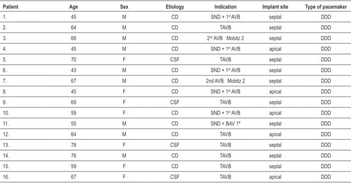
atrioventricular block corresponded to 62.5% of the sample. The electrode was implanted in the septal region in 75% of the cases. The clinical characteristics are shown in Table 1. All patients underwent a follow-up period of 8 months.
All patients started the protocol as Functional Class (FC) I; during the evolution, only one patient became FC II after 8 months (p>0.05). The percentage of ventricular stimulation in each patient was obtained through the telemetry system. The mean stimulation percentage was 99%. No statistically significant difference was obtained among the medians during the times t1, t2 and t3 (p>0.05) (Figure 1).
The width of the stimulated QRS complexes maintained a mean of 134 ms throughout the entire study, with no statistical difference during the 8 months (p>0.05).
The walk test showed a statistically significant difference among the means throughout time (p=0.0021), with a difference being observed between the means of the values between 4 and 8 months (p=0.0014), whereas no difference was observed between the initial time and 4 months (p>0,05) and between 10 days and 8 months (p>0.05) (Figure 2).
The BNP measurements did not show a significant difference between the means throughout time (p>0.05). The mean measurements were 29.75 at t1, 28.26 at t2 and 51.34 at t3 (Figure 3).
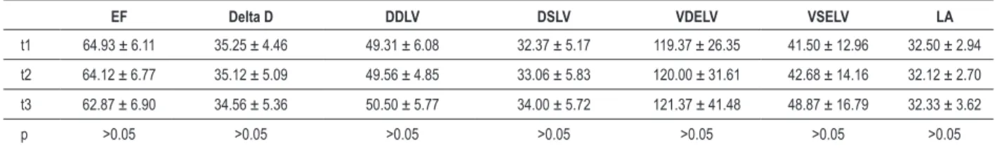
Table 2 shows the data related to the conventional echocardiographic parameters: left ventricular (LV) diastolic diameter, LV systolic diameter, LV end-diastolic volume, LV end-systolic volume, left atrium, ejection fraction (EF) and delta D. There were no statistical differences throughout time (p>0.05).
Table 3 shows the data related to the echocardiographic
Table 1 - Clinical characteristics
Patient Age Sex Etiology Indication Implant site Type of pacemaker
1. 45 M CD SND + 1st AVB septal DDD
2. 64 M CD TAVB septal DDD
3. 68 M CD 2nd AVB Mobitz 2 septal DDD
4. 45 M CD SND + 1st AVB apical DDD
5. 70 F CSF TAVB septal DDD
6. 43 M CD SND + 1st AVB septal DDD
7. 67 M CD 2nd AVB Mobitz 2 septal DDD
8. 45 F CD SND + 1st AVB apical DDD
9. 69 F CSF TAVB septal DDD
10. 59 F CD SND + 1st AVB apical DDD
11. 55 M CD SND + BAV 1º septal DDD
12. 64 M CD TAVB apical DDD
13. 78 F CSF TAVB septal DDD
14. 76 M CD TAVB septal DDD
15. 59 F CD TAVB septal DDD
16. 67 F CSF TAVB apical DDD
CD - Chagas’ disease, CSF - Conduction System Fibrosis, TAVB - total AV block, SND - sinus node disease, 1st AVB - irst-degree AV block, DDD - dual-chamber pacing.
Figure 2 -Walk test throughout time – Difference between t2 and t3 (p=0.0014).
Figure 3-BNP levels (p>0.05).
Table 2 - Echocardiographic variables measured throughout time
EF Delta D DDLV DSLV VDELV VSELV LA
t1 64.93 ± 6.11 35.25 ± 4.46 49.31 ± 6.08 32.37 ± 5.17 119.37 ± 26.35 41.50 ± 12.96 32.50 ± 2.94
t2 64.12 ± 6.77 35.12 ± 5.09 49.56 ± 4.85 33.06 ± 5.83 120.00 ± 31.61 42.68 ± 14.16 32.12 ± 2.70
t3 62.87 ± 6.90 34.56 ± 5.36 50.50 ± 5.77 34.00 ± 5.72 121.37 ± 41.48 48.87 ± 16.79 32.33 ± 3.62
p >0.05 >0.05 >0.05 >0.05 >0.05 >0.05 >0.05
EF – Ejection fraction; DDLV - left ventricular diastolic diameter, DSLV – left ventricular systolic diameter, VDELV – left ventricular end-diastolic volume, VSELV – left ventricular end-systolic volume, LA - left atrium.
Table 3 - Echocardiographic variables related to dyssynchrony
M Mode Pulsed Doppler Tissue Doppler
t1 39.68 ± 18.14 106.25 ± 18.96 43.81 ± 29.80
t2 50.81 ± 30.70 118.18 ± 26.45 45.25 ± 31.94
t3 52.06 ± 30.96 117.56 ± 20.48 45.87 ± 27.37
p >0.05 0.0302 >0.05
Table 4 shows the data related to the Quality of Life test (SF36) and no statistical difference was observed throughout time concerning the sub-items: physical aspects, emotional aspects and mental health. In the sub-item functional capacity, we observed an improvement (p=0.003) and this difference was observed between t1 and t2 (p=0.0002) as well as between t1 and t3 (p=0.0298). No difference was observed
between t2 and t3 (p>0.05). The general health status showed improvement only between t1 and t3 (p=0.0172). The item social aspects showed an improvement (p=0.190), which was observed between t1 and t2 (p=0.0084).
Discussion
The present study assesses a specific subgroup of patients: those with preserved ventricular function and those who present a high degree of ventricular stimulation in view of the type of block.
We observed that, during a period of 8 months, the right ventricular stimulation was not capable of producing significant deleterious effects, evaluated from a clinical and laboratory point of view.
Chagas’ disease was the main etiology of the present study. The conclusion whether the etiology of the block can determine a different evolution is uncertain and needs to be further analyzed. The complexity of the Chagasic patient can make it difficult to perform this analysis, as the block can be a marker of inflammatory reaction and the patient can develop HF regardless of the pacemaker, in addition to other risk markers23.
A statistically significant change was observed in the walk test. This is a method that objectively evaluates the degree of functional limitation and has a prognostic value in heart failure24. In the present study, the patients did not develop
HF and at the end of the 8 months, there was a 17-meter decrease in the walk test. Although there was a statistically significant difference, this information, from a clinical point of view, seems to have little importance, as there were no significant modifications in FC.
Table 4 – Quality of Life Test SF 36 (sub-items)
FC PAL Pain GHS Vitality SA EA Mental
Health t1 80± 22 54 ± 39 72 ± 25 72 ± 19 72 ± 18 76 ±19 74 ± 16 77 ± 19
t2 90 ± 16 75 ± 30 77 ± 23 74 ± 16 80 ± 12 94 ± 11 90 ± 13 87 ± 9
t3 86 ± 17 82 ± 30 84 ± 13 86 ± 11 83 ± 13 89 ± 14 94 ± 14 86 ± 7
p 0.0003 >0.05 >0.05 0.0143 >0.05 0.019 >0.05 >0.05
FC - functional capacity; PAL- physical aspect limitation; GHS - general health status; SA - social aspects, EA - emotional aspects.
The EF is acknowledged as an independent factor of mortality and it is widely applied in the management of patients with HF25. The stability of the EF, in the present study, indicates that
the ventricular stimulation during 8 months was not able to deteriorate the ventricular function.
Among the echocardiographic parameters used to evaluate the dyssynchrony, the main ones are those that evaluate intraventricular dyssynchrony. Of the three parameters assessed in the present study, only the one that measures intraventricular dyssynchrony by pulsed Doppler presented statistical alteration throughout time, with worsening of the parameter, from 106 ms at the start to 117 at the end of the study. However, these data must be analyzed with care, considering that the accepted value for the diagnosis of dyssynchrony is 140 ms; thus, one cannot affirm that the studied population presented dyssynchrony. The assessment of the intraventricular dyssynchrony through tissue Doppler has been considered an important parameter in the study of dyssynchrony26,27. Our sample did not show a statistical
difference. The study by Thambo et al28 assessed 23 patients
with total congenital atrioventricular block and a previously normal left ventricular function, with at least five years of cardiac stimulation. They analyzed the following parameters: time of ventricular filling, cardiac output, mitral failure severity, interventricular dyssynchrony and ergometric test. The results indicate that the prolonged ventricular stimulation was associated with ventricular remodeling, LV dilation, LV asymmetric hypertrophy and low physical capacity; however, the impact of these alterations from a clinical point of view has not been evaluated.
Our study showed a predominance of septal stimulation. This might have contributed to a better result, considering that the apical stimulation seems to be more deleterious29;
however, the best location inside the right ventricle has yet to be investigated30,31. Currently, the objective has
been to minimize the ventricular stimulation through new algorithms of stimulation. Ongoing studies (SAVEPACe, DAVID II, INTRINSIC, MVPtrial) investigate the role of the minimum ventricular stimulation. Nevertheless, the patients that need permanent ventricular stimulation do not benefit from this strategy, and therefore, new sites of stimulation have been researched32.
We did not observe a significant increase in BNP levels during the 8-month assessment, indicating preserved ventricular function33. Similar results were obtained by
Albertsen et al34 in their study, which compared the DDD
x biventricular stimulation and did not show a worsening of the pro-BNP levels with DDD stimulation. The DDD group showed only a decrease in EF of 2%, with no effects on the FC or walk test results. This study, however, included patients with and without ventricular dysfunction.
The analysis of the SF36 questionnaire showed an improvement in the following sub-items: functional capacity, social aspects and general health status. This improvement can be attributed to the effects of the artificial cardiac stimulation therapy, in the group of patients that were previously severely limited by bradycardia. Similar data were obtained in the MOST35 study, which evaluated 2015 patients, comparing
unichamber x bi-chamber stimulation. The authors observed a significant improvement in quality of life after the pacemaker implant in both groups; however, patients older than 75 years benefited less than younger ones.
The limitations of the present study refer to the assessed length of time; significant long-term clinical effects cannot be ruled out.
Conclusion
After 8 months, patients with normal ventricular function did not show significant clinical (functional class and quality of life function) or laboratory alterations (conventional echocardiography, dyssynchrony parameters and BNP levels); however, the patients presented a worsening in the walk test. New studies with long-term follow-up and a larger sample size will be necessary to discover risk markers that will help identify patients who will have an unfavorable evolution with an artificial cardiac pacemaker.
Potential Conflict of Interest
No potential conflict of interest relevant to this article was reported.
Sources of Funding
There were no external funding sources for this study.
Study Association
References
1. Furman S, Robinson G. Use of an intracardiac pacemaker in the correction of total heart block. Surg Forum. 1958; 9: 245-8.
2. Leung SK, Lau CP, Camm J. An overview of sensors: ideal characteristics sensor combination, and automaticity. In: Ellenbogen KA, Kay GN, Wilkoff BL (eds). Clinical cardiac pacing and defibrillation. 2nd. ed. Philadelphia: WB Saunders; 2000. p. 219-48.
3. Moss AJ, Hall WJ, Cannom DS, Daubert JP, Higgins SL, Klein H, et al. Improved survival with an implanted defibrillator in patients with coronary disease at high risk for ventricular arrhythmia. Multicenter Automatic Defibrillator Implantation Trial Investigators. N Engl J Med. 1996; 335 (26): 1933-40.
4. Buxton AE, Lee KL, Fisher JD, Josephson ME, Prystowsky EN, Hafley G. A randomized study of the prevention of sudden death in patients with coronary artery disease. Multicenter Unsustained Tachycardia Trial Investigators. N Engl J Med. 1999; 341 (25): 1882-90.
5. Moss AJ, Zareba W, Hall WJ, Klein H, Wilber DJ, Cannon DS, et al. Prophylactic implantation of a defibrillator in patients with myocardial infarction and reduced ejection fraction. N Engl J Med. 2002; 346 (12): 877-83.
6. Cazeau S, Leclercq C, Lavergne T, Walker S, Varma C, Linde C, et al. Effects of multisite biventricular pacing in patients with heart failure and intraventricular conduction delay. N Engl J Med. 2001; 344: 873-80.
7. Abraham WT, Fisher WG, Smith AL, Delurgio DB, Leon AR, Loh E, et al. Cardiac resynchronization in chronic heart failure. N Engl J Med. 2002; 346: 1845-53.
8. Cleland JG, Daubert JC, Erdmann E, Freemantle N, Gras D, Kappenberger L, et al. The effect of cardiac resynchronization on morbidity and mortality in heart failure. N Engl J Med. 2005; 352: 1539-49.
9. Aaronson KD, Schwartz JS, Chen TM, Wong KL, Goin JE, Mancini DM. Development and prospective validation of a clinical index to predict survival in ambulatory patients referred for cardiac transplant evaluation. Circulation. 1997; 95: 2660-7.
10. Grines CL, Bashore TM, Boudoulas H, Olson S, Shafer P, Wooley CF. Functional abnormalities in isolated left bundle branch block: the effect of interventricular asynchrony. Circulation. 1989; 79: 845-53.
11. Baldasseroni S, Opasich C, Gorini G. Left bundle branch block is associated with increased 1-year sudden and total mortality rate in 5517 outpatients with congestive heart failure: a report from the Italian network on congestive heart failure. Am Heart J. 2002; 143: 398-405.
12. Wonisch M, Lercher P, Scherr D, Maier R, Pokan R, Hoffman P, et al. Influence of permanent right ventricular pacing on cardiorespiratory exercise parameters in chronic heart failure patients with implanted cardioverter defibrillators. Chest. 2005; 127: 787-93.
13. Wilkoff BL, Cook JR, Epstein AE, Greene HL, Hallstron AP, Hsia H, et al. Dual-chamber pacing or ventricular backup pacing in patients with an implantable defibrillator: the Dual Chamber and VVI Implantable Defibrillator (DAVID) Trial. JAMA. 2002; 288: 3115-23.
14. Wu RC. Reynolds DW. Hemodynamics of cardiac pacing. In: Ellenbogen KA, Wood MA (eds). Cardiac pacing and ICD. 3rd. ed. New York: Wiley; 2002. p. 129-73.
15. Lamas GA, Orav EJ, Stambler BS, Ellenbogen KA, Sgarbossa EB, Huang SK, et al. Quality of life and clinical outcomes in elderly patients treated with ventricular pacing as compared with dual-chamber pacing. N Engl J Med. 1998; 338: 1097-104.
16. Connolly SJ, Kerr CR, Gent M, Roberts RS, Yusuf S, Gillis AM, et al. Effects of physiologic pacing versus ventricular pacing on the risk of stroke and death due to cardiovascular causes. N Engl J Med. 2000; 342: 1385-91.
17. Lamas GA, Lee KL, Sweeney MO, Silverman R, Leon A, Yel R, et al. Ventricular pacing or dual-chamber pacing for sinus-node dysfunction. N Engl J Med.
2002; 346: 1854-62.
18. Toff WD, Skehan JD, De Bono DP, Camm AJ. The United Kingdom pacing and cardiovascular events (UKPACE) trial. United Kingdom Pacing and Cardiovascular Events. Heart. 1997; 78: 221-3.
19. Sweeney MO, Prinzen FW. A new paradigm for physiologic ventricular pacing. J Am Coll Cardiol. 2006; 47: 282-8.
20. Martinelli Filho M, Zimerman LI, Lorga AM, Vasconcelos JTM, Rassi A Jr. Guidelines for implantable electronic cardiac devices of the Brazilian Society of Cardiology. Arq Bras Cardiol. 2007; 89 (6): e210-e238.
21. Schiller NB, Shah PM, Crawford M, DeMaria A, Devereux R, Feigenbaum H, et al. Recommendations for quantitation of the left ventricle by two dimensional echocardiography. American Society of Echocardiography Committee on Standards, Subcommittee on Quantitation of Two-Dimensional Echocardiograms. J Am Soc Echocardiogr. 1989; 2: 358-67.
22. Lane RE, Chow AWC, Mayet J. Selection and optimisation of biventricular pacing: the role of echocardiography. Heart. 2004; 90 (Suppl VI): 10-6.
23. Rassi A Jr, Rassi A, Rassi S. Predictors of mortality in chronic Chagas disease Circulation. 2007; 115: 1101-8.
24. Rubim VSM, Drumond NC, Romeo JLM, Montera MW. Prognostic value of the six-minute walk test in heart failure. Arq Bras Cardiol. 2006; 86 (2): 120-5.
25. Bristow MR, Lowes BD. Management of heart failure. In: Braunwald E, Zipes DP, Libby P, Bonow RO. Branwald´s heart disease. 7th. ed. Phladelphia: WB Saunders; 2005. p. 603-51.
26. Sun JP, Chinchoy E, Donal E, Popovic ZB, Perlic G, Asher CR, et al. Evaluation of ventricular synchrony using novel Doppler echocardiographic indices in patients with heart failure receiving cardiac resynchronization therapy. J Am Soc Echocardiogr. 2004; 17: 845-50.
27. Silva CES, Barretto ACP. Avaliação ecocardiográfica da terapia de ressincronização cardíaca. Arq Bras Cardiol. 2005; 84 (6): 503-7.
28. Thambo JB, Bordachar P, Garrigue S, Lafitte S, Sanders P, Reuter S, et al. Detrimental ventricular remodeling in patients with congenital complete heart block and chronic right ventricular apical pacing. Circulation. 2004; 110 (25): 3766-72.
29. Manolis AS. The deleterious consequences of right ventricular apical pacing: time to seek alternate site pacing. Pacing Clin Electrophysiol. 2006; 29: 298-315.
30. Stambler BA, Ellenbogen K, Zhang X, Porter TR, Xie F, Malik R, et al. and the ROVA Investigators. Right ventricular outflow versus apical pacing in pacemaker patients with congestive heart failure and atrial fibrillation. J Cardiovasc Eletrophysiol. 2003; 14: 1180-6.
31. Kypta A, Steinwender C, Kammler J. Long term outcomes in patients with atrioventricular block undergoing septal ventricular lead implantation compared with standard apical pacing. Europace. 2008; 10 (5): 574-9.
32. Zanon F, Bacchiega E, Rampin L, Aggio S, Baracca E, Pastore G, et al. Direct his bundle pacing preserves coronary perfusion compared with right ventricular pacing: a prospective, cross-over mid-term study. Europace. 2008; 10 (5): 580-7.
33. Silva LB, Ferreira CA, Blacher C, Leães P, Haddad H. Peptídeo natriurético tipo-B e doenças cardiovasculares. Arq Bras Cardiol. 2003; 81 (5): 529-34.
34. Albertsen AE, Nielsen JC, Pousen SH, Mortensen PT, Pedersen AK, Hansen PS, et al. Biventricular pacing preserves left ventricular performance in patients with high-grade atrio-ventricular block: a randomized comparison with DDD(R) pacing in 50 consecutive patients. Europace. 2008, 10: 314-20.