Instituto do Coração do Hospital das Clínicas – FMUSP
Mailing address: Edmar Atik – Incor – Av. Dr. Enéas C. Aguiar, 44 - 05403-000 São Paulo, SP
Objective – To assess neonates with aortic stenosis with early decompensation operated upon. (LCO) (CHF).
Methods – A and retrospective study analyzing 6 neonates with LCO, group I (GI), and 12 neonates with CHF, group II (GII). Clinical radiographic, electrocardiographic and echocardiographic findings also provided comparative bases for the study, as did surgical and evolutional findings.
Results – The mean ages at hospitalization and sur-gery (p = 0.0031) were 14.3 and 14.8 days in GI and 35.4 and 42.8 days in GII, respectively. Cardiac murmurs were more intense in GII (p = 0.0220). The aortic ring was smaller in GI (8.0 ± 2.5mm) as compared to GII (11.4±1.4mm) (p = 0.2882). Ventricular function was reduced to 18±5.5% and 33.3±7.6% in GI and GII, respectively (p = 0.0162). Aortic atresia, however, was present only in 2 neonates in GI. Five of 6 patients in GI died but all patients in GII survived (p=0.0007). In the latter group, 84.6% of the patients were in functional class I (FC-I) in the long-term follow-up, with moderate residual lesions in 6 neonates, discrete residual lesions in 4, and reoperation in 2.
Conclusion – Aortic stenosis is a severe anomaly of the neonate, whose immediate evolution depends on the pre-operative anatomic and functional findings, and the late evolution essentially depends on the anatomic featu-res of the valve.
Key words: aortic stenosis, neonate, congenital heart disease, surgery
Arq Bras Cardiol, volume 73 (nº 4), 354-358, 1999
Edmar Atik, Miguel Barbero-Marcial, Elisa R. Iwahashi, Jorge Y. Afiune, Munir Ebaid
São Paulo, SP- Brazil
Critical Aortic Stenosis in the Neonate. Clinical Assessment
and Surgical Outcome in 18 Patients
Aortic stenosis is a severe congenital defect when it be-comes manifest in the neonatal period. Therefore, it remains a challenge due to its high morbidity and mortality rate 1-3.
Inter-vention is mandatory even though controversy exists in regard to the most adequate treatment: surgery 2-6 or balloon 7-13
(table I).
According to a series of experiments, mortality seems to relate more to the unfavorable anatomical type of the left side of the heart, mainly the size of the aortic and mitral rings, the size of the left ventricle (LV), and any associated defects including endocardial fibroelastosis, rather than to the type of treatment chosen 1-4,11-13.
We provide here short- and long-term outcomes of our experience in the surgical treatment of aortic stenosis, con-tributing to the literature that shows different results in the use of one or the other type of treatment 2-13.
Methods
We retrospectively assessed 18 patients consecutively operated upon at INCOR-FMUSP, over the period from January ’90 to December ’98. Patients’ ages were under 2 months.
According to the clinical findings at the time of surgery, two groups were established: group I (GI) consisting of patients with low cardiac output (LCO), and group II (GII) consisting of patients with congestive heart failure (CHF). GI comprised 6 patients and was characterized by severe dyspnea, discomfort, cyanosis, inadequate peripheral perfusion, weak pulse, blood hypotension, and use of vasoactive drugs, such as dopamine, dobutamine, and prostaglandin E1. GII comprised 12 patients and was characterized by mild tachypnea, no cyanosis, good peripheral perfusion, weak pulse, blood hypotension, and no need for vasoactive drugs.
In each group, general data were analyzed comparati-vely as were the ages at the moment of the discovery of the heart disease, at hospitalization, and at surgery. We also compared the preponderant symptom, the significant symp-clinical data, such as the intensity of the cardiac murmur, and the results of complementary laboratory tests, in additi-on to the surgical and evolutiadditi-onal findings.
axis and the type of ventricular hypertrophy, in addition to in the T wave changes in the left precordial leads. On chest X-ray, the size of the cardiac area was evaluated. On echo-cardiogram, the aortic ring, the shortening fraction of the cardiac fiber, and the transvalvar aortic pressure gradient were assessed. Evaluations of the sizes of the LV and mitral ring were disregarded because they were considered normal in all patients (18mm and 14mm, respectively).
Qualitative variables of the groups of patients with LCO and CHF were compared using Fischer’s exact test. Quantitative variables were analyzed with the Student’s t test. When data distribution did not approach that which is considered normal, the Wilcoxon’s test was used for in– dependent samples. The Statistical Analysis System (SAS) was used for the statistical analysis 14.
Results
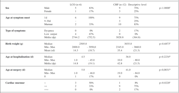
The male sex predominated in both groups: in GI, 5 (83%) out of the 6 patients were males, and in GII, 9 (75%) out of the 12 patients were males. Birth weight was lower in GI, where it varied from 2,000 to 3,950g (mean = 2,744±752.5g), as compared to GII, where it varied from 2,345 to 3,860g (mean = 3,028.8±364.8g). The median of birth weight was 2,505g in GI and 3,035g in GII (p = 0.683-NS).
Heart disease manifested itself on the first day of life in 15 out of the 18 patients (all the 6 patients in GI and 9 patients in
GII). The type of manifestation, however, was different because it resulted from the LCO present in 4 (67%) out of the 6 patients in GI and from the cardiac murmur present in the 2 other patients of GI and in 10 (83%) out of the 12 patients of GII.
The age at hospitalization was lower for the group with LCO, with a mean of 14.3±18.7 days, and in 4 out of the 6 patients it was only 2 days, as compared with 35.4 ± 21.3 days for the other group (p = 0.2234-NS). Earlier hospitalization is an indication of the major repercussion of the anomaly in the first group.
The same is true for the age at surgery. It was lower in the GI (mean = 14.8±19.1 days) and in 4 out of the 6 patients of this group it was only 4 days, as compared to 42.8±21.5 days for GII (p = 0.0031). The median ages at hospitalization and at surgery were 4.5 and 5.0 days for GI and 36 and 41 days for GII, respectively.
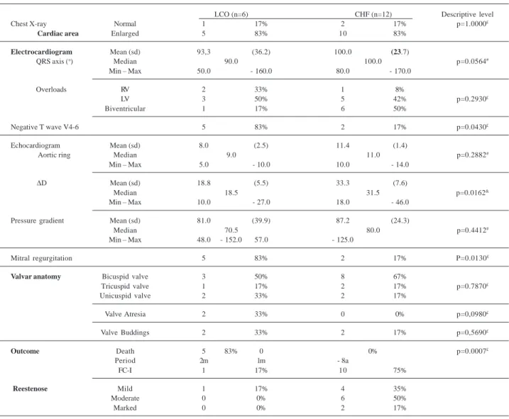
Intensity of the cardiac murmur showed an inverse relationship to the clinical repercussion, and it was proportio-nally less intense in the patients with LCO (p = 0.0220) (table II). Moderate cardiomegaly assessed on chest X-ray was evident in 83.3% of the patients. Biventricular hypertrophy was found in 7 patients and LV hypertrophy in 8 patients, while right ventricle hypertrophy was found in only 3. The median of the QRS Axis corresponded to +90 and +100 in the 2 groups (p = 0.0564). The negative T wave occurred in the left precordial leads in 83% of the GI and in 17% of the GII (p = 0.0430). On the echocardiogram, 2 out of the 6 patients in GI with aortic atresia and the unicuspid valve had a small aortic ring of 5mm. In this group, the mean value
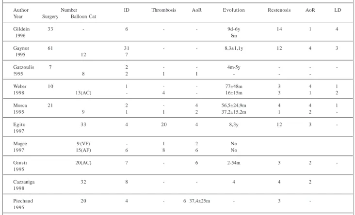
Table I – Critical aortic stenosis in the neonate. Immediate and late findings of the surgical intervention and balloon catheter dilation in the literature.
Author Number ID Thrombosis AoR Evolution Restenosis AoR LD
Year Surgery Balloon Cat
Gildein 33 - 6 - - 9d-6y 14 1 4
1996 8m
Gaynor 61 31 - - 8,3±1,1y 12 4 3
1995 12 7
Gatzoulis 7 2 - - 4m-5y - -
-!995 8 2 1 1 - -
-Weber 10 1 - - 77±48m 3 4 1
1998 13(AC) - 4 - 16±15m 3 1 2
Mosca 21 2 - 4 56,5±24,9m 4 4 1
1995 9 1 1 2 37,2±15,2m 1 2
-Egito 33 4 20 4 8,3y 12 3
-1997
Magee 9 (VF) - 1 2 No
1997 15(AF) 6 8 6 No
Giusti 20(AC) 7 - 6 2-54m 3 2
-1995
Cazzaniga 32 8 - - 4 4 2
1998
Piechaud 20 4 - 6 37,4±25m - 3
-1995
of the ring of the 6 patients corresponded to 8.0±2.5 mm, and in GII it corresponded to 11.4±1.4 mm (p = 0.2882).
The transvalvar aortic pressure gradient was equiva-lent in both groups: 81±39.9 mmHg and 87.2±24.3mmHg (p = 0.4412-NS). The shortening fraction of the myocardial fiber in GI varied from 10% to 27%, and its mean value (18.8±5.5%) was clearly lower than that of GII (33.3±7.6%), where the va-lues ranged from 18% to 46% (p = 0.0162).
According to the significant dysfunctions of the LV in GI, one can infer that the transvalvar aortic pressure gradient is underestimated. Mitral regurgitation occurred in 83% of the patients in GI and in only 17% of the patients in GII (p = 0.0130). Death at surgery occurred in 5 (83%) out of the 6 pati-ents with LCO and the post-operative evolution could be de-termined in the only survivor of this group and in the other 12 patients with CHF operated upon. From the latter, 10 patients were in functional class I (FC-I) over a follow-up period of 1 month to 8 years (mean = 4.2 years), and 2 patients were in FC-II. A mild lesion evaluated through the pressure gradient was found in 4 (35%) patients (mean = 35 mmHg); a moderate lesion was found in 6 (50%) patients (mean = 60 mmHg); and a marked lesion was detected in 2 (17%) patients (mean = 80 mmHg). These patients required re-operation and showed improvement after a new commissurotomy, and evolved to milder degrees of obstruction (table III).
Discussion
The reports about experiences with neonates with cri-tical aortic stenosis presented in the literature in a series of publications have stressed the high morbidity and mortality rates of this anomaly that occur irrespective of the type of treatment employed, either surgical 2-4 or through balloon
catheter 10-13. Published reports have emphasized that
mor-tality relates to unfavorable anatomy, mainly small aortic and mitral rings, and the LV hypoplasia and endocardial fi-broelastosis 1,4,11-13.
Our 2 patients with LCO and small aortic ring associa-ted with aortic atresia and hypoplasia of the ascending aorta died. The smallest aortic ring (8.0±2.5mm) in the group of patients with LCO as compared with that of the group of pa-tients with CHF (11.4±1.4mm) also favored the negative evo-lution.
Little emphasis in the literature is placed on the func-tional status of patients as a cause of death. According to our findings, there is a clear relation between the surgical mortality and the significant degree of ventricular dysfunc-tion assessed through the low shortening fracdysfunc-tion of the myocardial fiber on the echocardiogram and through the cli-nical features of the LCO. In these patients, peripheral per-fusion is maintained through the use of vasoactive drugs and prostaglandin E1.
Therefore, in the group with LCO and with anatomical findings similar to those in the group with CHF, except 2 ca-ses of aortic atresia and a severe hypoplastic aortic ring, 5 (83%) out of the 6 patients died. In GII, all patients survived. This relation is significant because the only variable was the degree of functional disorder, since the electrocar-diographic, raelectrocar-diographic, and anatomic and surgical fin-dings are all very similar in both groups. The negative T wave in the left precordial leads and the mitral regurgitation, more commonly found in the group with LCO, express the major repercussion of the anomaly in GI.
The earlier decompensation of some patients depends on the magnitude of the repercussion of the defect, which may already be felt during fetal life due to the smaller mean birth weight seen in the group with LCO. This group also showed a lower age at hospitalization and, mainly, at surgery. The hemodynamic instability of this more critical
Table II – Critical aortic stenosis in the neonatal period. General data and symptoms in the 2 groups: low cardiac output (LCO) and congestive heart failure (CHF)
LCO (n=6) CHF (n=12) Descriptive level
Sex Male 5 83% 9 75% p=1.0000£
Female 1 17% 3 25%
Age at symptom onset 1d 6 100% 9 75%
9-30d - 3 25%
Murmur 2 33% 10 83%
Type of symptoms Dyspnea 0 0% 2 17%
Low output 4 67% 0 0%
Média (dp) 2744.2 (752.5) 3028.8 (364.8)
Birth weight (g) Median 2505.0 3035.0 p=0.6873#
Min - Max 2000.0 - 3950,0 2345.0 - 3860.0
Mean (sd) 14.3 (18.7) 35.4 (21.3)
Age at hospitalization (d) Median 4.5 36.0 p=0.2234&
Min - Max 1.0 - 45.0 10.0 - 80.0
Média (dp) 14.8 (19.1) 42.8 (21.5)
Age at surgery (d) Median 5.0 41,0 p=0,0031&
Min - Max 1.0 - 46.0 19.0 - 84.0
0 1 17% 0 0%
Cardiac murmur + 3 50% 1 8% p=0.0220£
++ 2 33% 9 75%
+++ 0 0% 2 17%
group is such that the mechanisms of cardiac compensation fail, with a low threshold of significant modifications, even to minimally invasive procedures, to the anesthetic inducti-on itself, and to alteratiinducti-ons in envirinducti-onmental temperature. Any of those situations may trigger severe arrhythmias, such as ventricular fibrillation, and even cardiac arrest.
This is why the literature also reports such a high morta-lity rate for this group of patients undergoing interventionist therapy through dilation with balloon catheters. Considering the severity of the situation, the ideal management of these patients may be the most precocious intervention possible, still in the first hours of life. This management may be foreseen and programmed through prenatal echocardiographic assessment, aided by evaluation of low fetal body weight, which is an ex-pression of the systemic LCO in intrauterine life.
Even though the group of patients with CHF showed a good evolution in the long run, surgical reintervention was required in 2 out of the 12 patients. It may be required in the near future in an additional 6 out of the 12 patients because
they already show moderate aortic restenosis with a mean transaortic pressure gradient of 60mmHg. This occurred in the group operated upon and in the other that underwent dilation through balloon catheter, according to studies al-ready published 2,3,5,6,9,11.
The risk of aortic regurgitation may be higher in the group undergoing the interventionist procedure through balloon catheter, but general evolution has been shown to be similar for both therapeutic managements 3,5,6,7,9,11-13.
After the neonatal period some features, such as aortic restenosis, aortic regurgitation, arrhythmias, and syncopes occurring in patients who underwent either surgery or balloon catheter dilation, increase long-term morbidity 2,3,5-7,9,11-13. The reintervention indices are high in
any of the procedures, corroborating the severity of the valvar anomaly that becomes manifest in the neonatal period. In addition to the marked dysfunction, the morphological substrate of this anomaly that significantly influences the immediate evolution in the neonatal period
Table III – Critical aortic stenosis in the neonatal period: data of complementary laboratory examinations, anatomy and outcome in the 2 groups: low cardiac output (LCO) congestive heart failure (CHF).
LCO (n=6) CHF (n=12) Descriptive level
Chest X-ray Normal 1 17% 2 17% p=1.0000£
Cardiac area Enlarged 5 83% 10 83%
Electrocardiogram Mean (sd) 93,3 (36.2) 100.0 (23.7)
QRS axis (o) Median 90.0 100.0 p=0.0564#
Min – Max 50.0 - 160.0 80.0 - 170.0
Overloads RV 2 33% 1 8%
LV 3 50% 5 42% p=0.2930£
Biventricular 1 17% 6 50%
Negative T wave V4-6 5 83% 2 17% p=0.0430£
Echocardiogram Mean (sd) 8.0 (2.5) 11.4 (1.4)
Aortic ring Median 9.0 11.0 p=0.2882#
Min – Max 5.0 - 10.0 10.0 - 14.0
∆D Mean (sd) 18.8 (5.5) 33.3 (7.6)
Median 18.5 31.5 p=0.0162&
Min – Max 10.0 - 27.0 18.0 - 46.0
Pressure gradient Mean (sd) 81.0 (39.9) 87.2 (24.3)
Median 70.5 80.0 p=0.4412#
Min – Max 48.0 - 152.0 57.0 - 125.0
Mitral regurgitation 5 83% 2 17% P=0.0130£
Valvar anatomy Bicuspid valve 3 50% 8 67%
Tricuspid valve 1 17% 2 17% p=0.7870£
Unicuspid valve 2 33% 2 17%
Valve Atresia 2 33% 0 0% p=0,0980£
Valve Buddings 2 33% 2 17% p=0,5690£
Outcome Death 5 83% 0 0% p=0.0007£
Period 2m 1m - 8a
FC-I 1 17% 10 75%
Reestenose Mild 1 17% 4 35%
Moderate 0 0% 6 50%
Marked 0 0% 2 17%
1. Rhodes L, Colan S, Perry S, Jonas R, Sanders S. Predictors of survival in neonates with critical aortic stenosis. Circulation 1991; 84: 2325-35.
2. Gildein HP, Kleinert S, Weintraub RG, Wilkinson JL, Karl TR, Mee RB. Surgical commissurotomy of the aortic valve: outcome of open valvotomy in neonates with critical aortic stenosis. Am Heart J 1996; 131: 754-9.
3. Gaynor JW, Bull C, Sullivan ID, et al. Late outcome of survivors of intervention for neonatal aortic valve stenosis. Ann Thorac Surg 1995; 60: 122-5. 4. Gatzoulis MA, Rigby ML, Shinebourne EA, Redington AN. Contemporary
results of balloon valvuloplasty and surgical valvotomy for congenital aortic stenosis. Arch Dis Child 1995; 73: 66-9.
5. Weber HS, Mart CR, Kupferschmid J, Myers JL, Cyran SE. Transcarotid balloon valvuloplasty with continuous transesophageal echocardiographic guidance for neonatal critical aortic valve stenosis: an alternative to surgical palliation. Pediatr Cardiol 1998; 19: 212-17.
6. Mosca R, Iannettoni M, Schwartz S, et al. Critical aortic stenosis in the neonate. A comparison of balloon valvuloplasty and transventricular dilatation. J Thorac Cardiovasc Surg 1995; 109: 147-54.
7. Lababidi Z, Wu J, Walls T. Percutaneous balloon aortic valvuloplasty: results in 23 patients. Am J Cardiol 1984; 53: 194-7.
References
8. Lababidi Z, Winhaus L. Successful balloon valvuloplasty for neonatal critical aortic stenosis. Am Heart J 1986; 112: 913-16.
9. Egito ES, Moore P, O’Sullivan J, et al. Transvascular balloon dilation for neonatal critical aortic stenosis: early and midterm results. J Am Coll Cardiol 1997; 29: 442-7.
10. Magee AG, Nykanen D, McCrindle BW, Wax D, Freedom RM, Benson LN. Balloon dilation of severe aortic stenosis in the neonate: comparison of anterograde and retrograde catheter approaches. J Am Coll cardiol 1997; 30: 1061-6.
11. Giusti S, Borghi A, Radaelli S, et al. The carotid arterial approach for balloon dilation of critical aortic stenosis in neonates. Immediate results and follow-up. Cardiol Young 1995; 5: 155-60.
12. Cazzaniga M, Fernandez Pineda L, Abraira V, et al. Clinical and echocardiogra-phic impact of neonatal aortic valvuloplasty. Rev Esp Cardiol 1998; 51: 141-51. 13. Piechaud J, Delogu D, Kachaner J, Sidi D. Transcatheter valvotomy of aortic stenosis with dilated left ventricle in the newborns: long term results. J Am Coll Cardiol 1995; 25: 99A.
14. Rosner B. Fundamentals of Bioestatistics. (2nd ed.). Boston: PWS Publishers, 1986.
includes patients with aortic annular hypoplasia, valvar dysplasia, LV hypoplasia, mitral anomalies, and endo-cardial fibroelastosis.
In our service, we have chosen the surgical approach for all these anatomical changes, but we do not criticize management with cardiac catheterization, except in the
case of aortic regurgitation, which may be an aggravating evolutional factor.