• Nenhum resultado encontrado

Arq. Bras. Cardiol. vol.82 número1 en a09v82n1

N/A
N/A
Protected

Academic year: 2018

Share "Arq. Bras. Cardiol. vol.82 número1 en a09v82n1"

Copied!
4
0
0

Texto

(1)

9 8 9 8 9 8 9 8 9 8

Tumelero et al

Percutaneous balloon aortic valvuloplasty in a pregnant adolescent

Arq Bras Cardiol 2003; 81: 98-101.

Faculdade de Medicina da Universidade de Passo Fundo e Hospital São Vicente de Paulo - Passo Fundo

Mailing address: Rogério Tadeu Tumelero - Rua Teixeira Soares, 777 - S/705 Cep 99010-080 - Passo Fundo, RS, Brazil - E-mail: rttumelero@terra.com.br English version by Stela Maris C. e Gandour

Received: 4/10/02 Accepted: 01/14/03

Arq Bras Cardiol, volume 82 (nº 1), 98-101, 2003

Rogério T. Tumelero, Norberto T. Duda, Alexandre P. Tognon, Iselso Sartori, Silvane Giongo

Passo Fundo, RS - Brazil

Percutaneous Balloon Aortic Valvuloplasty in a

Pregnant Adolescent

Case Report

We report the case of a 16-year-old pregnant patient with severe aortic stenosis and pulmonary congestion cli-nically uncontrolled, in whom percutaneous balloon aor-tic valvuloplasty was used as the first choice of treatment in an emergency procedure. The clinical findings, patho-physiology, diagnostic features, and indications for per-cutaneous treatment are reported.

Severe congenital aortic stenosis is rare in children and young individuals. Bicuspid aortic valve occurs in 3% to 6% of patients with congenital heart disease; when associated with commissural fusion, significant stenosis may be present in childhood. The association of severe congenital aortic stenosis and pregnancy is difficult to control clinically, carrying a high risk of maternal and fe-tal morfe-tality, mainly when manifested with symptoms of pulmonary congestion 1,2.

Case Report

The patient is a 16-year-old pregnant white woman in the 27th gestational week, who was admitted with severe dyspnea and precordial pain. Her personal antecedent was a cardiac murmur without diagnosis or follow-up. When starting the prenatal follow-up 2 weeks before and complai-ning of dyspnea on exertion, the patient was referred for car-diac assessment. She evolved with symptoms at rest, being then hospitalized. She reported smoking and no familial his-tory of heart disease, diabetes mellitus, hypertension, or dyslipidemia.

On physical examination, the patient was in regular gene-ral condition, tachypneic, acyanotic, and her facies was not

characteristic. Her weight and height were, respectively, 40 kg and 1.50 m. Her blood pressure was 90/60 mmHg, and, on heart auscultation, she had a regular rhythm, tachycardia (heart rate=120 bpm), and an ejection systolic murmur (5+/6+) in the aortic region with irradiation to the carotid arteries. The pulmo-nary auscultation showed crepitant rales in both bases.

The electrocardiography revealed sinus rhythm, hy-pertrophy of the left chambers, and ventricular repolariza-tion changes secondary to overload (fig. 1).

The chest radiography showed a normal cardiac area, cranial diversion of the pulmonary circulation and Kerley B lines compatible with pulmonary congestion.

On admission, the echocardiogram showed a bicuspid aortic valve with commissural fusion of the right and left coronary leaflets, moderately reduced systolic mobility, systolic dynamics in cupule, and no calcification. In addi-tion, left ventricular (LV) concentric hypertrophy, left ventri-cle/aorta (LV/AO) gradient = 100 mmHg, and an aortic ring of 1.48 cm2 were observed.

The patient remained at the coronary unit under clini-cal treatment with digitalis, a diuretic, nitroglycerine, and oxygen therapy with a mask.

(2)

Arq Bras Cardiol 2003; 81: 98-101.

Tumelero et al Percutaneous balloon aortic valvuloplasty in a pregnant adolescent

9 9 9 99 9 9 9 9 9 On admission, the obstetric ultrasound showed the

presence of heart beats and fetal movements, as well as a Grannun zero-degree placenta with normal thickness. The amniotic fluid volume was normal. The fetus weighed ap-proximately 920 g and had no morphological peculiarities. The fetal biometry for the echographic gestational age of 27 weeks was average. A new ultrasound was performed every 48 hours for fetal follow-up.

In the 30th gestational week, the patient experienced progressive pulmonary congestion, which did not respond properly to clinical treatment. Percutaneous aortic valvulo-plasty was then indicated.

On cardiac catheterization, the findings were as fol-lows: bicuspid aortic valve with an LV/AO gradient = 105 mmHg (fig. 2), ascending aorta with normal angiogra-phic appearance, coronary circulation with normal origin and trajectory, and hypertrophic left ventricle.

Procedure – The patient was sedated and the cardio-fetal beats were monitored. Catheterization of the left ventri-cle was performed through puncture of the right femoral ar-tery with an 8F Sones catheter. A 0.35 guidewire (Amplatz “extra-stiff” 260 cm – Boston Scientific) was positioned in the left ventricle. Aortography was performed in left anteri-or oblique projection and left ventriculography was perfanteri-or- perfor-med in right anterior oblique projection (Multi-Track 5F an-giographic catheter, NuMed Inc) with radiographic nonio-nic low-osmolarity contrast medium. An 18x4-cm balloon (Z-MED – NuMed) was used (fig. 3). At the end of the proce-dure, the peak-to-peak ejection gradient LV/AO was 20 mmHg, and mild-to-moderate aortic valve insufficiency was observed (fig. 4).

After the procedure, the electrocardiogram showed a reduction in the ventricular rate and maintenance of the signs of hypertrophy in the left chambers.

The chest radiography showed a reduction in the signs of pulmonary congestion, and the echocardiogram showed a mild residual aortic stenosis (LV/AO gradient = 29 mmHg) and moderate aortic valvular insufficiency. The other parameters remained unchanged.

After the procedure, fetal monitoring with cardiotoco-graphy evidenced a reactive tracing. The obstetric echogra-phy and Doppler color velocimetry revealed good fetal vita-lity. On the 4th day after percutaneous aortic valvuloplasty, a new obstetric echography revealed severe oligohydram-nios with an amniotic fluid index (AFI) = 2.5 cm (normal Fig. 3 - Aortogram in right anterior oblique projection. Balloon dilation of the aortic valve. A constriction corresponding to valvular stenosis may be seen.

Fig. 4 - Aortogram in left anterior oblique projection. Final result showing mild aortic regurgitation and disappearance of the central jet.

(3)

1 0 0 1 0 0 1 0 0 1 0 0 1 0 0

Tumelero et al

Percutaneous balloon aortic valvuloplasty in a pregnant adolescent

Arq Bras Cardiol 2003; 81: 98-101.

AFI=8-18 cm). Due to the association of oligohydramnios and placental insufficiency, interruption of the pregnancy with a Cesarean delivery was indicated.

The male newborn infant weighed 1.425 g, had an Apgar score of 8 and 9, and a Capurro index of 32 weeks.

The mother was discharged from the hospital on the 4th day after the Cesarean delivery, in good general condi-tion and without dyspnea. The diuretic and angiotensin-converting-enzyme inhibitor were maintained for the clinical control of heart failure.

The premature newborn had hyaline membrane disea-se, extensive bronchopneumonia, septicemia, disseminated intravascular coagulation, and anemia. In addition, as his ductus arteriosus was patent, indomethacin was used until its closure on the 10th day of life. The infant was discharged from the hospital on the 46th day of life weighing 2.125 g, in good general condition and stable.

On the clinical 9-month follow-up, the patient remai-ned asymptomatic using an angiotensin-converting-enzy-me inhibitor and a diuretic. Due to the good clinical evolu-tion, the surgical valvular correction was postponed.

Discussion

Severe congenital aortic valve stenosis is rare in chil-dren and young individuals 1. Bicuspid aortic valve

accounts for 3% to 6% of congenital heart diseases 1,2. It is

more frequently found among males (male to female propor-tion of 4:1) and may be associated with cardiovascular ano-malies in up to 20% of the cases 2. The trauma produced by

the turbulence of the blood flow leads to thickening, fibro-sis, calcification, and stiffness of the valve, and the clinical manifestations occur from the 3rd decade of life onwards, mainly in the male sex 2,3. In some cases, when the bicuspid

valvular stenosis is associated with commissural fusion, it may already present itself as severe during childhood 3.

Some authors have considered aortic valve stenosis the most frequent congenital heart disease, because it is not usually detected during the first years of life. With the routi-ne use of echocardiographic techniques, the recognition of aortic valve stenosis has been facilitated 2.

The physiological changes induced by gestation cause the following 3 major hemodynamic changes in the heart: an increase in the cardiac output (30-40%); an increase in heart rate (10-20 bpm); and expansion of the blood volume (20-100%) 4,5. The association of these factors with obstruction of

the left ventricular outflow tract, which limits the variations in cardiac output, may lead to hemodynamic decompensation, which is frequently portrayed as symptoms and signs of pul-monary congestion, syncope, and sudden death.

According to Arias and Pineda 6, in a series of 38

ges-tations with 23 severe aortic stenoses, the natural history of this disease encompasses a maternal mortality rate during pregnancy of 17.4% for nontreated patients and a fetal mor-tality rate of 34%. An invasive cardiac intervention may be necessary in patients whose clinical condition has

deterio-rated during pregnancy, aiming at reducing the peak-to-peak ejection gradient by 60% to 70% 7,8. Surgical treatment

for severe bicuspid aortic stenosis has been recommended by the Brazilian Consensus on Heart Disease and Pregnan-cy at any time during gestation when the LV/AO gradient is greater than 70 mmHg 9. Cheitlin 10 recommends surgical

bal-loon valvuloplasty or even surgical valve replacement in the presence of symptoms, immediately if evidence of pul-monary congestion exists, and when the valvular area is 0.7 cm2 or lower, measured on the echocardiogram or during

cardiac catheterization. Balloon valvuloplasty successfully used in severe mitral stenosis during pregnancy and in aortic stenoses in nonpregnant, but at high risk for aortic valve replacement 11,12, patients was used in 2 pregnant

pa-tients with aortic stenosis 13,14. Both cases had favorable

maternal and fetal outcomes. A series of valvuloplasties performed in the 30th gestational week 15 has been

associa-ted with preterm birth of healthy babies 2 weeks later. In 2 studies 16,17 with 11 patients undergoing aortic valve

repla-cement, no maternal death was reported although the overall maternal surgical mortality rate with cardiopulmona-ry bypass was 1.5. On the other hand, the overall fetal surgi-cal mortality ranged from 16% 17 to 20% 18. Aortic valve

re-placement, in particular, seems to be associated with an ex-ceptionally high fetal mortality rate of 40% in the entire group and 57% in patients with aortic stenosis 17. Therefore,

open surgery for aortic valve stenosis during pregnancy should be considered as the last choice.

Percutaneous balloon aortic valvuloplasty began to be used in the mid 1980s. In 1984, based on the initial expe-rience with pulmonary valvuloplasty, Lababidi 19 and

Laba-bidi and Wu 20 extended the method for the treatment of

aortic stenosis. Currently, the refinement of materials provi-ded the use of that less invasive method in situations in which the surgical risk is unacceptable.

Balloon valvuloplasty and surgical valvuloplasty ha-ve been limited by their late results, which turn these proce-dures into palliative treatment 21. The immediate appearance

of aortic regurgitation or its progression, and the later ap-pearance of restenosis are the major complications of valvu-loplasty. Other complications during the procedure include bleeding, arrhythmias, stroke, iliac-femoral arterial complica-tions, injury to mitral valve, and death 7.

Exposure to X-rays during the procedure is another issue that deserves special attention, and for a short time it may be minimized by using radiological protection for the abdomen and pelvis of the pregnant female. This fully redu-ces the risks of congenital malformations 22.

(4)

Arq Bras Cardiol 2003; 81: 98-101.

Tumelero et al Percutaneous balloon aortic valvuloplasty in a pregnant adolescent

1 0 1 1 0 1 1 0 1 1 0 1 1 0 1 1. Roberts, WC. The structure of the aortic valve in clinically isolated aortic stenosis:

An autopsy study of 162 patients over 15 years of age. Circulation, 1970; 41(C):91. 2. Friedman WF. Congenital Heart Disease in Infancy and Childhood. In Braun-wald E (ed) Heart Disease: A Textbook of Cardiovascular Medicine. 5th ed.

Phila-delphia: WB Saunders Co, 1997; 29:877-962.

3. Martinez EE, Portugal OP. Valvopatia Aórtica. Em Barreto ACP, Souza AGMR (eds). Cardiologia Atualização e Reciclagem. Rio de Janeiro, Ateneu, 1994;44:447-54. 4. Elkayam U, Gleicher N. Hemodynamics and cardiac function during normal

preg-nancy and the puerperium. In Elkayam U, Gleicher N (eds): Cardiac problems in pregnancy: diagnosis and management of maternal and fetal disease. 2nd ed. New

York: Alan R. Liss, Inc, 1990, p5.

5. Machini IS, Albazzaz SJ, Fadel HE, et al. Serial noninvasive evaluation of car-diovascular hemodynamics during pregnancy. Am J Obstet Gynecol 1987;156: 1808-12.

6. Arias F, Pineda J. Aortic stenosis and pregnancy. J Reprod Med 1978;20:229-32. 7. McCrindle BW for the Valvuloplasty and Angioplasty of Congenital Anomalies (VACA) Registry Investigators. Independent predictors of immediate results of per-cutaneous balloon aortic valvotomy in childhood. Am J Cardiol 1996;772:286. 8. Sandhu SK, Silka MJ, Reller MD. Balloon aortic vasvuloplasty for aortic

steno-sis in neonatos, children and young adults. I Interven Cardiol 1995;8:477. 9. Consenso Brasileiro Sobre Cardiopatia e Gravidez. Arq Bras Cardiol

1999;72(supIII):5-26.

10. Cheitlin MD. The Timing of surgery in mitral and aortic valve disease. Curr Probl Cardiol 1987;12:112-23.

References

11. Letac B, Cribier A, Konin R, et al. Results of percutaneous transluminal valvoplasty in 218 adults with valvular aortic stenosis. Am J Cardiol 1988; 62:598-605. 12. Kuntz RE, Tosteson ANA, Berman A, et al. Predictor of event-free survival after

balloon aortic valvuloplasty. N Engl J Med 1991;325:17-23.

13. Angel JL, Chapman C, Knuppel RA, et al. Percutaneous balloon valvoplasty in pregnancy. Obstet Gynecol 1988;3:438-40.

14. McIvor RA. Percutaneous balloon aortic valvoplasty during pregnancy. Int J Cardiol 1991;32:1-4.

15. Colclough G. Epidural anesthesia for cesarean delivery in a parturient with aor-tic stenosis. Reg Anesthe 1990;15:273-4.

16. Ben-Ami M, Battino S, Rosenfeld T, et al. Aortic valve replacement during preg-nancy. Acta Obstet Gynecol Scand 1990;69:651-3.

17. Becker RM. Intracardiac surgery in pregnant women. Ann Thorac Surg 1983;36:453-8. 18. Bernal JM, Miralles PJ. Cardiac surgery with cardiopulmonary bypass during

pregnancy. Obstet Gynecol Surg 1986;41:1-6.

19. Lababidi Z. Aortic balloon valvuloplasty. Am Heart J 1983;106:751-5. 20. Lababidi Z, Wu JR. Percutaneous balloon aortic valvuloplasty: results in 23

pa-tients. Am J Cardiol 1984;53:194-7.

21. Fontes VF, Esteves CA, Braga SLN, et al. Cateterismo intervencionista das car-diopatias congênitas. In Barreto ACP, Souza AGMR (eds). Cardiologia Atuali-zação e Reciclagem. Rio de Janeiro: Ateneu, 1994;57:595-619.

Imagem

Fig. 1 - ECG at rest on hospital admission showing hypertrophy of the left chambers.
Fig. 2 - Aortogram in left anterior oblique projection. A bicuspid aortic valve with severe stenosis is seen (the decrease in opacity indicates flow through the stenotic valve).

Referências

Documentos relacionados

The increase of cardiac output during pregnancy is the cause of heart failure in women with severe aortic valvular stenosis.. Percutaneous aortic valvuloplasty

Influence of mitral valve morphology on double-balloon catheter balloon valvuloplasty in patients with mitral stenosis: analysis of factors predicting immediate and 3-month

The percutaneous valve replacement, which is a reality in valvular diseases (especially aortic stenosis), is indicated for patients with high or prohibitive surgical risk..

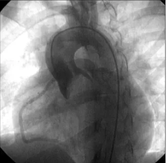
Transcatheter aortic valve implantation (TAVI) is a recent technique for the treatment of patients with severe symptomatic aortic stenosis (AS), who are at high risk for

The transcatheter aortic valve implantation has become a therapeutic option for patients with symptomatic severe aortic stenosis and high risk for conventional valve

Implantation was performed after valvuloplasty with balloon catheter in patients with native valve stenosis and without valvuloplasty in patients with pre-existing bioprosthesis

Transcatheter aortic valve implantation for the treatment of severe symptomatic aortic stenosis in patients at very high or prohibitive surgical risk: acute and late outcomes of

operation of a hemophilic patient with mitral and aortic valve disease of rheumatic etiology undergoing aortic valve replacement and mitral comissurotomy; in this case a VIIIf