3 7 8
Mattos et al.
Percutaneous mitral valvotomy in the young
Arq Bras Cardiol volume 73, (nº 4), 1999
Instituto Dante Pazzanese de Cardiologia - São Paulo - Brazil
Mailing address: Cláudia Mattos - Instituto Dante Pazzanese de Cardiologia-Av.Dr. Dante Pazzanese, 500 - 04012-180 - São Paulo, SP - Brazil
Received on 12/22/98 Accepted on 6/2/99
Objective – To analyze immediate and late results of
per-cutaneous mitral valvotomy (PMV) in patients ≤ 18 year.
Methods - Between August ’87 and July ’97, 48
procedures were performed on 40 patients. The mean age was 15.6 years; 68.7% were females four of whom were pregnant.
Results – Success was obtained in 91.7% of the
pro-cedures. Immediate complications were severe mitral re-gurgitation (6.3%) and cardiac tamponade (2.0%). Late follow-up was obtained in 88.8% of the patients (mean value=43.2±33.9 months). NYHA functional class (FC) I or II was observed in 96.2% of the patients and restenosis developed in five patients, at a mean follow-up of 29.7±11.9 months. Three patients presented with severe mitral insuf-ficiency and underwent surgery. Two patients died.
Conclusion - PMV represents a valid therapeutic
op-tion in young patients. In these patients, maybe because of subclinical rheumatic activity, restenosis may have a hi-gher incidence and occur at an earlier stage than in others persons.
Key words: mitral valvotomy, restenosis, teenagers.
Arq Bras Cardiol, volume 73 (nº 4), 378-381, 1999
Cláudia Mattos, Sérgio Luiz Navarro Braga, César Augusto Esteves, José Matos Brito Castello Branco, Nisia Lira Gomes, Mercedes Maldonado, Valmir Fernandes Fontes
São Paulo, SP - Brazil
Percutaneous Mitral Valvotomy in Patients Eighteen Years
Old and Younger. Immediate and Late Results
Original Article
Rheumatic valvar disease is still relatively frequent in developing countries, occurring in 25% to 40% of patients with cardiac diseases 1. Surgical treatment through closed
mitral commissurotomy, was initially proposed by Cutler et al 2 in 1923. As an alternative to surgical treatment, Inoue et
al3 in 1984, described a technique for percutaneous mitral
valvotomy using a balloon catheter created by themselves and an anterograde access. Using this via, a valvar opening is created by the separation of the commissures by the cen-trifugal force generated by the balloon at the level of the mi-tral valvar ring. Immediate and late results obtained with per-cutaneous treatment throughout the years have been found to be similar to those obtained with classical surgical treat-ment, with the advantage of having a lower rate of morbidity and mortality 4-6. Therefore, percutaneous mitral valvotomy
is now the first therapeutical choice for treating mitral steno-sis in selected patients.
Patients with rheumatic mitral stenosis who undergo surgery during childhood or adolescence, need to undergo other procedures during the follow-up of their disease due to the incidence of rheumatic attacks at this early age 7.
These new interventions may increase morbidity and mor-tality in this young group. Taking these data into conside-ration, we decided to use PMV as a first-choice therapy at our institution in patients ≤ 18 years.
The objective of this study was to describe early results and late clinical follow-up of patients at this young age who underwent PMV for treatment of rheu-matic mitral stenosis.
Methods
Arq Bras Cardiol volume 73, (nº 4), 1999
Mattos et al. Percutaneous mitral valvotomy in the young
3 7 9
(8.3%) were pregnant and did not respond to conventional clinical therapy, necessitating the intervention between the 13th and 34th weeks of pregnancy. Before percutaneous mitral
valvotomy, six patients (15%) were in NYHA functional class (FC) IV, 21 (52.5%) in FC III and 13 (32.5%) in FC II (table I).
The indications for the procedure were a mixture of cli-nical, radiological and echocardiographic findings, that is, symptomatic patients with a mitral valve area ≤1cm2 and a
score≤12 points by echocardiographic criteria 8. The
pre-sence of thrombus in the left atrium (LA) Ano/OR, associa-ted mitral regurgitation >2+/4+, according to Seller’s classification 9 were considered exclusion criteria.
A Doppler echocardiogram was performed in all pati-ents before the procedure, 48 hours afterwards and once a year during late follow-up. Valve anatomy, peak and mean diastolic gradients, and mitral valve area (MVA) calculated by planimetry were assessed in all patients. Morphology of the mitral valve and subvalvular apparatus was analyzed according to the criteria proposed by Wilkins et al 8, and
mi-tral regurgitation was quantitated when present.
All selected patients underwent a Cardiac Catheteriza-tion both before and after the procedure. This study included right and left heart catheterization performed through the puncture of the right femoral vein and left femoral artery. Manometry was obtained, including right chamber pressures and the gradient between mean LA pressure and left ven-tricular end diastolic pressure. Left ventriculography was per-formed at 30o in the oblique anterior view to analyze the mitral
subvalvular apparatus and to quantitate mitral regurgitation, when present. Aortography at 45o in the left anterior oblique
view was obtained to assess associated aortic lesions. After valvotomy and consequent opening of the mitral valve, left atriography at 30o in the left anterior oblique view was obtained
to analyze LA emptying and another left ventriculography was performed to detect mitral regurgitation at this view.
Valvotomy was always performed using the ante– rograde access through the puncture of the atrial septum, as described by Brockenbrough 10. Heparin (100U.i/kg) was
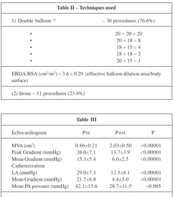
gi-ven to all patients after puncture of the atrial septum. Both the double balloon and the Inoue techniques were used (table II). The procedure was considered successful when the mitral valve area became ≥1.5cm2 and when no complications,
such as severe mitral regurgitation or left-to-right shunt >1.5:1 through the residual atrial septal defect (ASD), occurred.
Restenosis was defined as a loss of >50% of the mitral valve area obtained right after the procedure.
Results
Immediate Results - A transseptal puncture was perfor-med without complications in 47 of the 48 procedures, and the mitral valve was successfully dilated in 44 (91.7%) patients. In only one patient could the procedure not be completed due to a complication of the transseptal puncture with subsequent hemo-pericardium and signs of cardiac tamponade.
The mean mitral valve area assessed by planimetry and by the pressure half-time method increased from 0.86±0.21 to 2.03±0.50cm2 after percutaneous mitral valvotomy (p<0.00001).
Doppler echocardiography showed a decrease in peak and mean gradients of 26.0±7.1 to 13.7±3.9mmHg and from 15.3±5.4 to 6.0±2.5 (p<0.00001) respectively, right after the procedure.
Manometry data obtained at right and left catheterization showed a decrease in mean LA pressure: 29.0±7.3 to 12.5±-6.1mmHg (p<0.00001). The same happened to the mean diastolic gradient and the mean pulmonary pressure, which decreased respectively from 21.7±6.08 to 4.4±3.0mmHg (p<0.00001) and from 42.1±15.6 to 28.7±11.5mmHg (p<0.005) (table III).
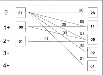
Regarding mitral regurgitation, which represents the most frequent complication of this procedure, of 37 patients with no previous mitral regurgitation, 28 did not have it at the control ventriculography. Five patients developed a 1+/4+ mitral regurgitation, according to Seller’s classification, one a 2+/4+, two a 3+/4+ and one 4+/4+. Of nine procedures with pre-vious 1+/4+ mitral regurgitation, six did not show any change while three increased their regurgitation to a 2+/4+ . In the only procedure with an initial 2+/4+ mitral regurgitation, no change occurred in the degree of regurgitation at a control
ventriculo-Table I – Methods (48 procedures)
Age (years) 10-18 (mean = 15.5±2.2)
Sex Female 33 (68.7%)
Male 15 (31.3%)
Sinus rhythm 48
Previous commissurotomy 1 (2.0%)
Pregnancy 4 (8.3%)
13º-34º week. (mean=27.7±8.5)
Echocardiographic score 5-10 (mean = 7.7±1.3) Functional Class (NYHA)
II 13 (32.5%)
III 21 (52.5%)
IV 6 (15.0%)
Table II – Techniques used
1) Double balloon * – 36 procedures (76.6%)
• 20 + 20 = 20
• 20 + 18 = 8
• 18 + 15 = 4
• 18 + 18 = 3
• 20 + 15 = 1
EBDA/BSA (cm2/m2) = 3.6 ± 0.29 (effective balloon dilation area/body
surface)
(2) Inoue – 11 procedures (23.4%)
Table III
Echocardiogram Pre Post P
MVA (cm2) 0.86±0.21 2.03±0.50 <0.00001
Peak Gradient (mmHg) 26.0±7.1 13.7±3.9 <0.00001 Mean Gradient (mmHg) 15.3±5.4 6.0±2.5 <0.00001 Catheterization
LA (mmHg) 29.0±7.3 12.5±6.1 <0.00001 Mean Gradient (mmHg) 21.7±6.8 4.4±3.0 <0.00001 Mean PA pressure (mmHg) 42.1±15.6 28.7±11.5 <0.005
3 8 0
Mattos et al.
Percutaneous mitral valvotomy in the young
Arq Bras Cardiol volume 73, (nº 4), 1999
graphy (fig. 1). Therefore, mitral regurgitation was found to develop to appear or increase in intensity in 12 (25.4%) of the 47 completed procedures, being 3+/4+ and 4+/4+ in 3 (6.3%).
The development and the measurement of the ASD we-re assessed by oximetry (QP/QS<1.5/1) and by Doppler echo-cardiogram. In 10 procedures where a left-to-right shunt was detected, the ASD was not greater than 3mm in diameter.
There were no embolic events or hospital deaths in this group of patients.
Success was not obtained in four procedures in one perforation of the LA free wall occurred during the atrial septal puncture with consequent cardiac tamponade that required emergent surgery. In the remaining three, after balloon valvotomy, severe mitral regurgitation was detected by left ventriculography, and all three patients underwent urgent mitral valve replacement surgery
Late results – Late follow-up was obtained in 32 (80%)
of the 40 patients who underwent percutaneous mitral val-votomy. Twenty-four (75%) were females and mean follow-up was 43.2±33.9 months.
Of these 32 patients (fig. 2), three were referred for elective valve replacement surgery (two because of mitral insufficiency and one because of restenosis). In the remaining 29 patients,
five developed restenosis in a mean period of 29.7±11.9 months and were successfully redilated. In three of these a third redilation was performed due to a new restenosis. Therefore, eight redilations were performed in five patients.
In this group of 40 patients, two late deaths occurred, one probably due to restenosis (acute pulmonary edema) and the other due to a non related cause (septicemia).
A better NYHA FC at follow-up was observed with significant improvement in the quality of life of these pati-ents. In the observed follow-up period, 24 (75%) patients were in NYHA FC I and only three (11.2%) were in FC II or III. At follow-up, 30 patients remained in sinus rhythm, and only two developed atrial fibrillation. All four pregnant wo-men had a term cesarean delivery while in FC I and no fetal deaths or associated malformations occurred.
Echocardiographic parameters of MVA and peak and mean gradients at follow-up are described in table IV.
Discussion
Data reporting the immediate results obtained in this group of patients do not differ significantly from those ob-served in the general population undergoing this type of procedure 11. The efficacy of this method in this age group
was demonstrated by the final MVA, the reduction of mean LA pressure, of the pulmonary artery pressure and of the mean diastolic transmitral gradient.
As for immediate complications, a residual ASD was observed in 21.2% of the patients by a Doppler echocardio-gram performed 48 hours after the procedure, a phenome-non also reported in the majority of published studies 10. In
our experience, the created defect was always small, and a significant residual shunt was not observed in any of the ca-ses. We think that the persistence of this defect occurs mainly in those patients where final results of the MVA are considered suboptimal (<1.5cm2), due to the persistence of
an elevated pressure in the left atrium 12.
The incidence of mitral regurgitation (27.2%) is similar to that observed by us in the general population(29.2 %) 11.
As is well known, the main predictors of this complication are oversizing of the balloon 13, commissural or leaflet
calci-fication or both and the intensity of the involvement of the mitral subvalvar apparatus 14. In our series of patients, the
development of severe mitral regurgitation was directly related to the degree of valvar involvement before valvoto-my, especially when MVA was ≤ 0.9cm2.
In this group, because patients were young, calcifica-tion was uncommon, and the involvement of the subvalvar apparatus was usually mild, our main concern was the exact diameter of the balloon, especially when MVA was <0.9 cm2.
Table IV
Echocardiogram Post Late P
Mitral valve area (cm2) 2.03±0.50 1.91±0.50 NS *
Peak gradient (mmHg) 13.7±3.9 11.5±4.9 NS Mean gradient (mmHg) 6.0±2.5 4.8±2.6 NS
*NS- nonsignificant.e Fig. 2 – Population (40 patients).
Arq Bras Cardiol volume 73, (nº 4), 1999
Mattos et al. Percutaneous mitral valvotomy in the young
3 8 1
Therefore, when the double balloon technique was em-ployed, we used an EBDA/BSA<3.5 relation and, when the Inoue balloon was the one chosen, we used the formula suggested by the author and subtracted 1mm. Even having done that, three procedures were followed by a 3+/4+ or 4+/4+ mitral regurgitation, which required urgent surgery. Some degree of involution of acute mitral insufficiency has been described in the literature in a certain percentage of patients15
and is probably related to the elastic retraction of the mitral ring, to commissural fusion and fibrosis and to the impro-vement of posttraumatic dysfunction (edema) of the papillary muscles caused by the balloon. However, this phenomenon was not observed in this group of patients.
Late follow-up data obtained at a mean period of 43.2±33.9 months showed that the great majority of the pati-ents (88%) were in NYHA FC I and atrial fibrillation occurred in only two patients.
No statistically significant difference occurred between immediate and late echocardiographic parameters, such as MVA, mean LA pressure, mean pulmonary artery pressure and, finally, mean transmitral diastolic gradient, which demonstrates the efficacy of the procedure in the medium-term follow-up.
We believe that the relatively high index of restenosis (15.6%) detected in five of the 32 patients in a relatively short period of time (29.7±11.9 months) is above the observed the index described in the literature for the general population, which is around 10% in five years, both for percutaneous or surgical treatment 15-17. The observed re-stenosis in the present
study is probably not directly related to the use of undersized balloons in both techniques, because our immediate results do not differ significantly from those previouly observed by us. Predisposing factors related to development of resteno-sis classically described are: old age, atrial fibrillation and a high echocardiographic score index (mainly the degree of
thi-ckening and the presence of calcium in the leaflets) 18. In this
group, these findings were uncommon because of the young age of the patients, and we think that restenosis may have been caused by recurrent subclinical rheumatic attacks, which usually can not be detected clinically or by laboratory tests.
Based on these results and considering the problem imposed by future re-interventions, we believe that percuta-neous treatment should be the primary intervention, becau-se its immediate and late results are similar to thobecau-se obtained by surgery 4-6, which has a higher morbidity. Another
advantage of the percutaneous procedure is that re-dilations can be performed without technical difficulties in the additional procedures and without a higher incidence of complications.
It is well known that rheumatic disease has a chronic and progressive course and young patients are prone to undergoing more then one valvular procedure during their lifetime. Therefore, when this becomes necessary, surgery can be performed without the inherent risks of a previous thoracotomy.
Analyzing these data, we believe that percutaneous mitral valvotomy is a valid therapeutic option for the treat-ment of severe rheumatic mitral stenosis in patients under 18. In this population, the incidence of restenosis is rela-tively higher and occurs earlier than in the general populati-on. This is probably related to the occurrence of subclinical attacks of rheumatic disease or to a more malign valvar in-volvement of the disease in this age group.
It has also been demonstrated that the technique can be repeated in cases of restenosis without additional tech-nical implications or more complications. We therefore con-clude that percutaneous mitral valvotomy should be the me-thod of choice for the treatment of severe rheumatic mitral stenosis in patients under 18 years of age.
1. Community control of rheumatic heart disease in developing countries. A major public health problem. Who Chron 1980; 34: 336.
2. Cutler EC, Levine As, Biy CS. Surgical, treatment of mitral stenosis: Experimen-tal and clinic studies. Arch Surg 1924; 689-821.
3. Inoue K, Owaki T, Nakamura T, Kitamura F, Miyamoto N. Clinical application of transvenous mitral commissurotomy by a new balloon catheter. J Thorac Cardio-vasc Surg 1984; 87: 395-402.
4. Can the long-term outcomes of percutaneous balloon mitral valvotomy and surgi-cal commissurotomy be expected to be similar? J Heart Valve Dis 1995; 4: 446-52. 5. Arora R, Nair M, Kalra G, et al. Immediate and long-term results of balloon and sur-gical closed mitral valvotomy. A randomized comparative study. Am Heart J 1993; 125: 1091-3.
6. Feldman T. Hemodynamic results, clinical outcomes and complications of Inoue balloon mitral valvotomy. Cathet Cardiovasc Diagn 1994; 2: 2-7.
7. Kirklin JW, Barrat-Boyes BG. Cardiac Surgery, 2nd edition, 1993; 440-51.
8. Wilkins GT, Weyman AE, Abaseal VM, Block PC, Palacios IF. Percutaneous bal-loon dilatation of the mitral valve: an analysis of echocardiography variables re-lated to outcome and the mechanism of dilatation. Br Heart J 1988; 60: 299-308. 9. Seller RD, Levy MJ, Amplatz K, Lillehei CW. Retrograde cardioangiography in
acquired cardiac disease: technique, indications and interpretation of 100 cases. Am J Cardiol 1964; 14: 437.
References
10. Mullins CE. Transeptal left heart catheterization: Experience with a new techni-que in 520 pediatric and adult patients. Pediatr Cardiol 1983; 4: 239-46. 11. Braga SLN, Esteves CA, Meneghelo Z, Sousa JEMR. Valvoplastias mitral e aórtica
por cateter balão. Socesp Cardiologia. Atualização e Reciclagem 1994; 466-75. 12. Palacios IF, Block PC, Wilkins GT, Weyman AE. Follow-up patients
undergo-ing percutaneous mitral balloon valvotomy. Analysis of factors determinundergo-ing res-tenosis. Circulation 1989; 79: 573-9.
13. Roth RB, Block PC, Palacios IF. Predictors increased mitral regurgitation after percutaneous mitral balloon valvotomy. Cath and Card Diag 1990; 20: 17-21. 14. Hung JS, Cherm MS, Wu JJ, et al. Short and long-term results of catheter balloon
per-cutaneous transvenous mitral commissurotomy. Am J Cardiol 1991; 67: 854-62. 15. Palacios IF, Block PC, Wilkins GT, Weyman AE. Follow-up of patients undergo-ing perutaneous mitral balloon valvotomy. Analysis factors determinundergo-ing reste-nosis. Circulation 1989; 79: 570-3.
16. Vahaniar A, Michiel PL, Cmier B, et al. Results of percutaneous mitral commissu-rotomy in 200 patients.Am J Cardiol 1989; 63: 847-52.
17. Desidiri A, Vanduperrer O, Serra A, et al. Long-term (9 to 33 months) echocardio-grafic follow-up after successful percutaneous mitral commissurotomy. Am J Car-diol 1992; 69: 1602-6.