Predictors of Viability in Patients with Negative Low-dose
Dobutamine Stress Echocardiography
Zainab Abdel-Salam and Wail Nammas
Cardiology Department, Faculty of Medicine, Ain Shams University, Cairo - Egypt
Mailing address: Wail Nammas •
Cardiology Department, Faculty of Medicine, Ain Shams University - Abbassia - Cairo - Egypt - P.O. 11381
E-mail: [email protected]
Manuscript received February 07, 2010; revised manuscript received September 15, 2010; accepted 21, 2010.
Abstract
Background: Low-dose dobutamine stress echocardiography is specific for predicting reversible contractility dysfunction, but its sensitivity is lower than ideal.
Objective: We sought to explore the predictors of myocardial contractile recovery following revascularization, in patients with no viability by low-dose dobutamine stress echocardiography.
Methods: We prospectively enrolled 30 consecutive patients with significant coronary stenosis/occlusion amenable for revascularization, regional wall motion abnormality in the distribution of the affected artery and absence of viability by low-dose dobutamine stress echocardiography. They underwent resting 99mTc-sestamibi imaging study, and then underwent successful coronary revascularization. Follow-up echocardiography was performed 3 months later. Patients were classified into 2 groups: group 1: with evidence of myocardial contractile recovery after revascularization at follow-up echocardiography and grofollow-up 2: with no such evidence of recovery. The two grofollow-ups were compared with respect to patients’ clinical, echocardiographic and scintigraphic data.
Results: The mean age was 52.3 ± 5.9 years, with 97% being males. The percentage of total 99mTc-sestamibi uptake was significantly higher in group 1 as compared to group 2 (p < 0.01), and it was the strongest independent predictor of myocardial contractile recovery at 3-month follow-up by multivariate regression analysis. Receiver operating characteristics curve revealed that a cutoff value of the percentage of total 99mTc-sestamibi uptake of 72% best predicted myocardial contractile recovery, with a sensitivity of 100% and specificity of 95.7%.
Conclusion: In patients with no viability by low-dose dobutamine stress echocardiography, the percentage of total 99m Tc-sestamibi uptake independently predicted myocardial contractile recovery following coronary revascularization. (Arq Bras Cardiol 2011;96(3):188-195)
Keywords: Predictors; viability; revascularization; dobutamine stress echocardiography.
was supported by the results of several studies, wherein only patients with severe left ventricular dysfunction who harbored viable myocardium, benefited from revascularization4.
Pharmacological stress echocardiography has gained wide acceptance for the identification of viable myocardium, chiefly because of its feasibility, safety, diagnostic accuracy and prognostic power5. Low-dose dobutamine stress
echocardiography (DSE) has emerged as an attractive and increasingly used method to identify viable myocardium through its ability to elicit a beta adrenoreceptor-mediated increase in myocardial contractility. Dobutamine-responsive wall motion was found to be specific for predicting reversible contractility dysfunction, but its sensitivity is lower than ideal6.
In a prospective study design, we sought to explore the predictors of contractile recovery after revascularization, in patients with absence of viability by low-dose DSE.
Methods
Patient selection
Prospectively, we enrolled 30 consecutive patients referred from our catheterization labs with significant coronary
Introduction
With the enormous progress in the field of myocardial revascularization over the last two decades, predicting the presence of viable myocardium has acquired paramount clinical importance, particularly in patients considered for interventional treatment1. Myocardial viability represents the
impairment of contractile function that is potentially reversible if blood supply is adequately restored2. Presumably, improving
stenosis/occlusion, during the period from November 2006 to October 2008. Patients were considered eligible for inclusion if they had regional wall motion abnormality in the anatomical distribution of the affected arteries as explained later, affected arteries amenable for revascularization, and absence of viability by low-dose DSE. Significant coronary
stenosis was defined as at least 70% luminal obstruction of at
least one sizable coronary artery (measuring 2.5 mm or more in diameter), seen in 2 different projections. Total coronary
occlusion was defined as 100% luminal obstruction with
Thrombolysis In Myocardial Infarction (TIMI) grade 0 forward flow distal to the site of obstruction. We excluded patients with recent myocardial infarction or unstable angina during the past 4 weeks, significant stenosis of the left main coronary artery
(defined as at least 50% luminal obstruction), decompensated
heart failure, protruding fresh left ventricular thrombus, significant valvular or congenital heart disease, any myocardial disease apart from ischemia, left ventricular ejection fraction
> 40%, bundle branch block, contraindication to dobutamine
(for example: history of complex ventricular arrhythmias,
uncontrolled hypertension with blood pressure > 180/110),
and patients with limited life expectancy due to coexistent disease (for example: malignancy). Before inclusion, an informed written consent was obtained from each patient after full explanation of the study protocol, the type of data collected, the way of data processing and the scope of data collection. The study protocol was reviewed and approved by the local institutional human research committee in our center according to the ethical guidelines of the 1964 Declaration of Helsinki, as revised in 2002.
Deinitions of risk factors
The presence of hypertension was defined as a systolic
blood pressure ≥ 140 mmHg and/or a diastolic blood pressure ≥ 90 mmHg, previously recorded by repeated non-invasive
office measurements, which lead to life-style modification or antihypertensive drug therapy. The presence of diabetes
mellitus was defined as a fasting plasma glucose ≥ 126 mg/ dl, and/or a 2-hour postload glucose ≥ 200 mg/dl, or specific
anti-diabetic drug therapy. Dyslipidemia was defined by a
low-density lipoprotein cholesterol > 100 mg/dl, and/or serum triglycerides > 150 mg/dl, and/or a high-density lipoprotein
cholesterol < 40 mg/dl in men and < 50 mg/dl in women.
Baseline echocardiographic assessment
Assessment of regional and global left ventricular systolic function was performed in all patients by transthoracic echocardiography within 48 hours of admission. Doppler echocardiography was performed using a Hewlett Packard Sonos 5500 cardiac ultrasound machine (Hewlett Packard, Andover, Massachusetts, USA) equipped with harmonic imaging capabilities. A 2.5 MHz phased array probe was used to obtain standard 2D, M-mode and Doppler images. Patients were examined in the left lateral recumbent position using standard parasternal and apical views. Global left ventricular systolic function was assessed in apical 4-chamber and 2-chamber views using the biplane modified Simpson’s method. Regional wall motion was assessed according to the standard 16-segment model recommended by the American
Society of Echocardiography7. Individual segments were then
sub-grouped based on the known vascular distribution into left anterior descending territory, left circumflex territory, right coronary artery territory, and overlap segments7.
Regional wall motion was visually assessed for each segment individually, considering both endocardial excursion and systolic thickening, and each segment was graded according to the semiquantitative scoring system described by Knudsen et al8. Segments with poorly defined endocardial borders for 50% or more of their length were considered non-visualized
and assigned a score of 09. Wall thickening was assessed at
a distance of at least 1 cm from the adjacent segment to minimize the effect of tethering10. Wall motion in a vascular
territory was considered abnormal if wall thickening was abnormal in at least two contiguous non-overlap segments7.
Wall motion score index (WMSI) was derived by dividing the sum of individual segment scores by the number of interpretable segments.
Stress echocardiographic protocol
All patients underwent low-dose DSE as follows: dobutamine (Dobutrex™, Lilly, Eli and Company, Indianapolis, USA) was infused intravenously starting at 5 µg/kg/minute, increased up to 20 µg/kg/minute, in 3-minute stages. Examinations were standardized and performed by the same operator. Standard views were recorded at baseline, during each stage of the infusion protocol, as well as during recovery. Images were digitized in cine-loop format and saved for subsequent playback and analysis. Views were analyzed by a single expert echocardiographist (Z.A.) employing the software program of the echocardiography machine. Analysis of viability was performed during all stages of the protocol. Visual assessment of wall motion and thickening was performed as before. Global left ventricular systolic function and wall motion score index were evaluated at rest and at the end of each stage. The presence of viability was defined by improvement in regional wall motion score by at least one grade in at least two contiguous non-overlap segments along with at least
20% reduction in global WMSI compared with baseline
evaluation10. The stress test was performed with the patients
on their full anti-ischemic and anti-failure medications.All patients were receiving beta-blockers, angiotensin converting enzyme inhibitors, statins and aspirin.
99mTc-sestamibi imaging protocol
Patients underwent resting 99mTc-sestamibi imaging study
with the administration of trimetazidine, using the standard imaging technique, within 4 days of coronary angiography, provided that no ischemic events were recorded during the time from the coro nary angiography to 99mTc-sestamibi imaging.
Trimetazidine (Vastarel™, Servier, France) was administered by the oral route the day before the study (60 mg divided in 3 equal doses 8 hours apart), and 1 hour before performing the study (60 mg in a single dose). Injection of 25-30 mCi of radioactive tracer was administered 45-60 minutes before image acquisition. Images were acquired using a rotating single-head gamma camera (GE Starcam 4000i, UK) equipped with
low-energy all-purpose collimators. Energy windows of 20% were
Thirty-two images were obtained over 180° extending from the 45° right anterior oblique to the 45° left posterior oblique projections. All studies were subjected to quality-control checks and cor rections when necessary for camera non-uniformity, center-of -rotation offsets, patient motion, and “upward creep”8.
99mTc-sestamibi image analysis
Two experienced nuclear cardiologists blinded to the clinical, echocardiographic and angiographic data, analyzed the 99mTc-sestamibi images. The vascular assignment of
myocardial segments to the vascular distribution of major coronary arteries was performed according to the 17 segments scoring system11. The percentage of 99mTc-sestamibi uptake
was assessed for each segment individually. The mean value of percent 99mTc-sestamibi uptake was calculated for all left
ventricular segments (total 99mTc-sestamibi uptake), as well as
separately, for each individual vascular territory.
Coronary revascularization
All patients underwent coronary revascularization either by percutaneous coronary angioplasty, or by surgical bypass grafting according to the decision of the attending physician. Revascularization was performed within 2 weeks of the index coronary angiography, provided that no ischemic events were recorded during the time from the coro nary angiography to revascularization. The decision was based on the clinical presentation, coronary anatomy and evidence of ischemia.
Echocardiographic follow-up
Follow-up echocardiographic re-assessment was performed 3 months after revascularization to evaluate regional and global left ventricular systolic function as described before. All evaluations were performed offline by the same echocardiographist (Z.A.) who was blinded to whether the images were obtained before or after revascularization. The occurrence of myocardial contractile recovery was defined by improvement in regional wall motion score by at least one grade in at least two contiguous non-overlap segments
along with at least 20% reduction in global WMSI compared
with baseline evaluation10. During follow-up, patients were
questioned regarding the occurrence of new myocardial infarction or congestive heart failure by means of clinical visits, telephone calls, hospital chart reviews, or personal communication with the referring physician.
Statistical analysis
All continuous variables were presented as mean ± SD, if they were normally distributed. Data were tested for normal distribution using the Kolmogorov-Smirnov test. Categorical variables were described with absolute and relative (percentage) frequencies. According to the above definition of myocardial contractile recovery, patients were classified into 2 groups: group 1 with evidence of actual myocardial contractile recovery after revascularization at follow-up echocardiography, and group 2 with no such evidence of recovery. The two groups were compared with respect to patients’ clinical characteristics, echocardiographic, scintigraphic and angiographic data, using the unpaired t test
for normally distributed continuous variables, and the Pearson chi-square test for categorical variables. Multivariate regression analysis was performed to identify the independent predictors of myocardial contractile recovery after revascularization, in which the dependent variable was the outcome variable of interest, whereas factors entered into the model included the mean value of percent 99mTc-sestamibi uptake by all
left ventricular segments (total 99mTc-sestamibi uptake), as
well as that assessed separately, for each individual vascular territory, the mean resting left ventricular ejection fraction and WMSI before revascularization. Eventually, we generated a receiver-operating characteristics (ROCs) curve to identify the cutoff value of the percentage of total 99mTc-sestamibi uptake
that best predicted myocardial contractile recovery after revascularization. The optimal cutoff value was defined as the value giving the largest area under the curve (AUC). Finally, twenty cases were randomly selected for analysis of intra-observer variability. Assessment of variability was performed using linear regression analysis. All analyses were 2-sided and a probability value of P < 0.05 was considered statistically significant. Analyses were performed with the SPSS, version 12.0 statistical package (SPSS Inc., Chicago, IL, USA).
Results
Baseline demographic characteristics
Of a total of 61 patients with significant coronary stenosis/ occlusion amenable for revascularization and regional wall motion abnormality in the anatomical distribution of the affected arteries, during the study period, 31 had evidence of viability by low-dose DSE, while 30 had no such evidence of viability. All patients with positive viability by low-dose DSE underwent coronary revascularization. According to the aforementioned definition of myocardial contractile recovery at the 3-month follow-up echocardiography, there were 27
patients (87.1%) with evidence of myocardial contractile recovery after revascularization, and 4 (12.9%) with no such evidence of
recovery. A total of 30 consecutive patients with no evidence of viability by low-dose DSE were included in the current study, who underwent coronary revascularization for significant coronary stenosis/occlusion. All patients completed the 3-month follow-up period, and no patient reported any clinical events during the period from revascularization to the follow-up echocardiography evaluation. According to the aforementioned definition of myocardial contractile recovery at follow-up echocardiography,
there were 7 patients (23.3%) with evidence of myocardial
contractile recovery after revascularization (group 1), and 23
(76.7%) with no such evidence of recovery (group 2). Of the
total cohort (61 patients), low-dose DSE predicted myocardial contractile recovery following revascularization with a sensitivity
of 79.4% and a specificity of 85.2%11.
Revascularization was successful and complete in all patients. All patients in group 1 were revascularized by surgical bypass
grafting, while in group 2, 5 patients (21.7%) were treated with percutaneous coronary angioplasty and 18 (78.3%) were
treated by surgical bypass grafting (p > 0.05). The demographic
characteristics of the entire cohort as well as of the 2 individual groups are shown in Table 1. The mean age was 52.3 ± 5.9
found more frequently in group 2 as compared to group 1
(69.6% versus 28.6% respectively, p < 0.05). Otherwise, no statistically significant differences were found between the two groups regarding any of the demographic characteristics.
Echocardiographic data
Table 2 shows the DSE data of the entire study cohort as well as the 2 individual groups. At baseline, the mean left ventricular ejection fraction of the entire study cohort was 24
± 4%, while the mean WMSI was 2.7 ± 0.15. No statistically
significant differences were found between the two groups regarding any of the DSE data (Table 2).At the 6-month
follow-up, the mean left ventricular ejection fraction was 30 ± 4% versus 24 ± 4%, while the mean WMSI was 2.3 ± 0.07 versus
2.6 ± 0.24, in group 1 as compared to group 2, respectively, (p < 0.05 for both).
The DSE protocol was well tolerated by all patients with no major side effects during or after the test.
Scintigraphic data
Table 3 shows 99mTc-sestamibi scintigraphy data of the entire
study cohort as well as of the 2 individual groups. The percentage of total 99mTc-sestamibi uptake was significantly higher in group 1 as compared to group 2 (78 ± 3% versus 64 ± 9% respectively, p < 0.01). Similarly, the percentage of 99mTc-sestamibi uptake
by segments in the left anterior descending coronary territory, was significantly higher in group 1 as compared to group 2 (90
± 12% versus 78 ± 19% respectively, p < 0.01).
Independent predictors of contractile recovery
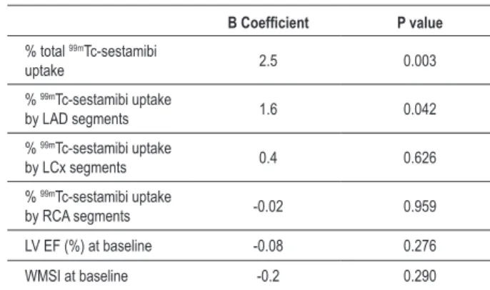
The multivariate regression analysis showed that the percentage of total 99mTc-sestamibi uptake, and the uptake
by segments in the left anterior descending coronary territory, independently predicted myocardial contractile recovery at 3-month follow-up after revascularization, with the former being the strongest independent predictor (Table 4).
Cutoff value for prediction of myocardial contractile recovery
The receiver operating characteristics (ROCs) curve revealed that a cutoff value of the percentage of total 99m Tc-sestamibi uptake of 72%, best predicted myocardial contractile
Table 1 - Baseline clinical characteristics of the whole cohort and the 2 individual groups
Total cohort (n = 30)
Group 1 (n = 7)
Group 2 (n = 23)
Age (years) 52.3 ± 5.9 52.3 ± 3.7 52.3 ± 6.6
Male gender 29 (97) 6 (85.7) 18 (78.3)
Diabetes mellitus 18 (60) 2 (28.6) 16 (69.6) *
Hypertension 18 (60) 4 (57.1) 14 (60.9)
Smoking 25 (83.3) 5 (71.4) 20 (86.9)
Dyslipidemia 7 (23.3) 2 (28.6) 5 (21.7)
Continuous variables are presented as mean ± SD, while categorical variables are presented as numbers (percentage). IHD indicates ischemic heart disease. * indicates p < 0.05.
Table 2 - Echocardiographic data of the whole cohort and the 2 individual groups
Total cohort (n = 30)
Group 1 (n = 7)
Group 2 (n = 23)
LV EF (%) at baseline 24 ± 4 24 ± 3 24 ± 4
WMSI at baseline 2.7 ± 0.16 2.7 ± 0.06 2.6 ± 0.25
LV EF (%) at low-dose
dobutamine 24 ± 4 24 ± 4 24 ± 4
SBP at baseline 119 ± 14 115 ± 16 123 ± 12
SBP at low-dose
dobutamine 125 ± 14 122 ± 14 128 ± 14
DBP at baseline 86 ± 8 89 ± 9 83 ± 7
DBP at low-dose
dobutamine 82 ± 6 84 ± 6 79 ± 6
HR at baseline 71 ± 9 73 ± 10 69 ± 9
HR at low-dose
dobutamine 77 ± 9 79 ± 10 75 ± 9
All variables are presented as mean ± SD. LV EF - indicates left ventricular ejection fraction; WMSI - wall motion score index; SBP - systolic blood pressure; DBP - diastolic blood pressure; HR - heart rate.
Table 3 - Scintigraphic data of the two individual study groups
Group 1 (n=7) Group 2 (N=23)
% total 99mTc-sestamibi
uptake 77.6 ± 2.6 64.4 ± 8.9 *
% 99mTc-sestamibi uptake
by LAD segments 90 ± 12.4 78.2 ± 19 *
% 99mTc-sestamibi uptake
by LCx segments 86 ± 5.9 89.2 ± 14.3
% 99mTc-sestamibi uptake
by RCA segments 62.1 ± 17.8 48.7 ± 20.7
All variables are presented as mean ± SD. LAD indicates left anterior descending artery; LCx, left circumlex artery; RCA, right coronary artery. * indicates p < 0.05.
Table 4 - Multivariate linear regression model demonstrating the independent predictors of myocardial contractile recovery following revascularization at 3-month follow-up
B Coeficient P value
% total 99mTc-sestamibi
uptake 2.5 0.003
% 99mTc-sestamibi uptake
by LAD segments 1.6 0.042
% 99mTc-sestamibi uptake
by LCx segments 0.4 0.626
% 99mTc-sestamibi uptake
by RCA segments -0.02 0.959
LV EF (%) at baseline -0.08 0.276
WMSI at baseline -0.2 0.290
LAD - indicates left anterior descending artery; LCx - left circumlex artery; RCA - right coronary artery, LV EF - left ventricular ejection fraction; WMSI - wall
recovery at 3 months following revascularization, with an area
under the curve (AUC) =0.957 (95% confidence interval
0.812 to 0.994, p < 0.001). Using this 72% cutoff value, the
percentage of total 99mTc-sestamibi uptake had a sensitivity of 100%, specificity of 95.7%, for predicting myocardial
contractile recovery following revascularization (Figure 1). The analysis of intra-observer variability revealed a close correlation between repeated assessments of regional wall motion by the single operator, with a correlation coefficient r =0.92.
Discussion
Assessment of myocardial viability is one of the most challenging areas of modern cardiology. The gold standard for the presence of viability is improvement in global and/or regional contractility following myocardial revascularization12.
Nevertheless, triaging a patient for revascularization depends on the ‘expected’ ensuing recovery of contractile function in the compromised area. In this regard, the ability of low-dose dobutamine to elicit a contractile response in dysfunctional, but viable myocardial segments supplied by occluded/critically stenosed arteries has been a controversial issue. Some previous studies reported the limited ability of even very low doses of dobutamine to unmask the presence of viable myocardium in the setting of severe coronary stenosis or total occlusion, where coronary flow reserve is exhausted and resting myocardial perfusion is severely reduced13. A meta-analysis of six studies
(287 patients) that used DSE to predict improvement in the left ventricular systolic function following revascularization showed a weighted mean sensitivity and specificity of 57
and 73%, with a PPV and NPV of 63 and 68%, respectively14.
Complex structural changes occur in viable dysfunctional myocardium at both cardiomyocyte and extracellular matrix levels15, which include ultrastructural abnormalities seen
by electron microscopy16. Moreover, indirect markers of
apoptosis have been recently demonstrated in more severely compromised hibernating cardiac myocytes17. Additionally,
reduced gap junction area in hibernating myocardium might interfere with local coordination of myocyte contraction18.
Other pathophysiological alterations in dysfunctional–but viable–myocardium include energy depletion19 and reduced
calcium responsiveness20. Overall, they may hamper the
contractile response to low-dose dobutamine, with a subsequent large proportion of false negative results to DSE, and a resultant suboptimal sensitivity6.
The issue of predicting myocardial contractile recovery following revascularization has long been controversial. Previously reported studies in literature have not provided consistent data to determine the specific predictors of potential contractile recovery. Some reports highlighted the importance of coronary collaterals to the infarct-related artery territory as a predictor of the presence of underlying myocardial viability and the potential myocardial contractile recovery following revascularization21,22. Generally speaking,
it is always recommended to search for viable myocardium before revascularization of an occluded coronary artery; however, there is no practical, yet sensitive, method for assessing myocardial viability in the cath lab21.
To the best of the authors’ knowledge, the current study was the first in literature to report that the percentage of total 99m
Tc-sestamibi uptake and the percentage of uptake by segments in the left anterior descending coronary territory, independently predicted ‘actual’ myocardial contractile recovery at the 3-month follow-up following revascularization, in a very specific group of patients with absence of viability by
low-dose DSE. Additionally, a cutoff value of 72%, of the former,
predicted contractile recovery, with an excellent sensitivity and specificity. Nevertheless, the current study does not compare the two methods (low-dose DSE and 99mTc-sestamibi
imaging) and an alleged superiority of 99mTc-sestamibi imaging
over low-dose DSE cannot be advocated. Furthermore, even when resting systolic function does not recover following revascularization, the presence of partial viability is likely to be beneficial for contractile reserve, exercise tolerance, prevention of remodeling and survival; revascularization can still be beneficial in this group of patients23.
In the current study, we employed a protocol of resting 99mTc-sestamibi imaging after oral administration
of trimetazidine. A robust body of evidence indicates that resting 99mTc-sestamibi may be a good marker for viability24-26.
Both trimetazidine and 99mTc-sestamibi share the same
intracellular target: the mitochondrion. As metabolic reserve does exist in the hibernating state, trimetazidine might exploit this reserve by increasing mitochondrial metabolism. One study demonstrated that trimetazidine was associated with an increase in 99mTc-sestamibi uptake in infarcted, but
viable myocardial areas and stated that this increase was probably related to improvement in mitochondrial oxidative metabolism, which is essential for 99mTc-sestamibi retention.
They concluded that coupling trimetazidine administration to Figure 1 -Receiver operating characteristics (ROCs) curve revealed that a cutoff
value of the percentage of total 99mTc-sestamibi uptake of 72% best predicted
myocardial contractile recovery at 3 months following revascularization, with
99mTc-sestamibi perfusion scintigraphy may represent a better
means of detecting viable myocardium27.
Our results suggested that the absence of diabetes mellitus also predicted the presence of viability, though it was not an independent predictor at the multivariate regression analysis. A previous study by Auerbach et al28 reported that, apart from
anginal symptoms, no statistically significant association was found between the presence of viability (detected by positron emission tomography) and any of the clinical characteristics, including diabetes28. Inconsistent findings would reflect the
heterogeneous nature of the underlying disease process, the lack of uniformity in patient selection and study protocols among different studies.
Limitations of the study
Our findings were based on a single-center study with a relatively small sample size, a fact that makes it difficult to generalize our results to all patients undergoing risk stratification for predicting contractile recovery after revascularization. Multicenter studies using the same protocol and examining a larger number of patients are needed. Additionally, the number of patients who recovered adequate contractility was very small; therefore the results of the current study should be considered with caution. Moreover, the follow-up period of 3 months might have been inadequate to allow recovery of some dyssynergic, but viable segments, which would otherwise translate to a better myocardial contractile recovery rate. Delayed recovery can further occur in a substantial number of segments up to a median of 14 months following revascularization, a fact that warrants repeated assessment after longer periods of follow-up. The fact that all patients were receiving beta blockers before DSE examination may have contributed to a substantial proportion of false negative results, since it is known that this drug interferes with the sensitivity of DSE. A possible limitation of the current study is that it does not provide a direct comparison between DSE and 99mTc-sestamibi perfusion scintigraphy. Given that
magnetic resonance imaging is considered the gold standard for detection of viability, the fact that the patients did not undergo this modality may constitute another limitation. Another limitation of the study is the lack of quantitative methods for measuring systolic thickening; instead, the operator adopted visual assessment only. Nevertheless, the problem of intra-observer variability can be minimized by stronger adherence to common and new methodological
standards. Finally, follow-up coronary angiography was not performed, therefore, restenosis or reocclusion cannot be definitely excluded, something that would hazard the initially achieved contractile recovery. However, no patient reported any clinical events during the period from revascularization to follow-up echocardiographic evaluation.
Conclusion
Our data suggest that myocardial contractile recovery after revascularization in patients with no evidence of viability by low-dose DSE could be independently predicted by the percentage of total 99mTc-sestamibi uptake and the uptake by
segments in the left anterior descending coronary territory. A cutoff value of the percentage of total 99mTc-sestamibi uptake of 72% best predicted myocardial contractile recovery at 3
months following revascularization.
Clinical implications
Patients with ischemic left ventricular dysfunction whose coronary arteries are amenable for revascularization and who have no evidence of viability by low-dose DSE, may still have a “glimpse of hope” in gaining significant myocardial contractile recovery following revascularization, if they have an ‘ample’ percentage of total left ventricular 99mTc-sestamibi
uptake, especially in the absence of diabetes mellitus. The other way around is also true, however: due to the relatively low specificity of perfusion scintigraphy to detect viability, DSE can be performed in patients with positive viability by perfusion scintigraphy. Yet, the routine use of both modalities together in all patients cannot be advocated, as it would not be cost-effective.
Potential Conflict of Interest
No potential conflict of interest relevant to this article was reported.
Sources of Funding
There were no external funding sources for this study.
Study Association
This study is not associated with any post-graduation program.
References
1. Dilsizian V, Bonow RO. Current diagnostic techniques of assessing myocardial viability in patients with hibernating and stunned myocardium. Circulation. 1993;87(1):1-20.
2. Hoffmann R. Stress echocardiography before and after interventional therapy. In: Marwick TH (ed.) Cardiac stress testing and imaging: a clinician’s guide. New York: Churchill Livingston; 1996. p. 355-67.
3. Beller GA. Comparison of 201Tl scintigraphy and low-dose dobutamine echocardiography for the noninvasive assessment of myocardial viability. Circulation. 1996;94(11):2712-9.
4. Jiménez Borreguero LJ, Ruiz-Salmerón R. Assessment of myocardial viability in patients before revascularization. Rev Esp Cardiol. 2003;56(7):721-33.
5. Marwick TH. Stress echocardiography. Heart. 2003;89(1):113-8.
6. Grayburn PA. Defining the threshold of myocardial viability by dobutamine echocardiography. Int J Cardiol. 2003;90(1):31-2.
Committee on Standards, Subcommittee on Quantitation of Two-Dimensional Echocardiograms. J Am Soc Echocardiogr. 1989;2(5):358-67.
8. Knudsen AS, Darwish AZ, Nørgaard A, Gøtzsche O, Thygesen K. Time course of myocardial viability after acute myocardial infarction: an echocardiographic study. Am Heart J. 1998;135(1):51-7.
9. Chaudhry FA, Singh B, Galatro K. Reversible left ventricular dysfunction. Echocardiography. 2000;17(5):495-506.
10. Meluzín J, Cerný J, Frélich M, Stetka F, Spinarová L, Popelová J, et al. Prognostic value of the amount of dysfunctional but viable myocardium in revascularized patients with coronary artery disease and left ventricular dysfunction. Investigators of this Multicenter Study. J Am Coll Cardiol. 1998;32(4):912-20.
11. American Heart Association Writing Group on Myocardial Segmentation and Registration for Cardiac Imaging. Standardized myocardial segmentation and nomenclature for tomographic imaging of the heart: a statement for healthcare professionals from the Cardiac Imaging Committee of the Council on Clinical Cardiology of the American Heart Association. Circulation. 2002;105(4):539-42.
12. Hendel RC, Chaudhry FA, Bonow RO. Myocardial viability. Curr Probl Cardiol. 1996;21(3):145-221.
13. Bonow RO. Contractile reserve and coronary blood flow reserve in collateral-dependent myocardium. J Am Coll Cardiol. 1999;33(3):705-7.
14. Schinkel AF, Bax JJ, Poldermans D, Elhendy A, Ferrari R, Rahimtoola SH. Hibernating myocardium: diagnosis and patient outcomes. Curr Probl Cardiol. 2007;32(7):375-410.
15. Vanoverschelde JL, Wijns W, Borgers M, Heyndrickx G, Depré C, Flameng W, et al. Chronic myocardial hibernation in humans. From bedside to bench. Circulation. 1997;95(7):1961-71.
16. Ausma J, Cleutjens J, Thoné F, Flameng W, Ramaekers F, Borgers M. Chronic hibernating myocardium: interstitial changes. Mol Cell Biochem. 1995;147(1-2):35-42.
17. Valen G. The basic biology of apoptosis and its implications for cardiac function and viability. Ann Thorac Surg. 2003;75(2):S656-60.
18. Saffitz JE, Yamada KA. Do alterations in intercellular coupling play a role in cardiac contractile dysfunction? Circulation. 1998;97(7):630-2.
19. Elsässer A, Müller KD, Skwara W, Bode C, Kübler W, Vogt AM. Severe energy deprivation of human hibernating myocardium as possible common pathomechanism of contractile dysfunction, structural degeneration and cell death. J Am Coll Cardiol. 2002;39(7):1189-98.
20. Heusch G, Rose J, Skyschally A, Post H, Schulz R. Calcium responsiveness in regional myocardial short-term hibernation and stunning in the in situ porcine heart. Inotropic responses to postextrasystolic potentiation and intracoronary calcium. Circulation. 1996;93(8):1556-66.
21. Kumbasar D, Akyürek O, Dincer I, Atmaca Y, Kiliçkap M, Erol C, et al. Good collaterals predict viable myocardium. Angiology. 2007;58(5):550-5.
22. Abdel-Salam Z, Nammas W. Predictors of myocardial contractile recovery after coronary revascularization in patients with prior myocardial infarction. Cardiovasc Revasc Med. 2010;11(1):2-7.
23. Balcells E, Powers ER, Lepper W, Belcik T, Wei K, Ragosta M, et al. Detection of myocardial viability by contrast echocardiography in acute infarction predicts recovery of resting function and contractile reserve. J Am Coll Cardiol. 2003;41(5):827-33.
24. Kauffman GJ, Boyne TS, Watson DD, Smith WH, Beller GA. Comparison of rest thallium-201 imaging and rest technetium-99m sestamibi imaging for assessment of myocardial viability in patients with coronary artery disease and severe left ventricular dysfunction. J Am Coll Cardiol. 1996;27(7):1592-7.
25. Dilsizian V, Arrighi JA, Diodati JG, Quyyumi AA, Alavi K, Bacharach SL, et al. Myocardial viability in patients with chronic coronary artery disease: comparison of 99mTc-sestamibi with thallium reinjection and (18F) fluorodeoxyglucose. Circulation. 1994;89(2):578-87.
26. Udelson JE, Coleman PS, Metherall J, Pandian NG, Gomez AR, Griffith JL, et al. Predicting recovery of severe regional ventricular dysfunction. Comparison of resting scintigraphy with 201Tl and 99mTc-sestamibi. Circulation. 1994;89(6):2552-61.
27. Ciavolella M, Greco C, Tavolaro R, Tanzilli G, Scopinaro F, Campa PP. Acute oral trimetazidine administration increases resting technetium 99m sestamibi uptake in hibernating myocardium. J Nucl Cardiol. 1998;5(2):128-33.