1
Adequacy of the cardiorespiratory function may be assessed through appropriate measurement of ventilatory gases. For such, tests with physical exercise provide the simultaneous study of the cellular, cardiovascular, and ventilatory systems responses under conditions of controlled metabolic stress 1.
The cardiopulmonary test assesses the cardiopulmonary and metabolic capacity through the measurement of maximum oxygen consumption. It determines the metabolic phases during progres-sive exercise based on the relation between oxygen consumption, pulmonary ventilation, and carbon dioxide production 2.
Estrogen stimulates vasodilation, as already shown by Lieberman et al 3. Gilligan et al 4 reported that physiological concentrations of estradiol potentiate endothelium-dependent vasodilation in heal-thy postmenopausal women and increase both endothelium-de-pendent and endothelium-indeendothelium-de-pendent vasodilation in women with risk factors. Tze et al 5 confirmed that the administration of estrogen is associated with a reduction in the pulsatility index and an increase in peripheral blood flow. De Meersman et al 6 reported that estrogen replacement increases the arteriolar distensibility, the sensitivity of baroreceptors, and the hemodynamic parameters in postmeno-pausal women.
Based on these studies and on the observation that postme-nopausal women have a smaller vasodilating capacity due to the reduction in estrogen levels, we developed the hypothesis that estradiol replacement would stimulate vasodilation with a conse-quent increase in peripheral muscle circulation, cardiac output, and oxygen consumption, and a decrease in blood pressure. This would result in an improvement in the cardiopulmonary and me-tabolic responses during physical activity.
The lack of studies about this issue was the reason for this research, which aimed at assessing the cardiopulmonary and me-tabolic responses of postmenopausal normotensive women receiving or not receiving estradiol for 90 days for estrogen replacement and undergoing maximum physical exercise during cardiopulmonary exercise testing.
Methods
This study was carried out at the Instituto do Coração of the Hospital das Clínicas of the Medical School of the Universidade de São Paulo from October 2001 to December 2002 and approved by the Committee on Ethics of the institution. The study comprised
Original Article
Effect of Estradiol on Cardiopulmonary and Metabolic
Responses of Postmenopausal Normotensive Women
Undergoing Cardiopulmonary Exercise Testing
Roberto Calvoso Júnior, José Mendes Aldrighi, Carlos Eduardo Negrão,
Ivani Credidio Trombetta, José Antonio F. Ramires
São Paulo, SP - Brazil
Instituto do Coração do Hospital das Clínicas - FMUSP Mailing address: Roberto Calvoso Júnior Rua Dr. Sodré, 122 -cj. 128 – Cep 04535-110 - São Paulo, SP, Brazil
E-mail: [email protected] Received for publication: 05/06/03 Accepted for publication: 01/26/04 English version by Stela Maris Costalonga
Objective
To assess the cardiopulmonary and metabolic responses of 30 postmenopausal women using estrogen during maximum physical activity during cardiopulmonary exercise testing. Twenty-five women completed the test.
Methods
A prospective, double-blind, randomized, placebo-controlled study was carried out to assess 2 groups of women: estradiol group - comprising 14 postmenopausal women (57.6±4.8 years) receiving oral estradiol at the dosage of 2 mg/day for 90 days; and placebo group - comprising 11 women (55.8±6.7 years) receiving placebo during the same period. Both groups underwent cardiopulmonary exercise testing on a cycloergometer, during which the following variables were assessed: volume of oxygen consumption per kilogram per minute during peak exercise (VO2peak); anaerobic threshold (AT); volume of oxygen con-sumption per kilogram per minute in the anaerobic threshold (VO2 in AT); point of respiratory decompensation (PRD); duration of exercise (DE); maximum load achieved (ML); maximum heart rate (HR); systolic blood pressure (SBP); and diastolic blood pressure (DBP) before and after drug administration.
Results
The following variables showed statistically significant reductions only in the group of women receiving estradiol: VO2peak (P=0.002); AT (P=0.01); VO2 in AT (P=0.001); and DE (P=0.05). The other variables did not change.
Conclusion
Estradiol did not improve the cardiopulmonary and metabolic responses when compared with placebo.
Keywords
2
30 patients who met the eligibility criteria and signed the written informed consent about the tests to be performed.
The inclusion criteria were as follows: women in menopause for at least 1 year; no hormone replacement therapy in the preceding 6 months; normotensive patients (blood pressure lower than or
equal to 130/85 mm Hg) 7,8; presence of uterus; endometrium
thickness, measured on transvaginal ultrasonography, lower than or equal to 5 mm 9.
The exclusion criteria were as follows: neoplasic diseases; chronic diseases, such as diabetes mellitus and liver disorders; valvular diseases, arrhythmias, coronary artery disease, and heart failure; smoking; antecedents of thromboembolic diseases; asth-ma, emphysema; and use of lipid-lowering or anticoagulant drugs. To establish the inclusion criteria, all patients underwent the following examinations: measurement of serum levels of total cholesterol and fractions, triglycerides, glucose, FSH, LH, prolactin, estradiol, antithyroid antibodies; mammography; transvaginal pelvic ultrasonography; Papanicolaou smear; bone densitometry (for detection of possible bone loss); and thyroid function tests (for diagnosing hypo- or hyperthyroidism).
All women had the following serum concentrations: estradiol, lower than 3 ng/dL; FSH, greater than or equal to 30 UI/L; and LH, greater than or equal to 15 UI/L.
Transvaginal pelvic ultrasonography was performed twice as follows: on admission, time zero (t0), aiming at ruling out the possibility of preexisting endometrial neoplastic disease, and after
90 days, time 1 (t1), aiming at assessing the occurrence of
endometrial hyperplasia.
Mammography and Papanicolaou smear were performed for screening neoplasias of the breast and the cervix of the uterus, respectively.
The following parameters were also assessed: age, weight, height, and body mass index (calculated by dividing weight, in kg, by height, in meters squared) 10.
This was a prospective, randomized, double-blind, placebo-controlled study of 30 postmenopausal women divided into 2 groups of 15 each. They were assessed in regard to their cardiopulmonary and metabolic responses during a maximum progressive exercise test 3 months after receiving oral estradiol at the dosage of 2 mg/ day (estradiol group – 57.55±4.78 years) or placebo (placebo group – 55.79±6.73 years).
All patients were assessed in regard to their cardiopulmonary and metabolic response during a maximum progressive exercise test prior to the beginning of the study.
The patients were followed up through gynecological and car-diological assessments on admission to the study (t0) and after 3 months (t1).
To assess the cardiopulmonary and metabolic responses during exercise, the patients underwent the cardiopulmonary exercise tests on an electromagnetic cycloergometer (Medfit). Analysis of the respiratory gases was performed by using a MedGraphics Car-diopulmonary Exercise System (Medical Graphics Corporation, St Paul, Minnesota, USA). The patients underwent electrocardio-graphy at rest and were electrocardiographically monitored during exercise up to 6 minutes after the activity. Noninvasive blood pressure measurements were manually obtained (Tyco sphygmo-manometer) at rest and every 2 minutes during physical exercise up to the sixth minute after the activity.
The cardiopulmonary tests were performed in an environment
with constant temperature and humidity. The tests consisted of a 2-minute adaptation of the patient to the gas measurement valve, a 2-minute warm-up with no load at a velocity of 60 rotations per minute, and, after that, the staged protocol was followed at a constant velocity and with load increases of 10 W per minute up to exhaustion, which was obtained when the patient could no longer cycle. This assessment was performed at t0 (admission) and t1 (after 3 months) of the experimental procedure. Of the 30 patients, 25 completed the study.
After 3 months, all patients underwent the progestogen test (medroxyprogesterone acetate at the dosage of 10 mg/day for 10 days), as an indirect way to cause endometrial shedding, in case endometrial stimulation had occurred. All patients using estradiol had uterine bleeding due to estrogen deprivation.
The cardiopulmonary and metabolic responses assessed during cardiopulmonary exercise testing on a cycloergometer comprised the following variables: 1)respiratory quotient (RQ), which is the ratio between carbon dioxide elimination volume (VCO2) and oxygen consumption volume (VO2), whose finality is the detection of
maximum exercise when the ratio exceeds 1.10 11; 2) maximum
oxygen consumption(VO2peak), measured in milliliters per kilogram per minute, is an important indicator of the exercise and/or aerobic capacity of the individual, defined as the greatest oxygen (O2) consumption 1,11,12; 3) oxygen and carbon dioxide ventilatory equivalents (VE/VO2 and VE/VCO2), useful for determining the anaerobic threshold (AT) and the point of respiratory decompen-sation (PRD), measured as a percentage of the total expired volu-me per minute 1,11,12; 4) anaerobic threshold (AT), defined as the greatest O2 consumption that can be maintained during prolonged exercise without a significant build-up of lactic acid, with the predominance of aerobic metabolism, reached between 40 and 60% of the maximum VO2. The anaerobic threshold may be detected by use of ventilatory variables, such as the loss of the linear relation between pulmonary ventilation (VE) and oxygen consumption (VO2), based on the oxygen ventilatory equivalent (VE/VO2) added to the tendency towards an elevation in the relation between the expired carbon dioxide volume per minute (VCO2) and oxygen consumption (VO2), called ratio of respiratory exchange (VCO2/VO2). The AT is expressed as a percentage of VO2 peak, ie, based on the observations above, VO2 is measured at a certain moment and the percentage in regard to peak is calculated 1,11,13; 5) VO2 in the anaerobic threshold (VO2 in AT), oxygen volume consumed at the moment of the anaerobic threshold, measured in milliliters per kilogram per minute; 6) load applied at the mo-ment of the anaerobic threshold (load in AT), measured in Watts; 7) point of respiratory decompensation (PRD), reached between 65 and 90% of maximum VO2, expressed as a percentage of VO2 peak, calculated in the same way as reported for AT, a moment at which a loss in the linear relation between VE and VCO2 occurs, observed based on the carbon dioxide ventilatory equivalent (VE/ VCO2) 1,2,12; 8) duration of exercise (DE), in minutes; 9) maximum load reached during exercise (ML), in Watts; 10) maximum heart rate (HR), in beats per minute (bpm); 11) maximum systolic blood pressure(SBPmax); and 12) maximum diastolic blood
pres-sure(DBPmax), measured in millimeters of mercury.
It is worth noting that all patients reached maximum physical exercise in both t0 and t1.
3
groups of patients who received placebo or estradiol. Assessment of the supposition of normal distribution of each continuous variable was not rejected by the nonparametric Kolmogorov-Smirnov test, in which all P values were greater than 5%. Due to this, repeated-measures analysis of variance was the statistical technique used to assess the group effects (placebo or estradiol) and moment of study (t0 and t1) in each variable 14-17.
Results
The characteristics of the patients in both groups were as follows: 1) placebo group: age, 57.8±9.6 years; weight, 63.8± 9.6 kg; and height, 1.5±0.1 m; 2) estradiol group: age, 55.8±6.7 years; weight, 62.7±7.8 kg; and height, 1.6±0.1 m. The Student
t test was used for comparing the means of the 2 groups for each variable. No significant difference was observed between the groups (P>0.45), therefore confirming that the placebo and estradiol groups were homogeneous in regard to the variables age, weight, and height (Tabs. I and II).
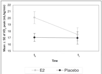
The group of patients receiving estradiol had a decrease in the mean in regard to t0 (P= 0.002) (Figure 1), and the mean difference between t0 and t1 was negative (P= 0.018) (Figure 2). It is worth noting that at t0, the mean VO2 peak of the patients in the estradiol group was greater than that in the placebo group (P= 0.018). The results of the analysis of variance with 2 factors (group and time) and repeated measures in 1 factor (time) for the variable VO2 peak are shown in Table III.
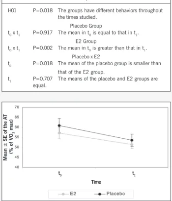
The groups had a decrease in the means in regard to t0
(P= 0.01) (Figure 3). The results of the analysis of variance with 2 factors (group and time) and repeated measures in 1 factor (time) for the variable AT are shown in Table IV.
The 2 groups had very similar behaviors regarding VO2 in the AT. Both groups showed a decrease in the mean in regard to t0 (P< 0.001) (Figure 4).
The estradiol group showed a decrease in the DE after 3 months (P= 0.047). It is worth noting that at t0, the mean of TE of the patients in the estradiol group was greater than that in the placebo group (P= 0.041).
The other variables studied showed no statistically significant changes.
Discussion
Our study aimed at assessing the effects of oral estradiol at the dosage of 2 mg/day on the functional capacity of postmeno-pausal women. Our rationale was that estrogen, favoring vasodi-lation, causes an increase in the muscular oxidative capacity, and, consequently, an increase in functional capacity. Our results, however, were totally different from that which was expected from the physiological point of view. They differed from the results of the study by Redberg et al 18, who reported an increase in the response to exercise – measured by use of VO2 – and in the AT of hormone replacement therapy users.
The results regarding AT, VO2, and load in the AT also did not differ with estradiol use.These results are similar to those reported by Snabes et al 19, McCole et al 20, Redberg et al 18, and Green et al 21. Similarly, estradiol use was supposed to cause an increase in
Fig. 1 - Mean profiles ± standard errors (SE) of VO2 peak in regard to the group
of patients and time of the study. *P=0.018
Mean ± SE of VO
2
peak (mL/kg/min)
t0 t1
Time
Difference between VO
2
peak in t
1
and t
0
(mL/kg/min) mean ± SE
Fig. 2 - Mean ± standard error (SE) of the difference (t0 – t1) of VO2 peak in regard to the group of patients. *p=0.018
1.5
1
0.5
0
-0.5
-1
-1.5
-2
Table I – Distribution of the patients according to the estradiol (E2) and placebo groups
Group Frequency
Absolute Relative
Placeb 11 44%
E2 14 56%
Total 25 100%
Table II - Descriptive statistics of the quantitative variables measured in t0 for characterization of the sample in regard to the group of patients
Variables Group Descriptive statistics P value
Mean Standard deviation Minimum Median Maximum (t test)
Age (years) Placebo 57.55 4.78 51.00 57.00 66.00 0.471
E2 55.79 6.73 47.00 54.50 69.00
Weight (kg) Placebo 63.76 9.57 44.50 62.00 78.90 0.762
E2 62.70 7.81 49.00 62.00 72.70
Height (m) Placebo 1.53 0.07 1.40 1.54 1.62 0.534
4
the maximum load reached during the cardiopulmonary exercise test, which was not observed.
In regard to DBP, we hypothesized that the estradiol group would have a lower exercise peak than would the placebo group, due to the steroid vasodilating response, but this was also not observed. This result is similar to that obtained by Green et al 21.
Table III - Results of the analysis of variance with 2 factors (group and time) and repeated measures with 1 factor (time) for the variable
VO2 peak.
H01 P=0.018 The groups have different behaviors throughout the times studied.
Placebo Group
t0 x t1 P=0.917 The mean in t0 is equal to that in t1.
E2 Group
t0 x t1 P=0.002 The mean in t0 is greater than that in t1. Placebo x E2
t0 P=0.018 The mean of the placebo group is smaller than that of the E2 group.
t1 P=0.707 The means of the placebo and E2 groups are equal.
Table IV - Results of the analysis of variance with 2 factors (group and time) and repeated measures with 1 factor (time) for
the variable AT.
H01 P=0.741 The groups have similar behaviors throughout the times studied.
H02 P=0.362 No difference exists between the placebo and estradiol groups.
H03 P=0.010 The times studied differ.
Times of the study
T0 x t1 P=0.010 The mean in t0 is greater than that in t1.
Fig. 3 – Mean profiles ± standard errors (SE) of AT in regard to the group of patients and the time of the study. *p=0.01 *p=0.01
Mean ± SE of the A
T
(% of VO
2
max)
t0 t1
Time
Difference of the A
T
in t
1
and t
0
(% of VO
2
max) mean ± SE
Fig. 4 - Mean ± standard error (SE) of the difference (t1 – t0) of the AT in regard to the group of patients.
0.00
-2.00
-4.00
-6.00
-8.00
-10.00
-12.00
-14.00
Although our results showed no improvement in the metabolic and cardiopulmonary responses, it is worth noting that the patients in the estradiol group reported an improvement in their major com-plaints, ie, hot flushes, insomnia, and vaginal atrophy. Unques-tionably, estrogen is the drug that best treats these symptoms. The sensation of well-being reported by these patients is noteworthy.
The following possibilities emerged as reasons for our result, which was so different from that which was expected: short-term estradiol use (3 months) and high steroid dosage, which could have impaired the oxidative capacity of the muscle fiber. The decrease in systolic volume and blood flow to the skeletal musculature may be other explanations.
Of all possibilities proposed to explain our results, we believe that the short duration of estradiol use is the best. Maybe a longer exposure to the steroid could trigger better cardiopulmonary and metabolic responses.
The clinical implication of these results is that women under-going estrogen replacement should be instructed that, at least in the first months of steroid therapy, a decrease in their functional capacity for physical activity may occur.
Although our results showed no improvement, we encourage adequate practice of regular exercise. On the contrary, exercise may potentiate the effects of estradiol or even prevent the appea-rance and development of chronic diseases in postmenopausal women. In addition, in patients with established diseases, the promotion of physical activity may improve the prognosis 22.
Another important aspect is the counterregulating effect of regular physical exercise on the functional capacity of postmeno-pausal women, because frequent physical activity is known to increase both muscular oxidative capacity and VO2 1.
Our results also do not invalidate hormone replacement in certain patients, because, in addition to the innumerable benefits to climacteric symptoms, the following cardiovascular effects also occur: direct antiatherosclerotic effects on the arteries; an increase in the catabolism of LDL-cholesterol, and in the number and activity of the receptors of that lipoprotein; an increase in the serum levels of HDL-cholesterol; the antiplatelet effect and vasodilation, which depend on the nitric oxide effect; endothelium-independent vasodilation; inotropic action of the heart and great vessels; an improvement in glycidic metabolism; antioxidant activity; inhibition of vascular smooth muscle cell growth; a favorable impact on fibrinolysis; a reduction in the levels of the renin and angiotensin-converting enzyme; and a reduction in homocysteine levels 1. It is also worth noting the important effect of hormone replacement therapy on bone remodeling, with resorption suppression, allowing bone mass stability or gain, or both, in addition to reducing the risk for Alzheimer’s disease 23-25.
Therefore, the adequate instruction of patients in regard to the possible interventions during climacteric is fundamental. Pro-motion of physical activity unequivocally contributes to improvement in climacteric symptomatology and prevention of osteoporosis, sleep disorders, breast, endometrial, and colon-rectal cancers, and cardiovascular disease26-31.
5
1. Wasserman K, Hansen JE, Sue DY, et al. Principles of Exercise Testing and Inter-pretation. 2nd ed. Pennsylvania: Lea & Febiger, 1994.
2. Yazbek P, Carvalho RT, Sabbag LMS, et al. Ergoespirometria. Teste de esforço car-diopulmonar, metodologia e interpretação. Arq Bras Cardiol 1998;71:719-24. 3. Lieberman EH, Gerhard MD, Uchata A et al. Estrogen improves
endothelium-de-pendent, flow-mediated vasodilatation in postmenopausal women. Ann Intern Med 1994;121:936-41.
4. Gilligan DM, Badar DM, Panza JA, et al. Acute vascular effects of estrogen in post-menopausal women. Circulation 1994;90:786-91.
5. Tze K, Lau MRCOG, Din Wan BS, et al. Prospective, randomized, controlled study of the effect of hormone replacement on peripheral blood flow velocity in postme-nopausal women. Fertil Steril 1998;70:284-8.
6. De Meersman RE, Zion AS, Giardina EG, et al. Estrogen replacement, vascular dis-tensibility, and blood pressures in postmenopausal women. Am J Physiol (USA) 1998;274(5 Pt 2):1539-44.
7. III Consenso Brasileiro de Hipertensão. Campos do Jordão (SP): Sociedade Bra-sileira de Hipertensão 1998.
8. Joint National Committee on prevention, detection, evaluation, and treatment of high blood pressure. The Sixth Report of the Joint National Committee on Preven-tion, DetecPreven-tion, EvaluaPreven-tion, and Treatment of High Blood Pressure (JNC VI). Arch Intern Med 1997;157:2413-46.
9. Menopause Core Curriculum Study Guide, Second Edition. The North American Menopause Society 2002.
10. Keys A, Fidanza F, Karvonen MJ, et al. Indices of relative weight and obesity. J Chron Dis 1972; 25:329-43.
11. Skinner JS, McLellan TH. The transition from aerobic to anaerobic metabolism. Res Q Exerc Sport 1980;51:234-48.
12. Zhang YY, Johnson MC 2nd, Chow N, et al. Effect of exercise testing protocol on
parameters of aerobic function. Med Sci Sports Exerc 1991; 23:625-630. 13. Wasserman K. The anaerobic threshold: definition, physiological significance and
identification. Adv Cardiol (Switzerland) 1986;35:1-23.
14. Bussab WO, Morettin PA. Estatística Básica – Métodos Quantitativos. 4a ed. São
Paulo 1987.
15. Siegel S. Nonparametric Statistics for the Behavioral Sciences. Kogakusha: McGraw-Hill 1956.
16. Andrade DF, Singer JM. Análise de Dados Longitudinais (VII Simpósio Nacional de
References
Probabilidade e Estatística). Campinas. Associação Brasileira de Estatística 1986. 17. Winer BJ. Statistical Principles in Experimental Design. 2nd ed. New York: Mc
Graw-Hill 1971.
18. Redberg RF, Nishino M, McElhinney DB, et al. Long-term estrogen replacement therapy is associated with improved exercise capacity in postmenopausal women without known coronary artery disease. Am Heart J 2000;139:739-744. 19. Snabes MC, Herd JA, Schuyler N, et al. In normal postmenopausal women
phy-siologic estrogen replacement therapy fails to improve exercise tolerance: a rando-mized, double-blind, placebo-controlled, crossover trial. Am J Obstet Gynecol 1996;175: 110-4.
20. McCole SD, Brown MD, Moore GE, et al. Enhanced cardiovascular hemodynamics in endurance-trained postmenopausal women athletes. Med Sci Sports Exerc 2000;32:1073-9.
21. Green JS, Stanforth PR, Gagnon J et al. Menopause, estrogen, and training effects on exercise hemodynamics: the HERITAGE study. Med Sci Sports Exerc 2002; 34: 74-82.
22. Castelli WP. Cardiovascular disease in women. Am J Obstet Gynecol 1988; 158:1553-60.
23. Aldrighi JM, Pires ALR. Climatério/TRH. Reprodução & Climatério, Suplemento 2001;16:24-30.
24. Bjarnason MH, Hassager C, Christiansen C. Postmenopausal bone remodelling and hormone replacement. Climateric. 1998;1:72-79.
25. Paganini – Hill A, Henderson VW. Estrogen deficiency and risk Alzheimer’s disease in women. Am J Epidemiol 1994;140:256261.
26. American College of Sports Medicine – Position Stand – Exercise and physical ac-tivity for older adults. Med Sci Sports Exerc 1998;30:992-1008.
27. American College of Sports Medicine – Position Stand - Exercise for patients with coronary artery disease. Med Sci Sports Exerc 1994;26:1-5.
28. American College of Sports Medicine – Position Stand – Osteoporosis and exercise. Med Sci Sports Exerc 1995;27: 1-7.
29. American College of Sports Medicine – Position Stand – Physical activity, physical and hypertension. Med Sci Sports Exerc 1993;25: 1-10.
30. Kalil LMP, Barreto ACP, Guimarães GV, et al. Capacidade física em idosos subme-tidos a programa de condicionamento físico. Rev Soc Cardiol SP 1996;1:68-76. 31. McTiernan A. Physical activity and the prevention of breast cancer. Medscape
Women’s Health eJournal 2000;5(5). in postmenopausal women by use of plethysmography and study
of sympathetic activity would be of great interest.
In conclusion, estradiol administration for 90 days to