1
Ventricular remodeling is an adaptive response of the heart to hemodynamic, neurohormone stimuli and to genetic factors. It is associated with a modification in the shape, size, composition, and function of the heart 1. Acute myocardial infarction is currently
one of the m ost im porta nt ca uses of ca rdia c rem odeling a nd significantly contributes to ventricular dilation, myocardial fibrosis, ventricular dysfunction, and development of congestive heart failure. Postinfarction remodeling is a phenomenon that begins right after coronary occlusion. In that phase, myocytic necrosis occurs, being followed by stretching of the infarcted area, which constitutes the expansion of the infarction. This may result in muscular rupture or formation of a ventricular aneurysm. Approximately 7 2 hours after the acute event, remodeling encompasses the entire heart. During that period, ventricular dilation, change in geometry, and hypertrophy of the remaining musculature is evident. The incapability of the heart to normalize wall stress results in progressive cardiac dilation, recruitment of the myocardium around the scar, and deterioration of the contractile function 1.
Although myocardial remodeling is a complex phenomenon involving several stimuli, the role of the renin-angiotensin-aldos-terone system has gained special attention in the last decade. From the therapeutic point of view, the benefits of angiotensin-converting-enzyme inhibitors and of AT1 receptor blockers are innumerous 2 -4. However, it has not been well defined whether
the effects of the 2 classes of drugs are comparable in regard to ventricular hypertrophy and interstitial fibrosis in the viable myo-cardium after acute coronary occlusion.
Lisinopril is an angiotensin-converting-enzyme inhibitor widely used in clinical practice for treating arterial hypertension, as well as an adjuvant drug for the treatment of acute myocardial infarction 5 -8. It
inhibits the formation of angiotensin II and aldosterone. That drug has a direct hemodynamic effect on the vessels, reducing the afterload, and a beneficial effect on ventricular remodeling.
Blockade of the angiotensin II activity may also be obtained by use of antagonists of angiotensin type 1 (AT1 ) receptors. Recent studies have shown the beneficial effects of AT1 antagonists on myocardial remodeling after myocardial infarction with improve-ment in cardiac function, as well as an increase in survival 9.
Losartan is an antagonist of angiotensin II receptors. It acts through direct competition with angiotensin II for only one class of receptors (AT1 ), has a significant role in reducing the process of interstitial fibrosis 3, and in reducing the mortality and morbidity associated
with heart failure after acute myocardial infarction 1 0.
Original Article
M yocardial Rem odeling Af t er Experim ent al Acut e
M yocardial Inf arct ion in Rat s. Ef f ect of
Renin-Angiot ensin-Aldost erone Syst em Blockade
Hindalis Ballest eros Epif anio, Leonardo Ant onio M amede Zornof f , Beat riz Bojikian
M at subara, Sergio Albert o Rupp de Paiva, Robert o M inoru Tani Inoue, Luiz Shiguero
M at subara
Bot ucat u, SP - Brazil
Faculdade de Medicina de Botucatu - UNESP Mailing address: Luiz Shiguero Matsubara
Depto. de Clínica Médica, Faculdade de Medicina de Botucatu, UNESP. Rubião Júnior s/n - 1 8 6 1 8 -0 0 0 - Botucatu - SP, Brazil
E-mail: lsmatsu@ cardiol.br Received for publication: 4 /1 6 /0 3 Accepted for publication: 3 /9 /0 4 English version by Stela Maris Costalonga
Objective
To assess the effect of lisinopril and losartan on myocardial remodeling in experimental infarction in rats.
M ethods
Male Wistar rats underwent myocardial infarction and were either treated with lisinopril [20 mg/kg/day (LIS, n= 13)] or lo-sartan [20 mg/kg/day (LOS, n= 11)], or kept without any treat-ment (NT, n= 11) for 3 months. Their results were compared with those of a control group (CONT, n= 11) comprising nonin-farcted rats. After euthanasia, the left ventricle was isolated and weighed. The following measurements were taken: the sec-tional area of myocytes (AC), interstitial collagen fraction (CVF), and myocardial hydroxyproline (HOP). The variables were com-pared by using 1-way ANOVA, with a significance level of P< 0.05.
Results
Ac u t e m yoc ard i al i n f arc t i on c au sed l ef t ven t ri c u l ar hypertrophy. The treatments with lisinopril or losartan could prevent hyper t rophy, whi ch was quant i f i ed by use of l ef t vent ri cul ar wei ght (LOS= 1 .0 6 ± 0 .1 2 g, LIS= 0 .9 7 ± 0 .1 8 g, N T= 1 . 2 6 ± 0 . 1 7 g, CON T= 1 . 0 2 ± 0 . 0 9 g; P< 0 . 0 5 ), of l ef t ventricular weight-to-body weight ratio LV/BW (LOS= 2 .3 7 ± 0 . 2 1 m g/g, LIS= 2 . 4 1 ± 0 . 3 8 m g/g, N T= 2 . 8 2 ± 0 . 3 7 m g/g, CONT= 2.27± 0.15mg/g), and of left ventricular AC measure-ment (LOS= 2 1 0 ± 3 9 µ2, LIS= 2 1 7 ± 3 5 µ2, NT= 2 5 6 ± 3 5 µ2,
CONT= 158± 06 µ2; P< 0.05). The CVF was significantly greater
in the left ventricle of the infarcted group, and its increase was prevented with treatment (LOS= 1.16± 0.4%, LIS= 1.27± 0.5%, NT= 1.8± 0.4%, CONT= 0.7± 0.5%). Myocardial hydroxyproline was greater in the infarcted group (NT= 6 .9 1 ± 2 .9 8 mg/g vs. CONT= 2.81± 1.21mg/g) and did not change with treatment.
Conclusion
Myocardial remodeling after infarction is characterized by an increase in the remaining ventricular mass and in interstitial col-lagen. The angiot ensin-convert ing-enzyme blockers and t he selective angiotensin II AT1 antagonist prevent hypertrophy of the myocyte and interstitial fibrosis.
Keyw ords
2
Myocardial Remodeling After Experimental Acute Myocardial Infarction in Rats. Effect of Renin-Angiotensin-Aldosterone System Blockade This study aim ed at assessing the role of an
angiotensin-converting-enzyme inhibitor (lisinopril) and an AT1 receptor blocker (losartan) in myocardial remodeling in the heart of rats experiencing experimental infarction.
M ethods
All procedures were submitted to and approved by the Ethical Committee on Animal Research and were in accordance with the ethical principles on animal experiments adopted by the Brazilian College of Animal Experiments.
This study used male Wistar rats from the Animal Facility of the University, whose weights ranged from 2 0 0 to 2 5 0 g. The animals were anesthetized with an intraperitoneal injection of sodium pentoba rbita l (5 0 m g/kg) a nd underwent left la tera l thoracotom y. After exteriorization of the heart, the left atrium was pushed aside and the left coronary artery was ligated with a 5 .0 0 mononylon thread between the emergence of the pulmonary artery and the left atrium. Then, the heart was replaced into the thorax, the lungs inflated with positive pressure, and the thoracic wall was sutured with # 1 0 cotton thread. After recovery from anesthesia, the animals were maintained in cages, fed a standard commercial food preparation, with free access to water. Three animals were placed in each cage, with control of light (1 2 -hour cycles), temperature (approximately 2 5 °C), and humidity.
The 3 5 surviving animals were divided into 3 groups: group NT (n= 1 1 ), comprising infarcted animals, which received no me-dication; group LIS (n= 1 3 ), comprising infarcted animals, which received lisinopril (2 0 mg/kg/day dissolved in drinking water); group LOS (n= 1 1 ), comprising infarcted animals, which received losar-tan (2 0 mg/kg/day dissolved in drinking water). The treatment was initiated 1 2 hours after surgery and maintained for 3 months. Noninfarcted animals of the same race, sex, and age, and sub-mitted to the same environmental conditions were used as controls (CONT, n= 1 1 ). After 3 months, the animals were weighed (BW), anesthetized with intraperitoneal injection of sodium pentobarbital (5 0 mg/kg), and underwent euthanasia. Their hearts were removed, dissected, and the right and left ventricles, including the ventricular septum, were isolated and weighed.
Samples of the cardiac tissue were fixed in a solution of 1 0 % formalin for 4 8 hours. After fixation, the tissue was embedded in paraffin blocks, and, then, 4 -micron coronal histological sections were obtained. The histological slides were stained with a solution of hematoxylin and eosin or Masson’s trichrome for assessing the sectional areas of m yocytes (AC). The m yocardial interstitium was assessed on 6 -micron coronal histological sections stained using the picrosirius red technique, specific for visualizing collagen. The histological sections stained with the hematoxylin and eosin solution or Masson’s trichrome were used for measuring the sectional areas of the myocytes with a DM LS LEICA microscope coupled with a video camera, which sends a digital image to a computer that has the Image Pro-plus analysis program (Media Cybernetics, Silver Spring, Maryland, USA). Fifty to 7 0 cells per ventricle analyzed were measured. The myocytes selected were transversely sectioned, had a round shape, a visible central nucleus, and were located in the subendocardial layer of the right and left ventricular muscular wall. This aimed at maximum uniformity of
the sets of myocytes of the different groups. The mean areas obtained for each group were used as an indicator of cell size.
The histological sections stained with the picrosirius red solution were used for assessing the collagen fractions (CVF) present in the interstitium of the cardiac tissues of the right and left ventricles. Reading was performed with polarized light in the LEICA DM LS m icroscope coupled with a video cam era that sends a digital image to a computer that has the Image Pro-plus analysis program (Media Cybernetics, Silver Spring, Maryland, USA). Thirty to 4 0 fields per ventricle were m easured with a 4 0 X objective. The fields chosen were located far from the infarcted area and from the perivascular region.
The biochemical quantification of hydroxyproline (HOP) was performed in a segment of myocardial tissue far from the infarcted area, according to a previously described method 1 1. The method
is based on oxidation of a cardiac tissue hydrolysate in 6 N HCl by chloramine T solution, followed by color obtainment by use of the reaction with the Ehrlich´ s solution and reading in optical density at a wave length of 5 6 5 nm.
The variables studied are presented as means and respective standard deviations, and the comparisons between the groups were perform ed using one-way analysis of variance (ANOVA), followed by the Tukey test, adopting a significance level of P< 0 .0 5 .
Results
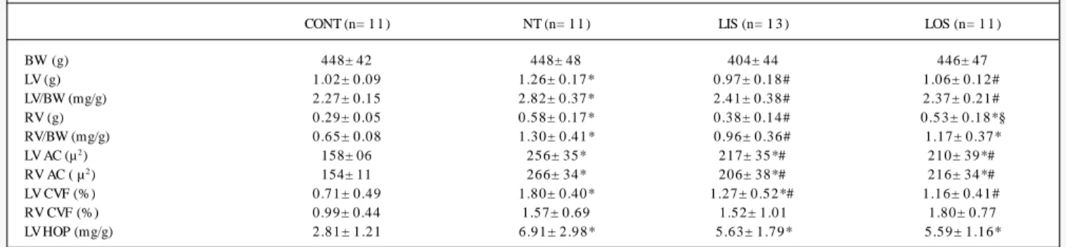
The mean values and respective standard deviations of the body weights, ventricular weights, ventricular weights corrected for body weights, sectional areas of the myocytes, and myocardial concentrations of HOP are shown in table I.
No statistical difference was found in mean body weight (BW) between the groups of infarcted rats 3 months after treatment and the control group. No difference was found in the experimen-tal groups (CONT: 3 6 ± 7 %; LIS: 3 6 ± 6 %; LOS: 3 9 ± 6 %) in regard to the infarcted area. The statistical analysis showed that the mean ventricular weights and the left ventricular weight-to-body weight ratio (LV/BW) were lower in groups LIS, LOS, and CONT when compared with those in group NT. No statistically significant difference was observed in the left ventricular weight and in the LV/BW ratio between the groups LIS and LOS. Acute myocardial infarction led to an increase in right ventricular mass, which was prevented by lisinopril, but not by losartan.
The statistical analysis showed that the sectional areas of the myocytes of the right and left ventricles in groups NT, LIS, and LOS were greater than those in group CONT (P< 0 .0 0 1 ). The areas of the myocytes in groups LOS and LIS were significantly smaller than those in group NT (P< 0 .0 5 ). No significant difference in the sectional area in both ventricles was observed between the groups LOS and LIS.
3
ventricular weight-to-body weight ratio is not a good parameter for assessing myocardial hypertrophy in the presence of infarction. Therefore, depending on the size of the infarcted area, the mea-surement of ventricular weight tends to underestimate the presence of ventricular hypertrophy. Based on the results obtained, we observed that, in the presence of myocardial infarction, the measurement of the cellular area of the myocyte is a better index of ventricular hypertrophy than the parameter of ventricular weight.
The measurement of left ventricular collagen fraction (CVF) showed an increase in interstitial fibrosis in remote areas of in-farction (tab. I, fig. 1 ). In the present study, a smaller CVF was observed in the infarcted groups treated with lisinopril or losartan. These results were similar to those found by other researchers, who reported prevention of interstitial fibrosis in animals treated with angiotensin-converting-enzyme inhibitors or with antagonists of the angiotensin II AT1 receptors 3 ,2 0 -2 2.
Greater levels of right ventricular CVF were found in infarcted rats. However, no significant differences were detected between the groups studied. We believe that the great variability in these data associated with the small size of the sample may have hindered the detection of the statistical difference. We have also observed smaller CVF values than those reported in the literature. The use of linear polarized light filter (fig. 1 ) seems to underestimate the CVF value, suggesting that the ideal method would be the use of a circular polarized light filter 2 3. As the same methodology was used in all
groups, the comparison between them could be established. The HOP value was significantly greater in group NT as com-pared with that in groups CONT, LIS, and LOS. Unlike CVF mea-surement, that of HOP was not sufficiently sensitive to detect the modifications in myocardial fibrosis due to treatment. However, we cannot exclude the possibility of including peri-infarction tissue in HOP quantification, which could falsify the effect of the treat-ment on interstitial fibrosis. Thus, in this model of experitreat-mental infarction and in regard to the analysis of myocardial fibrosis, we can assume that CVF measurement is more reliable than HOP quantification, because direct histological viewing allows the de-finitive exclusion of the scar area.
In conclusion, myocardial remodeling after acute myocardial infarction results mainly from hypertrophy of the remaining myo-cytes and interstitial fibrosis. Postinfarction hypertrophy of myocy-tes and interstitial fibrosis may be prevented by using angiotensin-converting-enzyme inhibitors (lisinopril) or an AT1 receptor anta-gonist (losartan). Further studies are required to establish whether
Discussion
Acute myocardial infarction was accompanied by left and right ventricular hypertrophy as shown by the ventricular weights and m ea surem ents of the cellula r a rea s. The results showed tha t m uscula r growth wa s highly com pensa tory, ie, the rem a ining myocytes had a hypertrophy that exceeded the area lost due to myocardial necrosis. According to the literature, this result suggests that hypertrophy is not due only to the mechanical stimulation of the elevation in the preload or afterload 1 2. Local m echanical
stimuli, through stretching and activation of the mechanotrans-ducers, associated with the inflammatory reaction due to myocytic necrosis cause release of growth factors that stimulate hypertrophy and fibrosis 1 3 ,1 4.
The right ventricle showed no infarcted area detected on optical microscopy, and the presence of right ventricular hypertrophy could be attributed to 2 factors: the mechanical stimulation triggered by pulmonary hypertension and activation of the systemic or local neurohormone system 1 5. After acute myocardial infarction, different
degrees of left ventricular diastolic dysfunction frequently occur. The consequences of that dysfunction are as follows: pulmonary arterial vasoconstriction, and right ventricular overload and hy-pertrophy. This hypertrophy could also result from the trophic action of the neurohormone mediators released after acute myo-cardial infarction, such as angiotensin II, which, by coupling with specific receptors activate intracellular mediators that cause the expression of proto-oncogenes, promoters of protein synthesis 1 6 ,1 7.
In the present study, the treatment of rats after acute myocardial infarction with lisinopril or losartan proved to be effective for pre-venting the increase in ventricular mass. The reductions in the right and left ventricular weights and in the ventricular weight-to-body weight ratio were significant in both measurements as com-pared with those in the nontreated infarcted group, and the para-meters were reduced to levels similar to those in the noninfarcted control group. These results were similar to those obtained by other authors 1 8 ,1 9, who reported a reduction in the ventricular
mass in infarcted hearts of rats treated with angiotensin-converting-enzyme inhibitor or with an antagonist of the angiotensin AT1 receptors. In the groups treated, the measurement of ventricular weight showed lack of mass gain; however, measurement of the m yocytic cellula r a rea s s h owed th e occurrence of m yocytic hypertrophy as compared with that in the noninfarcted control group. Therefore, the measurement of ventricular weight and of
Table I - M eans and standard deviations of the variables studied in the following groups of rats: noninfarcted (CONT). nontreated infarcted (NT). lisinopril-treated infarcted (LIS). and losartan-lisinopril-treated infarcted (LOS).
CONT (n= 1 1 ) NT (n= 1 1 ) LIS (n= 1 3 ) LOS (n= 1 1 )
BW (g) 448± 42 448± 48 404± 44 446± 47
LV (g) 1.02± 0.09 1.26± 0.17* 0.97± 0.18# 1.06± 0.12#
LV/BW (mg/g) 2.27± 0.15 2.82± 0.37* 2.41± 0.38# 2.37± 0.21#
RV (g) 0.29± 0.05 0.58± 0.17* 0.38± 0.14# 0 .5 3 ± 0 .1 8 *§
RV/BW (mg/g) 0.65± 0.08 1.30± 0.41* 0.96± 0.36# 1.17± 0.37*
LV AC (µ2) 158± 06 256± 35* 217± 35*# 210± 39*#
RV AC ( µ2) 154± 11 266± 34* 206± 38*# 216± 34*#
LV CVF (%) 0.71± 0.49 1.80± 0.40* 1 .2 7 ± 0 .5 2 *# 1 .1 6 ± 0 .4 1 #
RV CVF (%) 0.99± 0.44 1.57± 0.69 1.52± 1.01 1.80± 0.77
LV HOP (mg/g) 2.81± 1.21 6.91± 2.98* 5.63± 1.79* 5.59± 1.16*
4
Myocardial Remodeling After Experimental Acute Myocardial Infarction in Rats. Effect of Renin-Angiotensin-Aldosterone System Blockade
1. Sutton MGS, Sharpe N. Left ventricular rem odeling after m yocardial infarction -Pathophysiology and therapy. Circulation 2 0 0 0 ; 1 0 1 : 2 9 8 1 -8 .
2. Dahlof B. Effect of Angiotensin-II Blockade On Cardiac-Hypertrophy and Rem o-deling - a Review. J Hum Hypertens 1 9 9 5 ; 9 : S3 7 -S4 4 .
3. Frim m CD, Sun Y, Weber KT. Angiotensin II receptor blockade and m yocardial fi-brosis of the infarcted rat heart. J Lab Clin Med 1 9 9 7 ; 1 2 9 : 4 3 9 -4 6 .
4. Mill JG, Milanez MD, de Resende MM, Gom es MDS, Leite CM. Spironolactone prevents cardiac collagen proliferation after myocardial infarction in rats. Clin Exp Pharmacol Physiol 2 0 0 3 ; 3 0 : 7 3 9 -4 4 .
5. Brilla CG, Matsubara L, Weber KT. Advanced hypertensive heart disease in spon-taneously hypertensive rats - Lisinopril-m ediated regression of m yocardial fibro-sis. Hypertension 1 9 9 6 ; 2 8 : 2 6 9 -7 5 .
References
6. Hansson L, Lindholm LH, Ekbom T et al. Randomised trial of old and new antihy-pertensive drugs in elderly patients: cardiovascular m ortality and m orbidity the Swedish Tria l in Old Pa tients with Hypertension-2 study. La ncet 1 9 9 9 ; 3 5 4 : 1751-6.
7. Zornoff LAM, Matsubara BB, Matsubara LS, Paiva SAR, Spadaro J. Early rather than delayed administration of lisinopril protects the heart after myocardial infarc-tion in rats. Basic Res Cardiol 2 0 0 0 ; 9 5 : 2 0 8 -1 4 .
8. Wh ite CM. Angiotensin-converting-enzym e inh ibition in h ea rt fa ilure or a fter myocardial infarction. Am J Health-Syst Pharm 2 0 0 0 ; 5 7 : S1 8 -S2 5 .
9. Kots YI, Lebedyantsev LV, Saifutdinov RI, Bobylev VV. The use of angiotensin II receptor blockers for the treatment of left ventricular failure in acute period of myo-cardial infarction. Kardiologiya 2 0 0 1 ; 4 1 : 3 0 -3 .
the tissular response in the infarcted area is different from that in the noninfa rcted a rea for the m echa nica l a nd neurohorm one stimuli, and whether the remodeling response depends on the type of treatment.
Acknow ledgments
We thank Elenize Jamas Pereira, Jose Carlos Georgette, Vitor M. Souza, and Rogerio A. Monteiro for technical support, and FAPESP (n1 0 0 /1 3 7 9 8 process) for financial support.
Fig. 1 - Microphotograph of histological sections of rat myocardium (LV) stained with picrosirius red and analyzed under polarized light. The collagen fibers are shown in bright yellow (arrow) and the muscular fibers in brown.
Nt
Lis
Los
5
10. Sha rm a D, Buyse M, Pitt B, Rucinska EJ. Meta -a na lysis of observed m orta lity data from all-controlled, double-blind, m ultiple-dose studies of losartan in heart failure. Am J Cardiol 2 0 0 0 ; 8 5 :1 8 7 -9 2 .
11. Matsubara LS, Matsubara BB, Okoshi MP, Cicogna AC, Janicki JS. Alterations in m yocardial collagen content affect rat papillary m uscle function. Am J Physiol-Heart Circul Physiol 2 0 0 0 ; 2 7 9 : H1 5 3 4 -H1 5 3 9 .
12. Bolognese L, Cerisano G. Early predictors of left ventricular remodeling after acute myocardial infarction. Am Heart J 1 9 9 9 ; 1 3 8 : S7 9 -S8 3 .
13. Sadoshima J, Izumo S. Mechanotransduction in Stretch-Induced Hypertrophy of Cardiac Myocytes. J Recept Res 1 9 9 3 ; 1 3 : 7 7 7 -9 4 .
14. Suzuki Y, Ruiz-Ortega M, Lorenzo O, Ruperez M, Esteban V, Egido J. Inflammation and angiotensin II. International Journal of Biochemistry & Cell Biology 2 0 0 3 ; 3 5 : 881-900.
15. Anversa P, Leri A, Li B, Liu Y, Di Som m a S, Kajstura J. Ischem ic cardiom yopathy and the cellular reninangiotensin system. J Heart Lung Transplant 2 0 0 0 ; 1 9 : S1 -S11.
16. Unger T, Chung O, Csikos T et al. Angiotensin receptors. J Hypertens 1 9 9 6 ; 1 4 : S9 5 -S1 0 3 .
17. Lorell BH. Role of angiotensin AT(1 ) and AT(2 ) receptors in cardiac hypertrophy and disease. Am J Cardiol 1 9 9 9 ; 8 3 : 4 8 H-5 2 H.
18. Mulder P, Devaux B, Richard V et al. Early versus delayed angiotensin-converting enzym e inh ibition in experim enta l ch ronic h ea rt fa ilure - Effects on surviva l, hemodynamics, and cardiovascular remodeling. Circulation 1 9 9 7 ; 9 5 : 1 3 1 4 -9 . 19. Am brose J, Pribnow DG, Giraud FD, Perkins KD, Muldoon L, Greenberg BH.
An-giotensin type 1 receptor antagonism with irbesartan inhibits ventricular hypertro-phy and improves diastolic function in the remodeling post-myocardial infarction ventricle. J Cardiovasc Pharmacol 1 9 9 9 ; 3 3 : 4 3 3 -9 .
20. Thai HM, Van HT, Gaballa MA, Goldm an S, Raya TE. Effects of AT, receptor blo-ckade after m yocardial infarct on m yocardial fibrosis, stiffness, and contractility. Am J Physiol-Heart Circul Physiol 1 9 9 9 ; 4 5 : H8 7 3 -H8 8 0 .
21. Richer C, Gerva is M, Fornes P, Giudicelli JF. Com bined selective a ngiotensin II AT(1 )-receptor blockade and angiotensin I-converting enzyme inhibition on coro-na ry flow reserve in postischem ic hea rt fa ilure in ra ts. J Ca rdiova sc Pha rm a col 1 9 9 9 ; 3 4 : 7 7 2 -8 1 .
22. Cavasin MA, Yang XP, Liu YH et al. Effects of ACE inhibitor, AT(1 ) antagonist, and combined treatment in mice with heart failure. J Cardiovasc Pharmacol 2 0 0 0 ; 3 6 : 472-80.