493
Original Article
Effects of Oxidized LDL on In Vitro Proliferation
and Spontaneous Motility of Human Coronary
Artery Endothelial Cells
Hermes Toros Xavier, Dulcinéia Saes Parra Abdalla, Tania Leme da Rocha Martinez,
José Antonio Franchini Ramires, Antonio Ricardo de Toledo Gagliardi
São Paulo, SP - Brazil
Instituto do Coração da Faculdade de Medicina da USP and Faculdade de Ciências Farmacêuticas da USP
Mailing address: Hermes Toros Xavier - Av. Bartolomeu de Gusmão, 178/73 - Cep 11030-500 - Santos, SP, Brazil
E-mail: [email protected]
Received for publication: 06/11/2003 Accepted for publication: 04/14/2004 English version by Stela Maris Costalonga
Objective
To investigate the effects of low concentrations of oxidized LDL (oxLDL) on the proliferation and spontaneous motility of human coronary artery endothelial cells (HCAEC) in culture.
Methods
Cultures of HCAEC were treated with low concentrations of native LDL (nLDL) isolated from human plasma and with LDL minimally oxidized through different chemical methods; the effects were compared.
Results
Native LDL had no deleterious effects on in vitro proliferation and motility of HCAEC; however, at its highest concentration and for a longer exposure, nLDL inhibited cell proliferation. The LDL chemically oxidized by spermine nonoate (SNO) and 3-morpholinylsydnonimine (SYN-1) had significant inhibiting effects on in vitro proliferation and motility of HCAEC, which were proportional to the greatest concentrations and degrees of oxidation of LDL.
Conclusion
OxLDL has a cytotoxic effect, inhibiting the proliferation and spontaneous motility of HCAEC in culture. This effect is propor-tional to the concentration and degree of oxidation of LDL; native LDL is relatively innocuous.
Key words
oxidized LDL, coronary artery endothelial cells, cardiovascular atherosclerotic disease
High plasmatic levels of low-density lipoprotein (LDL) are con-sidered one of the major risk factors for the development of car-diovascular atherosclerotic disease. The hypothesis of response to injury to explain atherosclerosis proposes that the first step in atherogenesis is endothelial dysfunction induced by the action of risk factors, especially the exposure of the vascular endothelium
to oxidized LDL (oxLDL) 1.
Preliminary studies revealed that preparations of isolated human
LDL have toxic effects on endothelial cells in culture 2-4. Native
LDL (nLDL) undergoes chemical alterations related to peroxidation of polyunsaturated fatty acids, components of the lipoprotein, which result in a great increase in its susceptibility to phagocytosis and
break down by macrophages 5.
The major types of vascular wall cells, the endothelial cells, the smooth muscle cells, and the macrophages were shown in
vitro to be able to oxidize native LDL6. Oxidized LDL inhibits
endo-thelial cell migration, which is an essential mechanism in the processes of reestablishing vascular integrity after injury and an-giogenesis, proportionally to the concentration and degree of
oxi-dation of LDL, mediated by the formation of lipid hydroperoxides7.
Recent studies have shown that the lipid components of oxLDL 8,9
can cause a paradoxical increase in the production of vascular endothelial growth factor (VEGF) by endothelial cells in culture, which may be a protective mechanism in face of cellular injury. OxLDL was observed to have a double effect on cell cycle: to induce
cell proliferation at low concentrations (5 to 10 µg/mL), and apoptosis
at concentrations above 50 µg/mL10.
The endothelial cells constitute a heterogeneous population. Endothelial cells originating from different vascular beds express characteristic surface antigens, specific intracellular transporters,
and regulation of different intracellular enzymes 11.
Considering the importance of endothelial cell heterogeneity and the contradictory data in the literature, and aiming at contri-buting to a better understanding of the basic processes involved in the interaction between oxLDL and endothelial cell, we carried out the first systematized study using human coronary artery endo-thelial cells (HCAEC) in culture, whose role in coronary atheroge-nesis is primordial.
494
Methods
This study was performed with the cell line of human coronary artery endothelial cells obtained through CLONETICS (BioWhittaker Inc., Walkersville, MD, USA). The culture medium used was MCDB-131 (GIBCO) with the addition of the following: EGF (INTERGEN),
10µg/mL; hydrocortisone (SIGMA), 1.0 µg/mL; amphotericin B
(GIBCO), 50 µg/mL; penicillin, 100 U/mL; streptomycin (GIBCO),
100 U/mL; and 10% fetal calf serum (GIBCO) with 1 µM
L-glutamine (GIBCO). The experiments were performed in culture media with 5% fetal calf serum.
Cell count for assessing cell proliferation and migration was performed under direct microscopy (Nikon-TMS) with a 100X magnification in 3 different representative microscopic fields in each culture plate. The mean of the visualized cells was calculated. The assay of cell migration was performed according to the
model of in vitro healing established by Burk 12. Briefly, the
endo-thelial cells were cultivated in 60-mm culture plates (FALCON) with 5 mL of culture medium and 10% fetal calf serum. Two days after the culture became totally confluent, we created a lesion in the unicellular confluent layer with the aid of a razor blade pressed against the floor of the culture plate, cutting the cell layer and marking the plate. The razor blade was then carefully moved to the side, removing part of the unicellular layer. The cells were twice washed with PBS for removing the cell material pushed aside. Experimental culture medium (5% fetal calf serum) supple-mented with variable concentrations of nLDL and oxLDL was added. Four culture plates were used as control or experimental, for each phase of the experiment.
After 24 hours of incubation, the number of cell nuclei that crossed the line demarcated in the culture plate was counted, in 3 distinct microscopic fields representative of each culture plate. The mean number of cells that surpassed the demarcated line was calculated.
nLDL was extracted from human plasma and purified according
to the method reported by Sevanian et al 13. nLDL was stored at
a temperature lower than -70ºC until its use.
The LDL samples (0.5 µg/mL of protein) underwent oxidation
with 1.0 mM of 3-morpholinylsydnonimine (SYN-1) or 1.0 µM of
spermine nonoate (SNO) (SIGMA). The samples were incubated in a hot-water bath (37°C) under constant agitation at the following times: 1, 5, 10, 15, 30, and 60 minutes. The reaction was blocked with 100 mM of diethylene tetramine pentacetic acid, 100 mM of butylhydroxytoluene, 125 units/mL of superoxide dis-mutase, and 125 units/mL of catalase. All samples were
maintai-ned in the freezer at -20°C until the time of the experiments 14.
In statistical analysis, for comparing the concentrations, the Kruskal-Wallis nonparametric test was used, because it is consi-dered the most efficient test for independent samples, and the
Dunn test was used for multiple comparisons 15. Nonparametric
tests were used because the supposition of data normality was rejected. The significance level adopted for the tests was 5%.
Results
Figure 1 shows the effect of nLDL preparations on cell prolife-ration in partially confluent (60%) cultures of human coronary
artery endothelial cells. After 72 hours of exposure to varied concen-trations of lipoproteins, the cultures were washed twice with PBS (GIBCO) and stained according to the Giemsa method. Cell count was performed under microscopy (NIKON-TMS) with a 100X magnification in 3 different representative microscopic fields for each culture plate, and the mean number of cells was calculated. An inhibiting effect of nLDL was observed on cell proliferation at the maximum concentration used, which may be attributed to the longest exposure and oxLDL by the endothelial cells in culture.
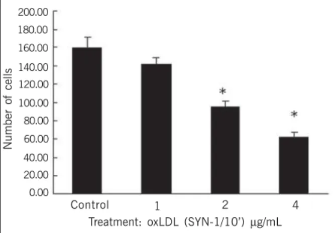
Figures 2 and 3 depict the effects of LDL oxidation by the SNO system for 1 and 10 minutes, respectively, on cell proliferation. An inhibitory effect proportional to the duration of oxidation is observed in both figures.
Figures 4 and 5 show the important inhibitory effects of oxLDL by the SYN-1 system for 1 and 10 minutes on cell proliferation. Briefly, the results of the treatment of partially confluent en-dothelial cell cultures for 72 hours with nLDL and oxLDL by the SNO and SYN-1 chemical systems on cell proliferation were as follows: oxLDL by SNO and SYN-1 had an inhibitory effect on cell proliferation proportional to the duration of oxidation and to the concentration of oxLDL; LDL oxidized by SYN-1 had a more intense toxic effect than the other treatments did; nLDL had an inhibitory effect only at the greatest concentration studied.
Number of cells
160.00
Control
140.00
120.00
100.00
80.00
60.00
40.00
20.00
0.00
1 2 4 8
Treatment: nLDL (µg/mL)
Fig. 1 - Effect of various concentrations of native LDL during 72 hours on the number of cells per microscopic field. Control differs from the treatment with 8µg/mL *(P < 0.05).
Number of cellss
60.00
Control
50.00
40.00
30.00
20.00
10.00
0.00
1 2 4 8
Treatment: oxLDL (SNO/1’)µg/mL
495
Figure 6 shows the effect of the nLDL preparations on thespon-taneous motility of human coronary artery endothelial cells accor-ding to the cell migration assay performed using the Burk technique. No inhibitory effect of nLDL on cell migration was observed.
Figures 7 and 8 show the effects of oxLDL by SYN-1 for 1 and 10 minutes, respectively. The effects were directly related to the concentration and duration of oxLDL.
Figure 9 is a microphotography representing the experiment of cell motility. A clearly smaller number of cell nuclei crossed the demarcated line in the treated field (B) as compared with that in the control field (A). This shows inhibition of cell migration.
Briefly, the effects of the treatment of endothelial cell cultures with native and SYN-1 system oxLDL for 24 hours on cell mo-tility, according to the Burk technique, were as follows: nLDL had no inhibitory effect on cell migration; SYN-1 oxLDL inhibited cell migration; the effect observed was proportional to the duration of oxidation and the concentration of oxLDL.
Discussion
Recent studies have shown that the products of cholesterol oxidation generated by the oxidative modification of LDL, the cho-lesterol hydroperoxides, provide cytotoxicity to the particle, are re-levant to the pathogenesis and progression of atherosclerosis, and
have been identified in atheroma plaques and human plasma 16,17.
The molecular mechanisms that initiate in vivo oxidation of LDL are yet to be identified; however, some chemical substances, such as peroxynitrite, participate effectively in the process of LDL
modification, the lipid peroxidation 18. The method of LDL chemical
oxidation through 2 systems, SYN-1 and SNO, was used. SYN-1 is known as a generator of peroxynitrite, and its use in the
expe-Number of cells
70.00
Control
60.00
50.00
40.00
30.00
20.00
10.00
1 2 4 8
Treatment: oxLDL (SNO/10’)µg/mL
Fig. 3 - Effect of various concentrations of oxidized LDL (SNO/10’) during 72 hours on the number of cells per microscopic field. Control differs from the treatments with 2, 4, and 8 µg/mL *(P < 0.05).
0.00
Number of cellss
70.00
Control
60.00
50.00
40.00
30.00
20.00
10.00
1 2 4 8
Treatment: oxLDL (SYN-1/1’)µg/mL
Fig. 4 - Effect of various concentrations of oxidized LDL (SYN-1/1’) during 72 hours on the number of cells per microscopic field. Control differs from the treatments with 1, 2, 4, and 8 µg/mL *(P < 0.05).
0.00
Number of cells
120.00
Control
100.00
80.00
60.00
40.00
20.00
0.00
1 2 4 8
Treatment: oxLDL (SYN-1/10’)µg/mL
Fig. 5 - Effect of various concentrations of oxidized LDL (SYN-1/10’) during 72 hours on the number of cells per microscopic field. Control differs from the treatments with 1, 2, 4, and 8 µg/mL *(P < 0.05).
Number of cells
250.00
Control
200.00
150.00
100.00
50.00
0.00
1 2 4
Treatment: nLDL (µg/mL)
Fig. 6 - Effect of various concentrations of native LDL on the number of cells that migrated beyond the demarcated line. No significant difference was observed between the treatments (P=0.8920).
Number of cells
160.00
Control
140.00
120.00
100.00
80.00
60.00
1 2 4
Treatment: oxLDL (SYN-1/1’) µg/mL
Fig. 7 - Effect of various concentrations of oxidized LDL (SYN-1/1’) on the number of cells that migrated beyond the demarcated line. The treatment with 4 µg/mL differs from those with other concentrations and from control *(P < 0.05).
40.00
20.00
496
Fig. 9 - Microphotography of cell fields representing cell migration. A: control; B: treatment: OxLDL (SYN-1/ 10’); (40X magnification).
A B
riments may be justified because the process is slow and constant, and, therefore, more similar to the path of peroxynitrite formation in physiological situations. SYN-1 has a high capacity of oxidation,
because it oxidizes both the protein and lipid portions of LDL 19,20.
On the other hand, SNO, generator of nitric oxide, has a low oxidation capacity, because it oxidizes only the lipid portion of LDL. Therefore, the generation of products of oxidation by the 2 systems produces an LDL particle with different degrees of oxidation
in different sites of the molecule 14.
The concentrations of oxLDL commonly used in the experiments
(25 to 100 µg/mL) are considerably more elevated than the levels
of oxLDL found in the human plasma (0.1 µg/mL)21,22. It is worth
nothing, however, that, in atherosclerotic lesions, oxLDL is located and concentrated in the subendothelial space, because it is retained in the extracellular matrix during the formation of the atheroma plaque. Therefore, large quantities of oxLDL, in much higher concen-trations than the plasmatic ones, accumulate in that region. The LDL retained in that microenvironment, ideal for oxidation, is more intensely oxidized and has characteristics similar to those of LDL
oxidized by the different systems used in experimental studies 23.
In this study, we chose to use low concentrations (1 to 8 µg/
mL) of LDL aiming at obtaining values closer to those of the real physiological situation, and, therefore, at investigating the biological effects of LDL on cell cultures.
Experimental data obtained with endothelial cells originating from a certain organ should not be automatically extrapolated to other systems, due to the heterogeneity of that cell line. A
consi-Number of cells
200.00
Control
180.00
160.00
140.00
120.00
100.00
1 2 4
Treatment: oxLDL (SYN-1/10’) µg/mL
Fig. 8 - Effect of various concentrations of oxidized LDL (SYN-1/10’) on the number of cells that migrated beyond the demarcated line. Control differs from the treatments with 2 and 4 µg/mL *(P < 0.05).
80.00
60.00
40.00
20.00
0.00
derable part of the information in the literature about the complex mechanisms involved in human atherosclerosis and angiogenesis was obtained with endothelial cells of the umbilical cord, which would, therefore, require certain care in interpreting the results. The endothelium of the umbilical cord is submitted to extremely high concentrations of the steroid hormones and low pressure regimen, differently from the endothelial bed that participates in
the coronary atherosclerotic process 11. Thus, in our study, we
chose to use the human coronary artery endothelial cell line. Totally confluent endothelial cell cultures may serve as a model for in vitro studies of the vascular endothelium, because the basic properties of the endothelial cell are preserved. The cells in partially confluent culture (60%) are proliferating, and in that phase they are much more sensitive to adverse agents than in the state of plain integrity of total confluence.
In our experiments, oxLDL inhibited in vitro proliferation and motility of human coronary artery endothelial cells, effects di-rectly related to the concentration and degree of oxidation of the lipoprotein. Our results may suggest the occurrence of similar effects of the oxLDL present in vascular walls, atherosclerotic plaques, and even circulating in the plasma, reinforcing the data in the literature that indicate the participation of oxLDL in
cardio-vascular events 24,25.
Recently, elevated oxLDL plasma levels were shown for the first time to directly relate to plaque instability in atherosclerotic lesions of human coronary arteries. OxLDL levels were measured in patients with acute myocardial infarction, unstable angina, stable angina, and controls, revealing a positive correlation with the severity of acute coronary syndrome. The serum levels of oxLDL were 4 times more elevated in patients with acute myocardial infarction when compared with those of controls, suggesting that circulating oxLDL may be a marker of severity in cardiovascular
events26.
Our results indicate that oxLDL concentrations similar to those
found in the acute phase of coronary syndromes inhibit the in
vitro proliferation and motility of human coronary artery endothelial cells. Considering that, one may suggest that elevated levels of oxLDL, due to its cytotoxicity, may negatively interfere not only with instability of the atherosclerotic plaque, but also with the reestablishment of the postinjury vascular integrity, worsening the prognosis of the patients.
497
1. Ross R, Glonset JA. The pathogenesis of atherosclerosis. N Engl J Med 1976;295:369-377.
2. Henriksen T, Evensen SA, Carlander B. Injury to human endothelial cells in culture induced by low density lipoproteins. Scand J Clin Lab Invest 1979; 39:361-8. 3. Henriksen T, Evensen SA, Carlander B. Injury to cultured endothelial cells in culture
induced by LDL: protection by HDL. Scand J Clin Lab Invest 1979; 39:369-75. 4. Hessler JR, Robertson AL, Chisolm GM. LDL-induced cytotoxicity and its inhibition
in human vascular smooth muscle and endothelial cells in culture. Atherosclerosis 1979; 32:213-29.
5. Morel DW, Dicorleto PE, Chisolm GM. Endothelial and smooth muscle cells alter low density lipoprotein in vitro by free radical oxidation. Arteriosclerosis 1984; 4:357-64.
6. Steinbrecher UP, Parthasarathy S, Leake DS et al. Modification of LDL by endo-thelial cells involves lipid peroxidation and degradation LDL phospholipids. Proc Natl Acad Sci USA 1984; 81:3883-7.
7. Murugesan G, Chisolm GM, Fox PL. Oxidized low density lipoprotein inhibits the migration of aortic endothelial cells in vitro. J Cell Biol 1993; 120:1011-19. 8. Dulak J, Jozkowicz A, Dichtl W et al. VEGF synthesis in vascular smooth muscle
cells is enhanced by 7-ketocholesterol and lysophospha tridylcholine independently of their effect on nitric oxide generation. Cardiovasc Res 2002; 5(3):487-8. 9. Dulak J, Jozkowicz A. Regulation of vascular endothelial growth factors synthesis
by nitric oxide: facts and controversies. Antiox Redox Signal 2003; 5(1):123-32. 10. Galle J, Heinloth A, Wanner C et al. Dual effect of oxidized LDL on cell cycle in hu-man endothelial cells trough oxidative stress. Kidney Int Suppl 2001; 78:s120-3. 11. Ribatti D, Nico B, Vacca A et al. Endothelial cell heterogeneity and specificity. J of
Hematotherapy & Stem Cell Research 2002; 11:81-90
12. Burk RR. A factor from a transformed cell line that affects cell migration. Proc Natl Acad Sci USA 1973; 70:368-372.
13. Sevanian A, Bittolo-Bon G, Cazollato G et al. LDL is a lipid hydroperoxide-enri-ched circulating lipoprotein. J Lipid Res 1997; 38:419-28.
14. Oliveira JMA. Efeito da suplementação com vitamina E sobre os níveis de LDL ele-tronegativa (LDL-), de auto-anticorpos anti-LDL oxidada e marcadores da modi-ficação oxidativa de lipídios e proteínas em pacientes ateroscleróticos. São Paulo,
References
2002. xxxp. Dissertação (Mestrado) – Faculdade de Ciências Farmacêuticas, Uni-versidade de São Paulo.
15. Rosner B. Fundamentals of Biostatistics - Boston, PWS Publishers, Second edition, 1986.
16. Crisby M, Kallin B, Thyberg J et al. Cell death in human atherosclerotic plaques involves both oncosis and apoptosis. Atherosclerosis 1997; 130(1-2):17-27. 17. Yasunobu Y, Hayashi K, Shingu T et al. Coronary atherosclerosis and oxidative
stress as reflected by autoantibodies against oxidized low-density lipoprotein and oxysterols. Atherosclerosis 2001; 155(2):445-53.
18. Eiserich JP, Patel RP, O’Donnell VB. Pathophysiology of nitric oxide and related spe-cies: free radical reactions and modification of biomolecule. Molec Aspect Med 1998; 19:221-357.
19. Feelisch M, Ostrowski J, Noack E. On the mechanism of NO release from sydnoni-mine. J Cardiovasc Pharmacol 1989; 14(suppl. 11):13-32.
20. Thomas S, Davies MJ, Stocker R. Oxidation and antioxidation of human low-density lipoprotein and plasma exposed to 3-morpholinoydnonimine and reagent peroxynitrite. Chem Res Toxicol 1998; 11:484-94.
21. Itabe H, Yamamoto H, Imanaka T et al. Sensitive detection of oxidatively modified low density lipoprotein using a monoclonal antibody. J Lipid Res 1996; 37:45-53. 22. Holvoet P, Donck J, Landeloos M et al. Correlation between oxidized low density lipoproteins and von Willebrand factor in chronic renal failure. Thromb Haemost 1996; 76:663-9.
23. Yla-Herttuala S, Palinski W, Rosenfeld ME et al. Evidence for the presence of oxi-datively modified low density lipoprotein in atherosclerotic lesions of rabbit and man. J Clin Invest 1989; 84:1086-95.
24. Chen CH, Henry PD. Atherosclerosis as a microvascular disease: impaired angio-genesis mediated by suppressed basic fibroblast growth factor expression. Proc Assoc Am Physicians 1997; 109:351-61.
25. Bucal M, Nguy J, Barrios R et al. Impaired adaptative vascular growth in hyper-cholesterolemic rabbit. Atherosclerosis 1998; 139:243-51.