Arq Bras Cardiol 2003; 81: 199-201.
Jacob & Salis Anomalous origin of the left coronary artery from the pulmonary trunk
1 9 9
Instituto de Moléstias Cardiovasculares (IMC) - São José do Rio Preto Mailing address: José Luiz Balthazar Jacob – IMC Rua Castelo D’Água, 3030 -15015-210 - São José do Rio Preto, SP, Brazil – E-mail: [email protected]
Arq Bras Cardiol, volume 81 (nº 1), 199-201, 2003
José Luiz Balthazar Jacob, Fernando Vilela Salis
São José do Rio Preto, SP - Brazil
Anomalous Origin of the Left Coronary Artery From the
Pulmonary Trunk in a 45-Year-Old Woman
Case Report
We report a rare case of anomalous origin of the left coronary artery from the pulmonary trunk in a 45-year-old woman. The approach and technique used for selective ca-theterization of an anomalous left coronary artery arising from the pulmonary trunk are described. Six years after diagnosis, echocardiography showed left ventricular dis-function, and surgical treatment was indicated again. The origin of the left coronary artery from the pulmonary trunk was closed, and the postoperative period was uneventful, with recovery of left ventricular function and disappea-rance of ischemic features on stress myocardial perfusion imaging with 99m Tc-sestamibi, performed 4 weeks after surgery.
Origin of the left coronary artery from the pulmonary trunk is a rare congenital anomaly, usually detected in infan-cy due to myocardial ischemia 1. Mortality is 85% during in-fancy, but some patients may present with the condition as adults 2,3. The diagnosis is suspected by clinical history, electrocardiographic features, ischemia revealed in exercise treadmill testing or stress myocardial perfusion imaging, but the diagnosis is really established by angiographic study. Currently, color Doppler flow mapping can make and simpli-fy the diagnosis in some cases. We report a case of this anomaly in a 45-year-old woman, diagnosed by coronary _arteriography, including selective catheterization of left coronary artery.
Case Report
A 45-year-old white woman was initially evaluated in 1972, due to palpitations. At the time, her physical examina-tion and chest radiogram were normal and her electrocardio-gram revealed an absent r wave in V1 and a small r in V2.
Se-ven years later, during pregnancy, the patient experienced atypical chest pain and mild dyspnea. The electrocardio-gram showed the same features, and the echocardioelectrocardio-gram disclosed a prolapsed mitral valve without regurgitation. The symptoms disappeared spontaneously. In 1981, the pa-tient experienced lymphatic leg edema that was followed by an angiologist. The patient did not have any cardiac symp-toms until 1991, when she again experienced palpitations. The electrocardiogram and echo-Doppler cardiogram sho-wed the same features described above, but an exercise treadmill test (Bruce protocol) revealed 2-mm ST depression after 9 minutes, and supraventricular ectopic beats. Chest pain was absent and these features were attributed to mitral valve prolapse. Propranolol 40 mg twice a day was started. In 1994, she was asymptomatic, but a new exercise treadmill test showed the same features described above. Left ventri-cular function was normal on the echocardiogram, with no abnormal echogenicity of the walls or papillary muscles. No abnormal flow to the pulmonary trunk was visible by the color Doppler mapping.
cathe-2 0 0
Jacob & Salis
Anomalous origin of the left coronary artery from the pulmonary trunk
Arq Bras Cardiol 2003; 81: 199-201.
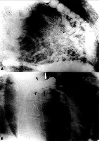
ter, which was advanced to the right atrium and ventricle, and arrived at the pulmonary artery across the pulmonary valve. The catheter tip was anchored in the posterior wall of the pulmonary trunk, just above the valve, and the catheter was forced into the pulmonary trunk, making a large loop and increasing the curvature of the tip. After this, the cathe-ter was slowly withdrawn with councathe-terclockwise rotation, allowing the selective catheterization of the left coronary artery. The selective injection in the left coronary showed a dilated short left main, small marginal branch of the left circumflex artery, and dilated proximal left anterior descen-ding artery, followed by a narrow short segment with a con-trast medium flow arrest, because the collateral supply was greater than the real coronary blood flow (fig. 2B). Surgical treatment was indicated, but not performed because the patient refused to have surgery. Six years after the diagno-sis, an echocardiogram revealed that the ejection fraction of the left ventricle had declined, and surgical treatment was indicated again. This time the patient agreed. The origin of the left coronary artery from the pulmonary trunk was clo-sed and the postoperative period was uneventful. Four weeks after surgery, left ventricular function was normal and stress myocardial perfusion imaging, performed with the same technique and same physical stress previously descri-bed, was normal (fig. 1C and D).
Discussion
Origin of the left coronary artery from the pulmonary artery, or Bland-White-Garland syndrome, is a rare anomaly that accounts for 0.5% of congenital malformations 2. It is fre-quently lethal in children and adults 3. Infants develop con-gestive heart failure due to myocardial ischemia and die 4. Those rare patients who survive to adulthood may be asymptomatic, if collateral circulation is adequate. In older asymptomatic children or adults, signs of infarction or ischemia are absent on the electrocardiogram 4. In our case, the pattern of r wave in V1 and V2 was a sign of ischemic injury in the past. Echocardiographic diagnosis of mitral valve prolapse misled the interpretation of the first stress electrocardiogram test. Although a minimal shunt was shown by angiography, an error occurred in our color Doppler study, because a report in the literature described
the accuracy of this method for detection of unsuspected abnormal flow to the pulmonary trunk, and the flow direction in the left coronary artery 5. After a stress myocardial perfu-sion test showing ischemic features was performed, coro-nary arteriography, which is the most accurate diagnostic method for this anomaly, was performed 6. In our patient, the cinecoronariographic features on the right coronary artery and collateral supply to the left coronary were the same described in other reports 1-3. Normal left ventriculogram and normal echocardiographic features of the left ventricle were in disagreement with the fixed apical defect shown in stress myocardial perfusion imaging, suggesting a viable myocardial area. Most clinicians recognize the need for sur-gical treatment, due to reports of recurrent arrhythmias, car-diac failure, or sudden death 3,7. Various surgical appro-aches have been described 1,3,5,8. Frequently, the conventio-nal angiographic study (right coronary arteriography or angiography in the pulmonary trunk) does not show the correct anatomy of the left coronary artery, due to opacifi-cation of large “collateral-like” vessels. For this reason, the surgical technique is frequently decided during the surgery, Fig. 1- A and B) Exercise (ex) and rest SPECT myocardial perfusion imaging with
99m Tc-sestamibi. On the exercise short-axis and horizontal long-axis slices, an ex-tensive anterior myocardial perfusion defect is present that shows only minimal re-versibility at rest; C and D) exercise and rest SPECT myocardial perfusion imaging with 99m Tc-sestamibi after the surgical procedure showing normal distribution of 99m Tc- sestamibi on the short-axis and horizontal long-axis slices.
A
B
Arq Bras Cardiol 2003; 81: 199-201.
Jacob & Salis Anomalous origin of the left coronary artery from the pulmonary trunk
2 0 1
increasing mortality and morbidity. Patients who died suddenly after different surgical approaches have been described, although several authors suggest that the post-operative deaths were caused by prepost-operative myocardial dysfunction 3. Of the 7 patients reviewed by Purut et al 1, only 1 died suddenly. Rarely, an asymptomatic adolescent or adult with this anomaly who presumably has had adequa-te collaadequa-teral circulation dies suddenly 5. Most of the patients who were operated on in adulthood had symptoms, either arrhythmias or ischemic signs, on the electrocardiogram, and myocardial dysfunction or papillary muscle damage with mitral regurgitation 2,3,7. We believe that the selective angiographic study is necessary, because it allows analysis of left coronary anatomy, flow balance between the left coro-nary and collateral vessels, and a more correct planning of the surgical approach. The catheterization, using the femo-ral vein approach, of coronary arteries arising from the aorta in children with congenital heart diseases has been pre-viously described 9 and is a simple procedure, because in
References
1. Purut CM, Sabiston DC. Origin of the coronary artery from the pulmonary artery in older adults. J Thorac Cardiovasc Surg 1991; 102: 566-70.
2. Saeed BT, Rosin MD, Murray RG. Successful operation in an old survivor of anomalous origin of the left coronary artery from the pulmonary trunk (Bland-White-Garland Syndrome). Br Heart J 1994; 71: 193-5.
3. Moodie DS, Fyfe D, Gill CC, et al. Anomalous origin of the left coronary artery from the pulmonary artery (Bland-White-Garland Syndrome) in adult patients: Long-term follow-up after surgery. Am Heart J 1983; 106: 381-8.
4. Takahashi M, Lurie PR. Abnormalities and diseases of the coronary vessels. In: Adams FH, Emmanouilides GC, Reimenschneider TA, eds. Heart Disease in Infants, Children and Adolescents, 4th ed. Baltimore: Williams and Wilkins,
1989: 627-35.
5. Houston AB, Pollock JCS, Doig WB, et al. Anomalous origin of the left coronary artery from the pulmonary trunk: elucidation with colour Doppler flow mapping. Br Heart J 1990; 63: 50-4.
6. Freedom RM, Culhem JAG, Moes CAF. Anomalies of the coronary arteries. In:
Freedom RM, Culhem JAG, Moes CAF, eds. Angiocardiography of Congenital Heart Diseases. New York: Macmillan, 1984: 405-11.
7. Wesselhoeft H, Fawcett JS, Johnson AL. Anomalous origin of the left coronary artery from the pulmonary trunk. its clinical spectrum, pathology, and pathophysiology. Based on a review of 140 cases with seven further cases. Circulation 1968; 38: 403-25. 8. Oliveira SA, Diament J, Carvalho VB, Arie S, Macruz R, Zerbini EJ. Anomalous origin of the left coronary artery from the pulmonary artery. Surgical repair of on unused form. J Cardiovasc Surg 1977; 18: 599-605.
9. Loya YS, Pinto RJ, Desai DM, Sundaram U, Bhagwat AR, Sharma S. Selective coronary angiography via antegrade venous route in congenital cyanotic heart disease. Cathet Cardiovasc Diagn 1993; 28: 179-82.
10. Jacob JLB. Selective catheterization of the anomalous left coronary artery arising from the pulmonary trunk. Cathet Cardiovasc Diagn 1996; 38: 102. 11. Vigneron M, Ninet J, Bernard Y, Nony P, Beaune J, Champsaur G. Abnormal
origin of the left coronary artery in the adult. Scintigraphic and surgical correlations. Arch Mal Coeur Vaiss 1991; 84: 113-16.
children the aorta is small in diameter. But difficulties exist for selective catheterization of the left coronary artery arising from the pulmonary trunk in adults, due to the large trunk diameter and the curve of the catheter at the tricuspid valve. The selective catheterization technique above described was first reported by Jacob 10. Surgery is always indicated for treatment of anomalous origin of the left coronary artery from the pulmonary trunk. The efficacy of the surgery with reimplantation of the left coronary artery in the aorta 8 or by internal mammary artery-left anterior descending artery by-pass are confirmed by myocardial scintigraphy 11.