online | memorias.ioc.fiocruz.br
Trypanosoma cruzi: ancestral genomes and population structure
Sérgio DJ Pena/+, Carlos Renato Machado, Andréa Mara Macedo
Departamento de Bioquímica e Imunologia, Universidade Federal de Minas Gerais, Av. Antônio Carlos 6627, 31270-901 Belo Horizonte, MG, Brasil
Although the genome of Trypanosoma cruzi has been completely sequenced, little is known about its population structure and evolution. Since 1999, two major evolutionary lineages presenting distinct epidemiological charac� teristics have been recognised: T. cruzi I and T. cruzi II. We describe new and important aspects of the population structure of the parasite, and unequivocally characterise a third ancestral lineage that we propose to name T. cruzi
III. Through a careful analysis of haplotypes (blocks of genes that are stably transmitted from generation to genera� tion of the parasite), we inferred at least two hybridisation events between the parental lineages T. cruzi II and T. cruzi III. The strain CL Brener, whose genome was sequenced, is one such hybrid. Based on these results, we pro�Based on these results, we pro� pose a simple evolutionary model based on three ancestral genomes, T. cruzi I, T. cruzi II and T. cruzi III. At least two hybridisation events produced evolutionarily viable progeny, and T. cruzi III was the cytoplasmic donor for the resulting offspring (as identified by the mitochondrial clade of the hybrid strains) in both events. This model should be useful to inform evolutionary and pathogenetic hypotheses regarding T. cruzi.
Key words:Trypanosoma cruzi - Chagas disease - genome - sex - evolution - pathogenesis
Two major anniversaries will dominate the biomedi-cal sciences in Brazil in 2009: the bicentennial of the birth of Charles Darwin (and 150 year anniversary of the publication of “On the Origin of Species”) and the 100th anniversary of Carlos Chagas’ first report of the disease that carries his name. There is one nexus between these two great scientists; Adler (1959) and others after him proposed that Charles Darwin suffered from Chagas disease, which he might have contracted in Mendoza, Argentina in 1853. Independent of the truth or fiction of this hypothesis (which probably will never be settled), Darwin and Chagas will be linked together in the pres-ent article, where Trypanosoma cruzi and Chagas dis-ease are analysed from an evolutionary point of view.
The ecobiology of T. cruzi
T. cruzi may be transmitted through wild hemiptera in a cycle that generally involves wild mammals (the “sylvatic” cycle) and may also be transmitted by home-dwelling hemiptera in a cycle primarily involving hu-mans and household animals (the so-called “domestic” cycle). The connection between the two ecosystems is made by infected rats, mice, bats, marsupials and other feral mammals. It is estimated that the parasite emerged as a species well over 150 million years ago, originally infecting primitive mammals dispersed throughout Lau-rasia and Gondwanaland, the regions that originated North and South America, respectively (Briones et al. 1999). Its first contact with humans occurred much more
Financial support: CNPq, PRONEX, FAPEMIG, WHO
+ Corresponding author: [email protected] Received 12 March 2009
Accepted 1 June 2009
recently, in the late Pleistocene (20,000-15,000 years ago), when humans first peopled the Americas. Thus,
Homo sapiens is a very recent new host for T. cruzi. Con-vincing molecular evidence indicates the presence of T. cruzi DNA in mummies exhumed in Northern Chile and Southern Peru dating as far back as 9,000 years BP (Auf-derheide et al. 2004).
The conventional mode of transmission of T. cruzi to humans is through the faeces of infected hematophagous triatomine bugs. Alternative modes of infection include blood transfusion, congenital transmission from in-fected mothers and the ingestion of contaminated foods. Thanks to intensive programmes of triatomine control, vectorial infection has been virtually abolished in Brazil and Argentina (Dias et al. 2002). Improved screening of blood-donors and early detection and treatment of con-genital cases have also contributed to a decrease in the number of new cases. However, it would be a mistake to think that Chagas disease has been controlled. High levels of vector-borne transmission are still apparent in many areas and several countries where T. cruzi is en-demic have yet to develop serious large-scale surveil-lance and intervention programmes (Dias et al. 2002). The migration of infected individuals presents the risk of new transmission in previously non-endemic regions, such as the United States (US) (Kirchhoff 1993). Fur-thermore, the ancient and wide-ranging sylvatic cycle maintains an enormous reservoir of the parasite that rep-resents a threat to humans.
raccoons, together with infected triatomine bugs in the state of Georgia (Pung et al. 1995). In certain areas of that state, up to 43% of the raccoons were infected (Pi-etrzak & Pung 1998). Closer to the human domestic en-vironment, Bradley et al. (2000) have shown that 3.6% of rural hunting dogs in Oklahoma were seropositive for
T. cruzi. Human infection from the sylvatic environment can occur either from sudden migration of hemiptera to the human environment, forced by the destruction of forests (Maguire et al. 1986) or by the ingestion of foods contaminated by hemipteran faeces or crushed in-sects (Shikanai-Yasuda et al. 1991, da Silva Valente et al. 1999). Thus, a complete understanding of the population structure of T. cruzi, especially the sylvatic cycle, will be indispensable for controlling the disease.
Genetics and genomics of T. cruzi
T. cruzi is diploid, with differentially-sized homolo-gous chromosome pairs (Pedroso et al. 2003). Its genome has been recently sequenced (El-Sayed et al. 2005) and is estimated to be between 106.4-110.7 Mb in size (diploid). At least 50% of the T. cruzi genome is repetitive sequence, consisting mostly of large gene families of surface pro-teins, retrotransposons and subtelomeric repeats.
T. cruzi exhibits extensive and well-characterised intraspecific genetic diversity (Macedo & Pena 1998, Tibayrenc 2003). Two major evolutionary lineages of the parasite, named T. cruzi I and T. cruzi II, have been iden-tified (Satellite Meeting 1999). These lineages are very divergent, as revealed by several biological and molecular markers including isozymes (Miles 1978) and
polymor-phism in 24Sα rDNA (GenBank accession nº L19411) and mini-exon gene (GenBank accession nº X62674) sequences
(Fernandes et al. 1999). T. cruzi I and T. cruzi II strains be-long predominantly to distinct ecological environments: the sylvatic and domestic transmission cycles of Chagas dis-ease, respectively (Briones et al. 1999, Zingales et al. 1999).
T. cruzi I strains are characterised as containing zymo-deme Z1 (a zymozymo-deme is a group of strains that have the
same isozyme profile), 24Sα rDNA and mini-exon group
2, and induce low parasitism in human Chagas patients. In contrast, T. cruzi II strains are characterised as
con-taining zymodeme Z2, 24Sα rDNA and mini-exon group
1, and cause human infections with high parasitemia in classic endemic areas (Zingales et al. 1999). In Brazil and Argentina, at least, T. cruzi II strains appear to be primarily responsible for the tissue lesions seen in Cha-gas disease (Di Noia et al. 2002, Freitas et al. 2005).
However, there are some parasite strains that can-not be properly grouped into any one of these two major lineages. Among these unclassified strains are those be-longing to zymodeme Z3 (23) and hybrid strains char-acterised as rDNA group 1/2 (Souto et al. 1996, Stolf et al. 2003). Using isozymes and random amplification of polymorphic DNA (RAPD) typing, Brisse et al. (2000, 2001) proposed an alternative subdivision of T. cruzi
strains into two major groups or DTUs (Discrete Typing Units) I and II. DTUI is a homogenous group correspond-ing to T. cruzi I. DTU II, however, is further subdivided into five sub-lineages (DTU IIa-e), each comprising one of the following reference strains: CanIII cl1 (IIa),
Es-meraldo cl3 (IIb), M5631 cl5 (IIc), MN cl2 (IId) and CL Brener (IIe). DTU IIb corresponds to the major T. cruzi
II lineage (Brisse et al. 2001). The extensive genetic di-versity within each of these clades or sub-lineages can be unraveled by analyses of microsatellites and several other genomic markers (Macedo et al. 2004).
Reproduction of T. cruzi
Although capable of recombination in vitro (Gaunt et al. 2003), T. cruzi reproduces predominantly by bi-nary fission. Consequently, its diploid nuclear geno-type is transmitted en bloc to its progeny. Thus, T. cruzi
presents extreme degrees of linkage disequilibrium, as shown through isozymes (Tibayrenc et al. 1986) and microsatellites (Oliveira et al. 1998), and exhibits a pre-dominantly clonal population structure. Indeed, T. cruzi
has been considered a prototypical clonal eukaryotic pathogenic microorganism (Tibayrenc & Ayala 2002).
The occurrence of hybridisation in natural popula-tions of T. cruzi was suggested by isozyme analyses (Bogliolo et al. 1996, Carrasco et al. 1996), restriction fragment length polymorphismof housekeeping genes (Higo et al. 2000), RAPD (Brisse et al. 2003) and geno-type variations observed at chromosomal level (Brisse et al. 2003, Pedroso et al. 2003, Sturm et al. 2003) and has been confirmed by nucleotide sequencing (Machado and Ayala 2001, Augusto-Pinto et al. 2003). This discovery proved that sexual events definitely have occurred and have shaped the genetic structure of current T. cruzi pop-ulations. However, such genetic exchange events seem to have been rare enough to allow the propagation of clonal genotypes over long periods of time and wide geograph-ical regions (Brisse et al. 2003). Genotyping of nuclear markers in T. cruzi has thus far been limited to the char-acterisation of multi-locus genotypes. To understand the evolutionary history of the species, it is desirable to dis-sect multi-locus genotypes into their constituent haploid genome blocks (haplotypes).
Haplotype studies in T. cruzi
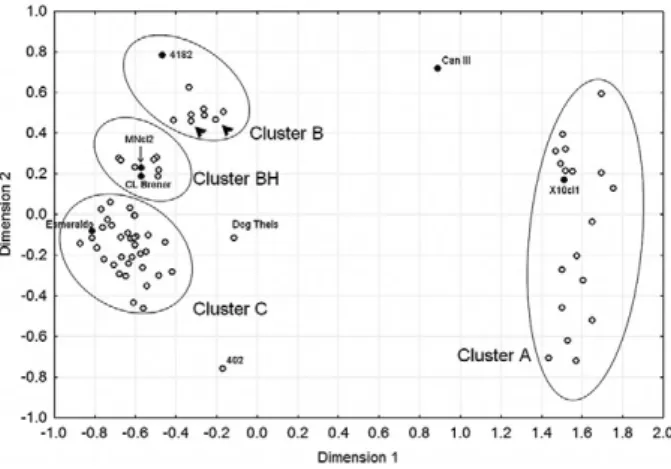
For our haplotype studies, we initially typed a large number of monoclonal strains of T. cruzi with five nu-clear CA-repeat microsatellites (Freitas et al. 2006). For our analysis we used as a metric the minimum number of mutationalsteps necessary to transform the microsatel-lite profile of one strain into that of another, assuming a stepwise mutation model for the evolution of the micro- satellites. From the pairwise distance between the strains, we built a geneticdistance matrix, which is pictorially de-picted by the multidimensional scaling (MDS) plot shown in Fig. 1. Four clusters are clearly visible and identified by ellipsoids in the MDS plot. The identity of the clusters is revealed by the presence of the prototypical strains of Brisse et al. (2000): MDS-cluster A corresponds to DTU I or T. cruzi I, MDS-cluster C to DTU IIb or T. cruzi II, MDS-cluster B to DTU IIc and MDS-cluster BH to the DTU IId and IIe. Only three strains fell outside the four clusters: CanIII (DTU IIa), Dog Theis and 402.
homologous chromosomes; in other words, this map is based on multi-local diploid genotypes of microsatellites. However, much more information could be acquired by separating the microsatellite alleles that are in each of the two homolog chromosomes, i.e., the two haplotypes.
If we were analysing a sexually reproducing organ-ism, we would obtain two haplotypes for each chromo-some pair. However, because of the predominant binary fission asexual reproduction of T. cruzi, extreme levels of linkage disequilibrium are present. Thus, the obtained haplotypes in fact represent whole haploid genomes, like the haploid gametes that unite during fertilisation to form a diploid organism.
To obtain the haplotypes, we ran the combined results
of the microsatellite genotypes and 24Sα rRNA gene
polymorphisms through PHASE software (Stephens et al. 2001). This powerful programme uses a Bayesian strategy to estimate haplotypes from population geno-type data. The programme identified no fewer than 141 different haplotypes in the 75 strains tested, correspond-ing to a haplotypic diversity of 0.993 (Freitas et al. 2006). The identified haplotypes were then subjected to a me-dian joining analysis using NETWORK 3.1 software (Bandelt et al. 1999). The resulting multitude of plausible trees is best expressed by a network that displays alter-native potential evolutionary paths (Fig. 2). Although the data are complex, three haplotypic clusters are clearly identifiable. We refer to these clusters as haplogroups X, Y and Z. The three haplogroups are connected by long and unique paths, emphasising the great genetic distance
between them. Seven haplotypes (numbers 33, 35, 58, 59, 60, 61 and 63) belong to these “bridges” and hence could not be assigned to any of the haplogroups. We assigned these haplotypes to a fourth haplogroup that we called “I” for indeterminate.
We then assigned two haplogroups to each of the 75 strains to constitute a haplogroup “genotype” (Table). The results were very simple. All strains belonging to the DTU I lineage (MDS-cluster A) (Fig. 1) proved to be Z/Z, i.e., had two haplotypes belonging to haplogroup Z. Likewise, all the strains of DTU IIb (MDS-cluster C) (Fig. 1) had Y/Y genotypes and those of DTU IIc (MDS-cluster B) (Fig. 1) had X/X genotypes (Freitas et al. 2006). Thus we were able to identify three large groups of an-cestral T. cruzi strains. The first had haplogroup “geno-type” Z/Z, corresponding to T. cruzi I. The second had haplogroup “genotype” Y/Y and clearly corresponded to
T. cruzi II. The most significant finding of our study was the identification of the X/X cluster, which corresponds exactly to the T. cruzi strains that belong to zymodeme Z3 found by Miles et al. (1978) and which do not fit into the dichotomous T. cruzi I/T. cruzi II model proposed in
Fig. 1:multidimensional scaling plot of 75 Trypanosoma cruzi strains genotyped for five microsatellites. Only outliers and the prototypical strains of Brisse et al. (2000) 11 are named in the plot. Arrowheads indicate strains 222 and 115. The strains in the regions delimited by ellipsoids are: multidimensional scaling (MDS)-cluster A: 1001, 1004, 1006, 1502, 1523, A83, A87, Col18/05, Colombiana, Cuíca, Cutia, D7, Gambacl1, Rb1, Rb2, Rb6, SE, X10cl1, 402, Mas1cl1, 84, 207, 209, 239, 577, 578, 580, 581, 803, 1005, 1014, 1043, 1931, 183744, 169/1; MDS-cluster B: 115, 222, 226, 231, 3663, 3869, 4182, M5631cl5; MDS-cluster BH: M6241cl6, 167, 1022, c182, CL Brener, MNcl2, NR, SC43 cl1, SO3, Tulacl2; MDS-cluster C: 200pm, 84Ti, Be62, CPI11/94, CPI95/94, Esmeraldo, Gil, GLT564, GLT593, GMS, GOCH, Ig539, JAF, JG, JHF, JSM, MPD, OPS27/94, Tu18 cl11, Y. Modified from Freitas et al. (2006).
1999. We suggest that this is a third phylogenetic lineage of T. cruzi and should be designated T. cruzi III.
Interestingly, the strains in cluster BH all had X/Y genotypes, confirming their hybrid nature and indicat-ing that they were derived from recombination events between ancestral types Y/Y and X/X, i.e., T. cruzi II and
T. cruzi III. Due to the way that PHASE identifies hap-lotypes, proximity of haplotype numbers is highly cor-related with genetic proximity. Hybrid strains 167, 1022, 182, CL Brener and Tulacl2 have genotypes 4/99, 2/102, 5/108, 5/100 and 3/103, respectively, and form one group, while strains MNcl2, NR, SC43cl1, and SO3 have geno-types 52/133, 55/130, 54/129 and 54/130 and form another group. Notably, these groups are equivalent to sub-lin-eages IIe and IId of Brisse et al. (2000). This suggests that at least two independent hybridisations occurred, presumably followed by clonal microdifferentiation.
The classification of T. cruzi into three major phylo-genetic trunks does not exhaust all ancestral possibili-ties. The strains Can III [genotype I/I, cytochrome oxi-dase subunitII (COII) B], Dog Theis (genotype I/I, COII C), 402 and Mas1cl1 (both genotype I/Y, COII C) share haplotypes with haplogroup I. Three of these four strains are located outside MDS clusters in Fig. 1A, which may suggest the existence of yet other phylogenetic lineages in T. cruzi.
Mitochondrial DNA studies
The extreme levels of linkage disequilibrium in the genome of T. cruzi not only cause the association of all nuclear markers into haploid genotypes, but also cre-ate strong associations between nuclear markers and mitochondrial genotypes. The latter are known to be
TABLE
Nuclear and mitochondrial markers of Trypanosoma cruzi
Strains COIIa Clustersb Haplotypesc Haplogroupsd
A83 A A 25/26 Z/Z
A87 A A 27/28 Z/Z
Col18/05 A A 21/56 Z/Z
Colombiana A A 24/56 Z/Z
Cuíca A A 31/49 Z/Z
Cutia A A 44/47 Z/Z
D7 A A 6/29 Z/Z
Gamba cl1 A A 30/46 Z/Z
Rb1 A A 36/57 Z/Z
Rb2 A A 16/36 Z/Z
Rb6 A A 38/41 Z/Z
SE A A 48/53 Z/Z
SilvioX10cl1 A A 32/50 Z/Z
1001 A A 43/43 Z/Z
1004 A A 23/37 Z/Z
1006 A A 22/40 Z/Z
1502 A A 39/45 Z/Z
1523 A A 42/51 Z/Z
115 B B 7/19 X/X
222 B B 1/20 X/X
226 B B 9/18 X/X
231 B B 11/17 X/X
3663 B B 8/10 X/X
3869 B B 13/14 X/X
4182 B B 15/62 X/X
M5631cl5 B B 12/12 X/X
M6241cl6 B B 33/34 X/X
167 B BH 4/99 X/Y
182 B BH 5/108 X/Y
1022 B BH 2/102 X/Y
CLBrener B BH 5/100 X/Y
Mncl2 B BH 52/133 X/Y
NR B BH 55/130 X/Y
SC43 cl1 B BH 54/129 X/Y
SO3 B BH 54/130 X/Y
Tula cl2 B BH 3/103 X/Y
CanIII cl1 B outlier 58/59 I/I
Be62 C C 88/105 Y/Y
CPI95/94 C C 110/114 Y/Y
Esmeraldo C C 109/135 Y/Y
Gil C C 77/79 Y/Y
GLT564 C C 97/98 Y/Y
GLT593 C C 74/131 Y/Y
GMS C C 90/131 Y/Y
GOCH C C 81/126 Y/Y
Ig539 C C 123/125 Y/Y
JAF C C 67/92 Y/Y
JG C C 82/92 Y/Y
JHF C C 83/84 Y/Y
JSM C C 65/95 Y/Y
Mas1 cl1 C C 63/136 Y/I
MPD C C 64/76 Y/Y
OPS27/94 C C 134/141 Y/Y
Tu18 cl11 C C 137/138 Y/Y
Y C C 86/87 Y/Y
84 C C 66/78 Y/Y
84Ti C C 71/73 Y/Y
Strains COIIa Clustersb Haplotypesc Haplogroupsd
169/1 C C 85/94 Y/Y
200pm C C 112/115 Y/Y
207 C C 75/139 Y/Y
209 C C 69/101 Y/Y
239 C C 80/96 Y/Y
577 C C 89/124 Y/Y
578 C C 122/127 Y/Y
580 C C 117/118 Y/Y
581 C C 113/116 Y/Y
803 C C 72/119 Y/Y
1005 C C 100/106 Y/Y
1014 C C 68/93 Y/Y
1043 C C 107/128 Y/Y
1931 C C 91/104 Y/Y
183744 C C 120/121 Y/Y
CPI11/94 C C 111/140 Y/Y
Dog Theis C outlier 60/61 I/I
402 C outlier 35/70 Y/I
a: restriction fragment length polymorphismtyping of the T. cruzi cytochrome oxidase subunitII (COII) gene; b: clusters of strains generated by multidimensional scaling analysis; c: haplotypes inferred by PHASE; d: clusters of haplotypes in the Network. Modified from Freitas et al. (2006).
uniparental (by convention, the maternal gamete is the mitochondrial donor) and functionally haploid. Indeed, Gaunt et al. (2003) have shown that the hybridisation of
T. cruzi strains involves only nuclear genomes and that mitochondrial fusion does not occur.
We studied the sequences of large portions of the max-icircle-encoded COII gene (Freitas et al. 2006) as well as cytochrome b (Brisse et al. 2003) and nicotinamide adenine dinucleotide dehydrogenase subunit 1 (Machado & Ayala 2001). The neighbour-joining trees generated for each of these regions are shown in Fig. 3. All trees was had very similar topologies, consisting of three tightly clustered sets of strains, separated by very large genetic distances, permitting the straightforward allocation of T. cruzi strains into three mitochondrial clades. For simplic-ity, we called these clades clusters A, B, and C.
It was most rewarding to observe that our MDS clus-ters (Fig. 1) corresponded perfectly to their homonymous mitochondrial clades, i.e., MDS cluster A was composed of the same strains that belonged to haplogroup Z/Z and to mitochondrial clade A, MDS cluster C was composed of the same strains that belonged to haplogroup Y/Y and to mitochondrial clade C and MDS cluster B was com-posed of the same strains that belonged to haplogroup X/X and to mitochondrial clade B. Thus, the results of the mitochondrial sequencing strongly confirm the ex-istence of the T. cruzi III phylogenetic group.
The MDS cluster BH contained the strains belonging to the hybrid sub-lineages IId and IIe (Brisse et al. 2000), all of which fall within mitochondrial clade B. Thus, the strains of sub-lineages IId and IIe are not only produced by hybridisations between T. cruzi II and T. cruzi III, but both have the same mitochondrial donor, T. cruzi III.
An evolutionary model
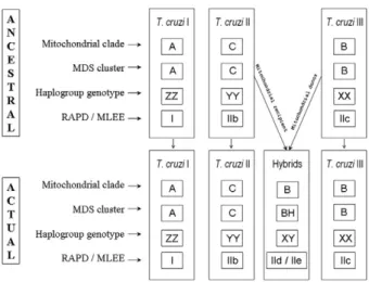
Based on our results, we propose that three ancestral genomes, T. cruzi I, T. cruzi II and T. cruzi III (Fig. 4), existed in the distant past. It is interesting to note that this proposal matches the initial suggestion made by Miles et al. (1978) almost 30 years ago on the basis of isozyme studies. It is likely that T. cruzi II and T. cruzi
III had overlapping ecological niches and thus the
condi-tions necessary for hybridisation were in place. At least two hybridisation events produced evolutionarily viable progeny. In both events, the cytoplasmic donor for the resulting offspring (as identified by the mitochondrial clade of the hybrid strains) was T. cruzi III.
The existence of strains that cannot be accommo-dated into this scenario, i.e., CanIII [sub-lineage IIa of Brisse et al. (2000)] and Dog Theis, suggests additional complexity in the evolutionary history of T. cruzi. Other alternative models have been proposed for its population structure(Westenberger et al. 2005), but we will not dis-cuss them fully here as they are not based on the novel high resolution haplotypic analysis presented in our work (Freitas et al. 2006).
The fact that the same population structure of T. cruzi
is predicted with different molecular markers, such as isozymes (Miles et al. 1978), RAPD (Brisse et al. 2000, 2003), microsatellites (Oliveira et al. 1998) and several other nuclear sequences (Fernandes et al. 1999, Zingales et al. 1999, Machado & Ayala 2001, August-Pinto et al. 2003), and mitochondrial markers (Machado & Ayala 2001, Brisse et al. 2003, Freitas et al. 2006), bears wit-ness to its extreme stability. Although it has been shown conclusively in our study and also by others (Machado & Ayala 2001, Brisse et al. 2003) that hybridisation events have occurred in the evolutionary history of T. cruzi, they seem to have been subsequently stabilised by strong clon-al propagation (Macedo & Pena 1998, Tibayrenc 2003).
REFERENCES
Adler S 1959. Darwin’s illness. Nature 184: 1102-1104.
Aufderheide AC, Salo W, Madden M, Streitz J, Buikstra, Guhl F, Ar-riaza B, Renier C, Wittmers LE Jr, Fornaciari G, Allison M2004. A 9,000-year record of Chagas’ disease. Proc Natl Acad Sci USA 101: 2034-2039.
Augusto-Pinto L, Teixeira SM, Pena SD, Machado CR 2003. Single-nucleotide polymorphisms of the Trypanosoma cruziMSH2 gene support the existence of three phylogenetic lineages presenting differences in mismatch-repair efficiency. Genetics 164: 117-126. Fig. 3: median joining network of the haplotypes identified by the
PHASE software.
Fig. 4: diagram depicting the proposed model for the evolution of Try�
Bandelt HK, Forster P, Rohl A 1999. Median-joining networks for inferring intraspecific phylogenies. Mol Biol Evol 16: 37-48. Bogliolo AR, Lauria-Pires L, Gibson WC 1996. Polymorphisms in
Trypanosoma cruzi: evidence of genetic recombination. Acta
Trop 61: 31-40.
Bradley KK, Bergman DK, Woods JP, Crutcher JM, Kirchhoff LV 2000. Prevalence of American trypanosomiasis Chagas disease among dogs in Oklahoma. J Am Vet Med Assoc 217: 1853-1857. Briones MR, Souto RP, Stolf BS, Zingales B 1999. The evolution of
two Trypanosoma cruzi subgroups inferred from rRNA genes can
be correlated with the interchange of American mammalian fau-nas in the Cenozoic and has implications to pathogenicity and host specificity. Mol Biochem Parasitol 104: 219-232.
Brisse S, Dujardin JC, Tibayrenc M 2000. Identification of six Try�
panosoma cruzi phylogenetic lineages by random amplified
polymorphic DNA and multilocus enzyme eletrophoresis. Int J Parasitol 30: 35-44.
Brisse S, Henriksson J, Barnabe C, Douzery EJ, Berkvens D, Serrano M, De Carvalho MR, Buck GA, Dujardin JC, Tibayrenc M2003. Evidence for genetic exchange and hybridization in Trypanosoma
cruzi based on nucleotide sequences and molecular karyotype.
Infect Genet Evol 2: 173-183.
Brisse S, Verhoef J, Tibayrenc M 2001. Characterization of large and small subunit rRNA and mini-exon genes further supports the distinction of six Trypanosoma cruzi lineages. Int J Parasitol 31: 1218-1226.
Carrasco HJ, Frame IA, Valente SA, Miles MA 1996. Genetic ex-change as a possible source of genomic diversity in sylvatic popu-lations of Trypanosoma cruzi.Am J Trop Med Hyg 54: 418-224. da Silva Valente SA, de Costa Valente V, Neto HF 1999.
Consider-ations on the epidemiology and transmission of Chagas disease in the Brazilian Amazon. Mem Inst Oswaldo Cruz 94 (Suppl. I): 395-398.
Dias JC, Silveira AC, Schofield CJ 2002. The impact of Chagas dis-ease control in Latin America: a review. Mem Inst Oswaldo Cruz 97: 603-612.
Di Noia JM, Buscaglia CA, De Marchi CR, Almeida IC, Frasch AC 2002. A Trypanosoma cruzi small surface molecule provides the first immunological evidence that Chagas’ disease is due to a single parasite lineage. J Exp Med 195: 401-413.
El-Sayed NM, Myler PJ, Bartholomeu DC, Nilsson D, Aggarwal G, Tran AN, Ghedin E, Worthey EA, Delcher AL, Blandin G, Wes-tenberger SJ, Caler E, Cerqueira GC, Branche C, Haas B, Anu-pama A, Arner E, Aslund L, Attipoe P, Bontempi E, Bringaud F, Burton P, Cadag E, Campbell DA, Carrington M, Crabtree J, Darban H, da Silveira JF, de Jong P, Edwards K, Englund PT, Fazelina G, Feldblyum T, Ferella M, Frasch AC, Gull K, Horn D, Hou L, Huang Y, Kindlund E, Klingbeil M, Kluge S, Koo H, Lac-erda D, Levin MJ, Lorenzi H, Louie T, Machado CR, McCulloch R, McKenna A, Mizuno Y, Mottram JC, Nelson S, Ochaya S, Osoegawa K, Pai G, Parsons M, Pentony M, Pettersson U, Pop M, Ramirez JL, Rinta J, Robertson L, Salzberg SL, Sanchez DO, Seyler A, Sharma R, Shetty J, Simpson AJ, Sisk E, Tammi MT, Tarleton R, Teixeira S, Van Aken S, Vogt C, Ward PN, Wick-stead B, Wortman J, White O, Fraser CM, Stuart KD, Andersson B2005. The genome sequence of Trypanosoma cruzi, etiologic agent of Chagas disease. Science 309: 409-415.
Fernandes O, Santos S, Junqueira A, Jansen A, Cupolillo E, Campbell D, Zingales B, Coura JR1999. Populational heterogeneity of Bra-zilian Trypanosoma cruzi isolates revealed by the mini-exon and ribosomal spacers. Mem Inst Oswaldo Cruz 94: 195-197.
Freitas JM, Augusto-Pinto L, Pimenta JR, Bastos-Rodrigues L, Gon-çalves VF, Teixeira SM, Chiari E, Junqueira AC, Fernandes O, Macedo AM, Machado CR, Pena SD 2006. Ancestral genomes, sex and the population structure of Trypanosoma cruzi. PLoS Pathog 2: e24.
Freitas JM, Lages-Silva E, Crema E, Pena SD, Macedo AM 2005. Real time PCR strategy for the identification of major lineages
of Trypanosoma cruzi directly in chronically infected human
tis-sues. Int J Parasitol 35: 411-417.
Gaunt MW, Yeo M, Frame IA, Stothard JR, Carrasco HJ, Taylor MC, Mena SS, Veazey P, Miles GA, Acosta N, de Arias AR, Miles MA 2003. Mechanism of genetic exchange in American trypanosomes.
Nature 421: 936-939.
Higo H, Yanagi T, Matta V, Agatsuma, Cruz-Reyes A, Uyema N, Mon-roy C, Kanbara H, Tada I2000. Genetic structure of Trypanosoma
cruzi in American continents: special emphasis on sexual
repro-duction in Central America. Parasitology 121: 403-408.
Jansen AM, Santos de Pinho AP, Lisboa CV, Cupolillo E, Mangia RH, Fernandes O 1999. The sylvatic cycle of Trypanosoma cruzi: a still unsolved puzzle. Mem Inst Oswaldo Cruz 94 (Suppl. I): 203-204.
Kirchhoff LV 1993. American trypanosomiasis Chagas’ disease. A tropical disease now in the United States. N Engl J Med 329: 639-644.
Lisboa CV, Mangia RH, De Lima NR, Martins A, Dietz J, Baker AJ, Ramon-Miranda CR, Ferreira LF, Fernandes O, Jansen AM 2004. Distinct patterns of Trypanosoma cruzi infection in Leontopith� ecus rosalia in distinct Atlantic coastal rainforest fragments in Rio de Janeiro - Brazil. Parasitology 129: 703-711.
Macedo AM, Machado CR, Oliveira RP, Pena SDJ 2004. Trypano�
soma cruzi: genetic structure of populations and relevance of
ge-netic variability to the pathogenesis of Chagas disease. Mem Inst
Oswaldo Cruz 99: 1-12.
Macedo AM, Pena SDJ 1998. Genetic variability of Trypanosoma
cruzi: implications for the pathogenesis of Chagas’ disease. Para�
sitol Today 14: 119-124.
Machado CA, Ayala FJ 2001. Nucleotide sequences provide evidence of genetic exchange among distantly related lineages of Trypano�
soma cruzi.Proc Natl Acad Sci 98: 7396-7401.
Maguire JH, Hoff R, Sleigh AC, Mott KE, Ramos NB, Sherlock IA 1986. An outbreak of Chagas’ disease in Southwestern Bahia, Brazil. Am J Trop Med Hyg 35: 931-936.
Miles MA, Souza A, Povoa M, Shaw JJ, Lainson R, Toyle PJ 1978. Isozymic heterogeneity of Trypanosoma cruzi in the first autoch-thonous patients with Chagas’ disease in Amazonian Brazil. Na� ture 272: 819-821.
Oliveira RP, Broude NE, Macedo AM, Cantor CR, Smith CL, Pena SD 1998. Probing the genetic population structure of Trypano�
soma cruzi with polymorphic microsatellites. Proc Natl Acad Sci
USA 95: 3776-3780.
Pedroso A, Cupolillo E, Zingales B 2003. Evaluation of Trypanosoma cruzi hybrid stocks based on chromosomal size variation. Mol
Biochem Parasitol 129: 79-90.
Pietrzak SM, Pung OJ 1998. Trypanosomiasis in raccoons from Geor-gia. J Wildl Dis 34: 132-136.
Pung OJ, Banks CW, Jones DN, Krissinger MW 1995. Trypanosoma
cruzi in wild raccoons, opossums and triatomine bugs in
south-east Georgia, USA. J Parasitol 81: 324-326.
Satellite Meeting 1999. Recommendations from an international sym-posium to commemorate the 90th anniversary of the discovery of Chagas disease, April 11-16 1999, Rio de Janeiro, Brazil. Mem
Shikanai-Yasuda MA, Marcondes CB, Guedes LA, Siqueira GS, Bar-one AA, Dias JC, Amato Neto V, Tolezano JE, Peres BA, Arruda Júnior ER 1991. Possible oral transmission of acute Chagas’ dis-ease in Brazil. Rev Inst Med Trop São Paulo 33: 351-357. Souto RP, Fernandes O, Macedo AM, Campbell DA, Zingales B
1996. DNA markers define two major phylogenetic lineages of
Trypanosoma cruzi.Mol Biochem Parasitol 83: 141-152.
Stephens M, Smith NJ, Donnelly P 2001. A new statistical method for haplotype reconstruction from population data. Am J Hum Genet 68: 978-989.
Stolf BS, Souto RP, Pedroso A, Zingales B 2003. Two types of ri-bosomal RNA genes in hybrid Trypanosoma cruzi strains. Mol
Biochem Parasitol 126: 73-80.
Sturm NR, Vargas NS, Westenberger SJ, Zingales B, Campbell DA 2003. Evidence for multiple hybrid groups in Trypanosoma cruzi. Int J Parasitol 33: 269-279.
Tibayrenc M 1996. Towards a unified evolutionary genetics of
micro-organisms. Annu Rev Microbiol 50: 401-429.
Tibayrenc M 2003. Genetic subdivisions within Trypanosoma cruzi
discrete typing units and their relevance for molecular epidemiol-ogy and experimental evolution. Kinetoplastid Biol Dis 28: 2-12. Tibayrenc M, Ayala FJ 2002. The clonal theory of parasitic protozoa:
12 years on. Trends Parasitol 18: 405-410.
Tibayrenc M, Ward P, Moya A, Ayala FJ 1986. Natural populations of
Trypanosoma cruzi, the agent of Chagas’ disease, have a complex
multiclonal structure. Proc Nat Acad Sci USA 83: 115-119. Westenberger SJ, Barnabé C, Campbell DA, Sturm NR 2005. Two
hybridization events define the population structure of Trypano� soma cruzi. Genetics 171: 527-543.
Zingales B, Stolf BS, Souto RP, Fernandes O, Briones MR 1999. Epidemiology, biochemistry and evolution of Trypanosoma cruzi
lineages based on ribosomal RNA sequences. Mem Inst Oswaldo