Ancestral Genomes, Sex, and the Population
Structure of
Trypanosoma cruzi
Jorge M. de Freitas1[, Luiz Augusto-Pinto1[, Juliana R. Pimenta1[, Luciana Bastos-Rodrigues1, Vanessa F. Gonc¸alves1, Santuza M. R. Teixeira1, Egler Chiari2, Aˆ ngela C. V. Junqueira3, Octavio Fernandes3, Andre´a M. Macedo1,
Carlos Renato Machado1, Se´rgio D. J. Pena1*
1Departamento de Bioquı´mica e Imunologia, Universidade Federal de Minas Gerais, Belo Horizonte, Brazil,2Departamento de Parasitologia, Universidade Federal de Minas Gerais, Belo Horizonte, Brazil, 3Departamento de Medicina Tropical, Fundac¸a˜o Oswaldo Cruz, Rio de Janeiro, Brazil
Acquisition of detailed knowledge of the structure and evolution ofTrypanosoma cruzi populations is essential for control of Chagas disease. We profiled 75 strains of the parasite with five nuclear microsatellite loci, 24SaRNA genes, and sequence polymorphisms in the mitochondrial cytochrome oxidase subunit II gene. We also used sequences available in GenBank for the mitochondrial genes cytochrome B and NADH dehydrogenase subunit 1. A multidimensional scaling plot (MDS) based in microsatellite data divided the parasites into four clusters corresponding toT. cruzi I (MDS-cluster A),T. cruzi II (MDS-cluster C), a third group ofT. cruzi strains (MDS-cluster B), and hybrid strains (MDS-cluster BH). The first two clusters matched respectively mitochondrial clades A and C, while the other two belonged to mitochondrial clade B. The 24SarDNA and microsatellite profiling data were combined into multilocus genotypes that were analyzed by the haplotype reconstruction program PHASE. We identified 141 haplotypes that were clearly distributed into three haplogroups (X, Y, and Z). All strains belonging toT. cruziI (MDS-cluster A) were Z/Z, theT. cruziII strains (MDS-cluster C) were Y/Y, and those belonging to MDS-cluster B (unclassifiedT. cruzi) had X/X haplogroup genotypes. The strains grouped in the MDS-cluster BH were X/Y, confirming their hybrid character. Based on these results we propose the following minimal scenario forT. cruzievolution. In a distant past there were at a minimum three ancestral lineages that we may call, respectively,T. cruziI, T. cruziII, and T. cruziIII. At least two hybridization events involvingT. cruziII and T. cruziIII produced evolutionarily viable progeny. In both events, the mitochondrial recipient (as identified by the mitochondrial clade of the hybrid strains) was T. cruzi II and the mitochondrial donor wasT. cruziIII.
Citation: Freitas JM, Augusto-Pinto L, Pimenta JR, Bastos-Rodrigues L, Gonc¸alves VF, et al. (2006) Ancestral genomes, sex, and the population structure ofTrypanosoma cruzi.
PLoS Pathog 2(3): e24.
Introduction
The parasite protozoan Trypanosoma cruzi causes Chagas disease, a malady that afflicts almost 20 million people in South America and Central America, with more than 20,000 deaths reported each year [1,2]. Two different ecosystems exist forT. cruzi:one related to wild hemiptera and generally involving wild mammals (the‘‘sylvatic’’ cycle), and another dependent on home-dwelling hemiptera and primarily involving humans and household animals (the so-called
‘‘domestic’’cycle). The connection between the two ecosys-tems is made by infected rats, mice, bats, marsupials, and other feral mammals. It is estimated that the parasite emerged as a species well over 150 million years ago, originally infecting primitive mammals dispersed throughout Laurasia and Gondwanaland, regions that originated North and South America, respectively [3]. The first contact with humans occurred much more recently, in the late Pleistocene, 15,000–20,000 years ago, when humans first peopled the Americas—thus,Homo sapiensis a very recent new host forT. cruzi. There is convincing molecular evidence for the presence ofT. cruziDNA in mummies exhumed in Northern Chile and Southern Peru and dating as far back as 9,000 years before the present day. [4].
The conventional mode of transmission of T. cruzi to humans is by the feces of infected hematophagous triatomine bugs. Alternative modes of infection include blood trans-fusion, congenital transmission from infected mothers, and
ingestion of contaminated foods. Thanks to intensive programs of triatomine control, vectorial infection has been virtually abolished in Brazil, Chile, Uruguay, and Argentina [5]. Moreover, improved screening of blood donors to reduce the likelihood of transfusional transmission and early detection and treatment of congenital cases have added to this success. It would be, however, a mistake to think that Chagas disease has been controlled. High levels of vector-borne transmission are still apparent in many areas, and several of the endemic countries have yet to develop serious large-scale surveillance and intervention programs [5]. Also, migrations of infected individuals offer a risk of new
Editor:John Boothroyd, Stanford University, United States of America
ReceivedOctober 13, 2005;AcceptedFebruary 21, 2006;PublishedMarch 31, 2006
DOI:10.1371/journal.ppat.0020024
Copyright:Ó2006 Freitas et al. This is an open-access article distributed under the
terms of the Creative Commons Attribution License, which permits unrestricted use, distribution, and reproduction in any medium, provided the original author and source are credited.
Abbreviations:COII, cytochrome oxidase subunit II;CYb, cytochrome b; MDS, multidimensional scaling plot;ND1, NADH dehydrogenase subunit 1; NJ, neighbor-joining; RAPD, random amplified polymorphic DNA; RFLP, restriction fragment-length polymorphism
* To whom correspondence should be addressed. E-mail: [email protected]
transmission in previously nonendemic regions, such as the United States [6].Furthermore, the ancient and wide-ranging sylvatic cycle constitutes an enormous reservoir of parasites that represents a threat for humans.
Recent studies have shown that in a nonendemic area of the Brazilian Atlantic coastal rainforest 50% of the triato-mine vectors and of the marsupialsDidelphis marsupialisand Philander opossum [7] as well as 52% of the golden lion tamarins and several other species of New World primates [8] were naturally infected withT. cruzi. Moreover, in the United StatesT. cruzihas been found in 11.4% of opossums and 22% of the raccoons, together with infected triatomine bugs in the state of Georgia [9]. In certain areas of that state 43% of the raccoons were infected [10]. Closer to the human domestic environment, Bradley et al. [11] have shown that 3.6% of the rural hunting dogs in Oklahoma were seropositive forT. cruzi. Human infection from the sylvatic environment can occur either from sudden migration of hemiptera to the human environment, forced by the destruction of forests [12] or by the ingestion of foods contaminated by the feces of hemi-pterae or by crushed insects [13,14]. Thus, a complete understanding of the population structure of T. cruzi, especially the sylvatic cycle, will be indispensable for controlling the disease.
T. cruzi is diploid, with different-sized homologous chro-mosome pairs [15]. Its genome has been recently sequenced [16], and its size (diploid) has been estimated between 106.4 and 110.7 Mb. At least 50% of theT. cruzigenome is made up of repetitive sequences, consisting of large gene families of surface proteins, retrotransposons, and subtelomeric repeats. There is extensive and well-characterized intraspecific genetic diversity inT. cruzi (reviewed in [17,18]). Two major evolutionary lineages of the parasite, namedT. cruziI andT. cruzi II, have been identified [19]. These lineages are very divergent as revealed by several biological and molecular markers, including isozymes, 24SarDNA, and mini-exon gene polymorphisms [20].T. cruzi I andT. cruzi II strains belong predominantly to distinct ecological environments: respec-tively, the sylvatic and domestic transmission cycles of Chagas
disease [3,21].T. cruziI strains are characterized by zymodeme Z1 (a zymodeme is a group of strains that have the same isozyme profile), 24SarDNA group 2, and mini-exon group 2, and induce low parasitism in human chagasic patients. In contrast,T. cruziII strains are characterized by zymodeme Z2, 24Sa rDNA and mini-exon group 1, and cause human infections with high parasitemia in classic endemic areas [21]. At least in Brazil, T. cruzi II strains appear to be exclusively responsible for tissue lesions in Chagas disease [22]. Additionally, there are some parasite strains that cannot be properly grouped into any one of these two major lineages. Among these unclassified strains are those identified as belonging to zymodeme Z3 [23] and other hybrid strains characterized as rDNA group 1/2 [24,25]. Using isozymes and random amplified polymorphic DNA (RAPD) typing, Brisse et al. [26] proposed thatT. cruziII strains could be partitioned into five phylogenetic sublineages (IIa-e), each comprising one of the following reference strains: CanIII cl1 (IIa), Esmeraldo cl3 (IIb), M5631 cl5 (IIc), MN cl2 (IId), and CLBrener (IIe). In contrast, T. cruzi I strains could not be further subdivided. Within each of these clades or subline-ages, there is extensive genetic diversity that can be unraveled by analyses with microsatellites and several other genomic markers (reviewed in [27]).
Although capable of recombination in vitro [28], T. cruzi reproduces predominantly by binary fission and conse-quently its diploid nuclear genotype is transmitteden blocto the progeny. Thus, the parasite presents extreme degrees of linkage disequilibrium, as shown through isozymes [29] and microsatellites [30], and exhibits a predominantly clonal population structure. Indeed, T. cruzi still has been consid-ered the paradigm for clonal eukaryotic pathogenic micro-organisms [31]. The occurrence of hybrid strains in natural populations of T. cruzi was suggested by isozyme analyses [32,33], restriction fragment-length polymorphism (RFLP) of housekeeping genes [34], RAPD [35], and genotype variations observed at chromosomal level [15,35,36], and has been confirmed using nucleotide sequences [37,38]. Their discov-ery proved that sexual events definitely have taken place in the past and have shaped the genetical structure of currentT. cruzi populations. However, such genetic exchange events seem to have been rare enough to allow the propagation of clonal genotypes over long periods of time and wide geographical regions [35]. Because of the linkage disequili-brium, genotyping of nuclear markers inT. cruzihas thus far been limited to characterization of multilocus genotypes. Therefore, to understand the evolutionary history of the species it would be desirable to dissect the multilocus genotypes into their constituent haploid genome blocks. We wish to report that we have achieved this, revealing the existence of ancestral haplogroups and repeated hybrid-ization events inT. cruzi.
Results
We have typed 75 strains of T. cruzi (Table 1) with five nuclear CA-repeat microsatellites (Table S1). We assumed a stepwise mutation model for the evolution of microsatellites and used the minimum number of mutational steps necessary to transform one strain microsatellite profile into another to build a genetic distance matrix. The multidimensional scaling (MDS) plot shown in Figure 1 provided, with excellent fit
Synopsis
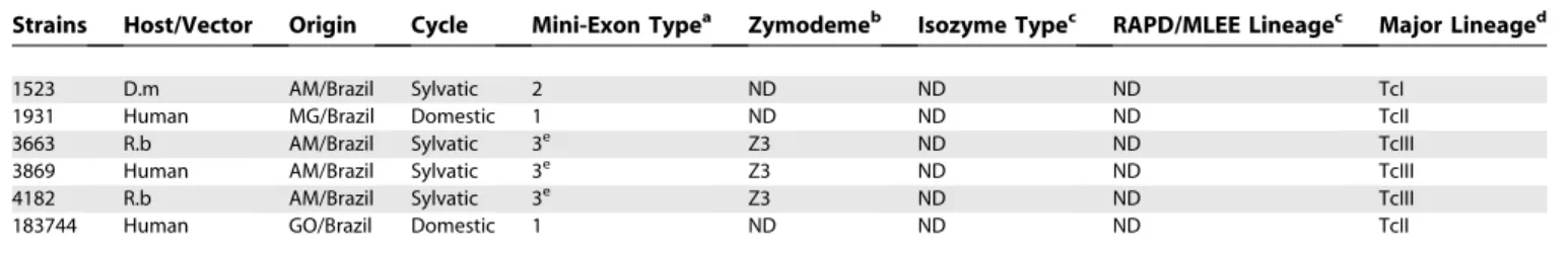
Table 1.Characteristics ofT. cruziStrains Analyzed
Strains Host/Vector Origin Cycle Mini-Exon Typea Zymodemeb Isozyme Typec RAPD/MLEE Lineagec Major Lineaged
A83 D.m F.G. Sylvatic 2 ND ND ND TcI
A87 D.m F.G. Sylvatic 2 ND ND ND TcI
Be62 Human MG/Brazil Domestic 1 ND ND ND TcII
CanIII cl1 Human PA/Brazil Domestic 3e Z3 27 IIa Hybridf
CLBrener T.i RS/Brazil Domestic 1 Z2 43 IIe Hybridf,g
Colombiana Human Colombia Domestic 2 ND ND ND TcI
Col18/05 Human Colombia Domestic 2 ND ND ND TcI
CPI11/94 Human Colombia Domestic 1 ND ND ND TcII
CPI95/94 Human PI/Brazil Domestic 1 ND ND ND TcII
Cutia D.a ES/Brazil Sylvatic 2 Z1 19 I TcI
Cuı´ca P.o. SP/Brazil Sylvatic 2 Z1 19 I TcI
Dog Theis ND ND ND 1 ND ND ND TcII
D7 D.m RJ/Brazil Sylvatic 2 ND ND ND TcI
Esmeraldo Human MG/Brazil Domestic 1 Z2 30 IIb TcII
Gamba cl1 D.g SP/Brazil Sylvatic 2 Z1 19 I TcI
Gil Human MG/Brazil Domestic ND ND ND ND TcIIg
GLT564 L.r RJ/Brazil Sylvatic 1 ND ND ND TcII
GLT593 L.r RJ/Brazil Sylvatic 1 ND ND ND TcII
GMS Human MG/Brazil Domestic 1 ND ND ND TcII
GOCH Human GO/Brazil Domestic 1 ND ND ND TcII
Ig539 Human MG/Brazil Domestic 1 ND ND ND TcII
JAF Human MG/Brazil Domestic ND ND ND ND TcIIg
JG Human MG/Brazil Domestic 1 ND ND ND TcII
JHF Human MG/Brazil Domestic ND ND ND ND TcIIg
JSM Human MG/Brazil Domestic ND ND ND ND TcIIg
Mas1 cl1 Human MG/Brazil Domestic 1 Z2 32 IIb TcII
Mn cl2 Human Chile Domestic 1 Z2 39 IId Hybridf,g
MPD Human MG/Brazil Domestic 1 ND ND ND TcII
M5631 cl5 D.n PA/Brazil Domestic 3e Z3 36 IIc TcIII
M6241 cl6 Human PA/Brazil Domestic 3e Z3 35 IIc TcIII
NR cl3 Human Chile Domestic 1 Z2 39 IId Hybridf,g
OPS27/94 Human PI/Brazil Domestic 1 ND ND ND TcII
Rb1 R.b AM/Brazil Sylvatic 2 ND ND ND TcI
Rb2 R.b AM/Brazil Sylvatic 2 ND ND ND TcI
Rb6 R.b AM/Brazil Sylvatic 2 ND ND ND TcI
SC43 cl1 T.i MG/Brazil Domestic 1 Z2 39 IId Hybridg
SE Human AM/Brazil Sylvatic 2 ND ND ND TcI
SilvioX10 cl1 Human PA/Brazil Domestic 2 Z1 17 I TcI
SO3 T.i Bolivia Domestic 1 Z2 39 IId Hybridf,g
Tu18 cl2 T.i MG/Brazil Domestic 1 Z2 32 IIb TcII
Tula cl2 Human Chile Domestic 2 Z2 43 IIe Hybridf,g
Y Human SP/Brazil Domestic 1 ND ND ND TcII
84Ti Human MG/Brazil Domestic ND ND ND ND TcIIg
84 Human MG/Brazil Domestic 1 ND ND ND TcII
115 Human MG/Brazil Domestic 1 ND ND ND TcIII
167 Human MG/Brazil Domestic ND ND ND ND Hybridg
169/1 Human MG/Brazil Domestic 1 ND ND ND TcII
182 Human MG/Brazil Domestic 1 ND ND ND Hybridg
200pm Human MG/Brazil Domestic 1 ND ND ND TcII
207 Human MG/Brazil Domestic ND ND ND ND TcIIg
209 Human MG/Brazil Domestic 1 ND ND ND TcII
222 Human MG/Brazil Domestic ND ND ND ND TcIII
226 Human MG/Brazil Domestic 1 ND ND ND TcIII
231 Human MG/Brazil Domestic ND ND ND ND TcIII
239 Human MG/Brazil Domestic 1 ND ND ND TcII
402 ND ND ND ND ND ND ND ND
577 Human GO/Brazil Domestic 1 ND ND ND TcII
578 Human GO/Brazil Domestic 1 ND ND ND TcII
580 Human GO/Brazil Domestic 1 ND ND ND TcII
581 Human GO/Brazil Domestic ND ND ND ND TcIIg
803 Human MG/Brazil Domestic 1 ND ND ND TcII
1001 D.m MG/Brazil Sylvatic 2 ND ND ND TcI
1004 T.i MG/Brazil Domestic 2 ND ND ND TcI
1005 T.i MG/Brazil Domestic 1 ND ND ND TcII
1006 T.i MG/Brazil Domestic 2 ND ND ND TcI
1014 P.m MG/Brazil Domestic 1 ND ND ND TcII
1022 P.m MG/Brazil Domestic 1 ND ND ND Hybridg
1043 Human MG/Brazil Domestic 1 ND ND ND TcII
(stress¼0.08), a visual representation of the distance matrix.
Four clusters are clearly visible and identified by ellipsoids in the MDS plot. The identity of the clusters is revealed by the presence of the prototypical strains of Brisse et al. [26]: MDS-cluster A corresponds toT. cruziI, MDS-cluster C toT. cruzi IIb, MDS-cluster B toT. cruziIIc, and MDS-cluster BH to the IId and IIe sublineages. Only three strains fell outside the four clusters: CanIII (sub-lineage IIa), Dog Theis, and 402.
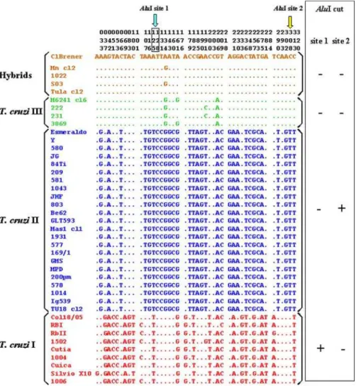
In 41 of the 75 strains we sequenced a 290–base pair region of the maxicircle-encoded cytochrome oxidase subunit II (COII) gene encompassing 44 variable positions (Figure 2). The sequenced data were used to generate a neighbor-joining (NJ) tree that is shown in Figure 3. It is clear that there are
three tightly clustered sets of strains, separated by very large genetic distances, permitting straightforward allocation ofT. cruzistrains into three mitochondrial clades that can also be simply identified by variation in just two AluI RFLP sites (Figure 2), which were then scored for all 75 strains (Table 2). Our MDS clusters corresponded perfectly to these mitochon-drial clades, with the exceptions of MDS-clusters B (subline-age IIc) and BH (thus called because it contains the hybrid sublineages IId and IIe), both of which fall within mitochon-drial clade B. To confirm our finding, we also built NJ trees for sequences obtained from GenBank of two other mito-chondrial genes, cytochrome b (CYb) [35] and NADH dehydrogenase subunit 1(ND1)[37]. TheCYbandND1trees Table 1.Continued.
Strains Host/Vector Origin Cycle Mini-Exon Typea Zymodemeb Isozyme Typec RAPD/MLEE Lineagec Major Lineaged
1523 D.m AM/Brazil Sylvatic 2 ND ND ND TcI
1931 Human MG/Brazil Domestic 1 ND ND ND TcII
3663 R.b AM/Brazil Sylvatic 3e Z3 ND ND TcIII
3869 Human AM/Brazil Sylvatic 3e Z3 ND ND TcIII
4182 R.b AM/Brazil Sylvatic 3e Z3 ND ND TcIII
183744 Human GO/Brazil Domestic 1 ND ND ND TcII
a
Revised by Zingales et al. [21].
b
Described by Miles et al. [23].
c
Described by Tibayrenc et al. [43] and Brise et al. [26].
d
TcI, TcII, and TcIII are abbreviations for the major lineages ofT. cruzi.TcII was characterized in this work.
e
Mini-exon type 3 associated with Z3 strains.
f
The hybrid characteristics of these strains were based on the data described by Brisse et al. [35] and Machado and Ayala [37].
g
The hybrid characteristic of these strains were based on data described in this work.
D.a,Dasyprocta aguti;D.g,Didelphis azarae; D.m,Didelphis marsupialis;D.n,Didelphis novemcinctus;F.G., French Guyana; L.r, Leonthopitecus rosalia; MLEE, multilocus enzyme electrophoresis; P.o.,Philander opossum;P.m,Panstrongylus megistus;R.b,Rhodnius brethesi;T.i,Triatoma infestans;and ND, not determined.
DOI: 10.1371/journal.ppat.0020024.t001
Figure 1.Multidimensional Scaling Plot of 75T. cruziStrains Genotyped for Five Microsatellites
Only outliers and the prototypical strains of Brisse et al. [26] are named in the plot. Arrowheads indicate strains 222 and 115. The strains in the areas delimited by ellipsoids are the following: MDS-cluster A: 1001, 1004, 1006, 1502, 1523, A83, A87, Col18/05, Colombiana, Cuı´ca, Cutia, D7, Gambacl1, Rb1, Rb2, Rb6, SE, X10cl1, 402, Mas1cl1, 84, 207, 209, 239, 577, 578, 580, 581, 803, 1005, 1014, 1043, 1931, 183744, 169/1; MDS-cluster C: 200pm, 84Ti, Be62, CPI11/94, CPI95/94, Esmeraldo, Gil, GLT564, GLT593, GMS, GOCH, Ig539, JAF, JG, JHF, JSM, MPD, OPS27/94, Tu18 cl11, Y; MDS-cluster B: 115, 222, 226, 231, 3663, 3869, 4182, M5631cl5; MDS-cluster BH: M6241cl6, 167, 1022, c182, CLBrener, MNcl2, NR, SC43 cl1, SO3, Tulacl2.
had very similar topology to that of theCOII tree (all with extremely high bootstrap values for the three main branches), confirming that sublineages IIc, IId, and IIe indeed belong to the same mitochondrial clade (Figure 3). We tested this notion further using analysis of molecular variance [39]. By partitioning the variability within and between mitochon-drial clades we found that for COII, CYb, and ND1, respectively, 97%, 91%, and 68% of the genetic variability was found among clades.
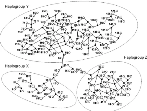
We also typed all strains for the polymorphism of the D7 divergent domain of the 24Sa rRNA gene (Table S1) and combined the results with the microsatellites into multilocus genotypes that were analyzed with the PHASE software [40]. We identified 141 different haplotypes corresponding to a haplotypic diversity of 0.993. The identified haplotypes were then subjected to a median joining analysis using the NETWORK 3.1 software [41]. The resulting multitude of plausible trees is best expressed by a network that displays alternative potential evolutionary paths (Figure 4). Three haplotypic clusters are clearly identifiable: we called them haplogroups X, Y, and Z. Within these haplotypic clusters
there is extensive reticulation because of the stepwise recurrent nature of microsatellite mutations [42]. However, the three haplogroups are connected by long and unique paths, emphasizing the great genetic distance between them. Seven haplotypes (numbers 33, 35, 58, 59, 60, 61, and 63) belong to these‘‘bridges’’and hence could not be assigned to any of the haplogroups—they were lumped into a haplogroup
‘‘I’’(for indeterminate). We could then assign to each of the 75 strains a haplogroup genotype (Table 2). All strains belonging to theT. cruzi I lineage (MDS-cluster A in Figure 1) proved to be Z/Z (i.e., had two haplotypes belonging to haplogroup Z). Likewise, all the strains in MDS-cluster C (Figure 1) had Y/Y genotypes and those in MDS-cluster B had X/X genotypes. The strains in cluster BH all had X/Y genotypes confirming their hybrid nature. Strains Can III (genotype I/I,COIIB), Dog Theis (genotype I/I,COIIC), 402, and Mas1cl1 (both genotype I/Y, COII C), and M6241cl16 (genotype I/X,COIIB) presented haplotypes of haplogroup I. It is noteworthy that three of these five strains are the ones outside MDS clusters in Figure 1A.
Figure 2.Sequence of a 290–Base Pair Fragment of the MitochondrialCOIIGene of 41 Strains ofT. cruzi
Only the variable nucleotides are shown.T. cruziIII refers to sublineage IIc, and‘‘Hybrids’’indicate the strains belonging to sublineages IId and IIe of Brisse et al [26]. Two AluI restriction sites are indicated. RFLP analysis of these two sites allows unambiguous classification ofT. cruzistrains to the three mitochondrial clades as shown on the right hand side.
Discussion
The population structure of T. cruzi is far from being completely understood. Although the existence of two major lineages in this species is well accepted, uncertainties about the existence or not of a third major ancestral group have been raised [15,35,36]. For instance, strains belonging to zymodeme Z3 or to rDNA group 1/2 could not be classified into eitherT. cruziI orT. cruziII [19]. Likewise, other strains (such as SC43) that present incongruities between the rDNA (group 2) and mini-exon (group 1) typing cannot be allocated into any of the two major lineages [24]. One of the major goals of this work was to investigate the genetic relationships among these‘‘unclassifiable’’strains.
Our first strategy was to perform the phylogenetic analysis of T. cruzi populations by using microsatellite data. Albeit extremely variable, these DNA markers allowed us to reliably identify four significant major clusters of strains (MDS clusters A, B, C, and BH in Figure 1). MDS-cluster A corresponds to T cruzi I and MDS-cluster C to classical T. cruziII or T. cruzi IIb as named by Brisse et al. [26]. MDS-cluster B contains strains classified as Z3 and assigned to the IIc sublineage [26]. Finally, the strains within MDS-cluster BH were known to belong to the putative hybrid isozyme clonets 39 or 43 as proposed by Tibayrenc [43] and later classified as IId and IIe sublineages by Brisse et al. [26] (see Table 1).
Nucleotide sequencing and AluI RFLP analysis of a 290-bp stretch of the mitochondrialCOIIgene demonstrated that all strains enclosed in our microsatellite clusters B and BH (Z3 and hybrid strains) belonged to the same mitochondrial clade B. Sequences of two other mitochondrial genes,CYb[35] and ND1 [37], obtained from GenBank, amply confirmed this observation by showing that indeed hybrid strains (subline-ages IId and IIe) and Z3 strains (sublineage IIc) were grouped
together into the same mitochondrial clade B. This same conclusion had been reached earlier [35,37].
Gaunt et al. [28] have shown that the hybridization ofT. cruzi strains involves only nuclear genomes, without mitochondrial fusion. Here, we clearly demonstrated that the mitochondrial clade B is a third major phylogenetic division of T. cruzi, distinct fromT. cruziI (mitochondrial clade A) andT. cruziII (mitochondrial clade C) major lineages. We have also shown that the strains with hybrid molecular markers in their nuclear genomes have a distinct mitochondrial genome (genotype B). The analyses with all studied nuclear markers identified 141 different haplotypes that could be clustered into three haplogroups. All strains belonging to the T. cruzi I major lineage (MDS-cluster A in Figure 1) proved to be Z/Z (i.e., had two haplotypes belonging to haplogroup Z). Likewise, all the strains in MDS-cluster C (Figure 1) had Y/Y genotypes and those in MDS-cluster B had X/X genotypes. Thus, our data do not corroborate the suggestion made by Sturm et al. [36] that sublineage IIc (MDS-cluster B) is a hybrid. In contrast, the strains in MDS-cluster BH all had X/Y genotypes, confirming their hybrid character. Because of the way that PHASE identifies haplotypes, proximity of haplotype numbers is highly correlated with genetic proximity. Hybrid strains 167, 1022, 182, CLBrener, and Tulacl2 have, respectively, genotypes 4/99, 2/102, 5/108, 5/100, and 3/103, forming one group, while strains MNcl2, NR, SC43cl1, and SO3 have genotypes 52/133, 55/130, 54/129, and 54/130, and form another (notice equiv-alence with sublineages IIe and IId of Brisse et al. [26]). This indicates that at least two independent hybridizations occurred, presumably followed by clonal microdifferentiation. Based on these results we propose the following minimal scenario for the evolution ofT. cruzipopulations (Figure 5). In the distant past there were at least three ancestral clades (MDS clusters A, C, and B in Figure 1) that we may call,
Figure 3.NJ Trees Obtained from the Sequences of Three Mitochondrial Genes ofT. cruzi: COII, CYb,andND1
respectively, T. cruzi I, T. cruzi II, and T. cruzi III. It is interesting to note that this proposal matches the initial suggestion made by Miles et al. [23] almost 30 years ago on the basis of isozyme studies. Most likely,T. cruziII andT. cruziIII had overlapping ecological niches, and thus the conditions necessary for hybridization were in place. At least two hybridization events produced evolutionarily viable progeny. In both events, the cytoplasmic donor for the resulting offspring (as identified by the mitochondrial clade of the hybrid strains) was T. cruzi III. From the haplotype recon-stitutions we can estimate the parentage of a hybrid strain. For instance, CLBrener, the reference strain for the recently completedT. cruzi genome sequencing [16], has genotype 5/ 100. Its most likely mitochondrial recipient was a strain proximate to 1005 (genotype 100/106), while the most likely mitochondrial donor was a close relative of strains 222 and 115, which are very near each other in Figure 1 (arrowheads). The existence of strains that cannot be accommodated into this scenario (i.e., CanIII [sublineage IIa of Brisse et al. [26]] and Dog Theis) indicates that the evolutionary history had additional complexities. However, our simple model (de-picted in Figure 5) should be useful for proposing and testing evolutionary and pathogenetic hypotheses.
The fact that the same population structure ofT. cruzican be envisaged with different molecular markers, such as isozymes [23], RAPD [26,35], microsatellites [30], and several sequence-based nuclear [20,21,37,38] and mitochondrial ([35,37], this study) markers, bears witness to its extreme stability. Although, as shown conclusively in our study and also by others [35,37], hybridization events clearly did occur in the evolutionary history ofT. cruzi,they seem to have been only occasional and to have been subsequently stabilized by strong clonal propagation (reviewed in [17,18]).
Materials and Methods
T. cruziisolates.T. cruzistocks (75) isolated from both domestic and sylvatic transmission cycles were analyzed (Table 1). DNA from the parasites were kindly provided by Dr. E´gler Chiari from the Departamento of Parasitologia, Universidade Federal de Minas Gerais (Belo Horizonte, Brazil); Dr. Jose´ Rodrigues Coura and Dr. Ana Maria Jansen-Franken, from the Departamento de Medicina Tropical and the Departamento de Protozoologia, Fundac¸a˜o Oswaldo Cruz (Rio de Janeiro, Brazil), respectively; and Dr. M. Tibayrenc from the Centre d’E´tudes sur le Polymorphisme des Microorganismes (Montpellier, France).
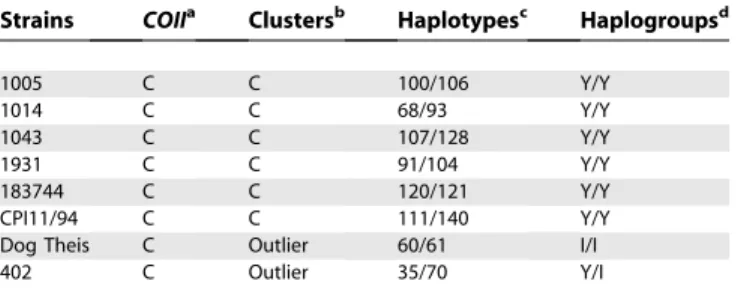
Table 2.Nuclear and Mitochondrial Markers ofT. cruziInferred in This Work
Strains COIIa Clustersb Haplotypesc Haplogroupsd
A83 A A 25/26 Z/Z
A87 A A 27/28 Z/Z
Col18/05 A A 21/56 Z/Z
Colombiana A A 24/56 Z/Z
Cuı´ca A A 31/49 Z/Z
Cutia A A 44/47 Z/Z
D7 A A 6/29 Z/Z
Gamba cl1 A A 30/46 Z/Z
Rb1 A A 36/57 Z/Z
Rb2 A A 16/36 Z/Z
Rb6 A A 38/41 Z/Z
SE A A 48/53 Z/Z
SilvioX10cl1 A A 32/50 Z/Z
1001 A A 43/43 Z/Z
1004 A A 23/37 Z/Z
1006 A A 22/40 Z/Z
1502 A A 39/45 Z/Z
1523 A A 42/51 Z/Z
115 B B 7/19 X/X
222 B B 1/20 X/X
226 B B 9/18 X/X
231 B B 11/17 X/X
3663 B B 8/10 X/X
3869 B B 13/14 X/X
4182 B B 15/62 X/I
M5631cl5 B B 12/12 X/X
M6241cl6 B B 33/34 X/X
167 B BH 4/99 X/Y
182 B BH 5/108 X/Y
1022 B BH 2/102 X/Y
CLBrener B BH 5/100 X/Y
Mncl2 B BH 52/133 X/Y
NR B BH 55/130 X/Y
SC43 cl1 B BH 54/129 X/Y
SO3 B BH 54/130 X/Y
Tula cl2 B BH 3/103 X/Y
CanIII cl1 B Outlier 58/59 I/I
Be62 C C 88/105 Y/Y
CPI95/94 C C 110/114 Y/Y
Esmeraldo C C 109/135 Y/Y
Gil C C 77/79 Y/Y
GLT564 C C 97/98 Y/Y
GLT593 C C 74/131 Y/Y
GMS C C 90/131 Y/Y
GOCH C C 81/126 Y/Y
Ig539 C C 123/125 Y/Y
JAF C C 67/92 Y/Y
JG C C 82/92 Y/Y
JHF C C 83/84 Y/Y
JSM C C 65/95 Y/Y
Mas1 cl1 C C 63/136 Y/I
MPD C C 64/76 Y/Y
OPS27/94 C C 134/141 Y/Y
Tu18 cl11 C C 137/138 Y/Y
Y C C 86/87 Y/Y
84 C C 66/78 Y/Y
84Ti C C 71/73 Y/Y
169/1 C C 85/94 Y/Y
200pm C C 112/115 Y/Y
207 C C 75/139 Y/Y
209 C C 69/101 Y/Y
239 C C 80/96 Y/Y
577 C C 89/124 Y/Y
578 C C 122/127 Y/Y
580 C C 117/118 Y/Y
581 C C 113/116 Y/Y
803 C C 72/119 Y/Y
Table 2.Continued.
Strains COIIa Clustersb Haplotypesc Haplogroupsd
1005 C C 100/106 Y/Y
1014 C C 68/93 Y/Y
1043 C C 107/128 Y/Y
1931 C C 91/104 Y/Y
183744 C C 120/121 Y/Y
CPI11/94 C C 111/140 Y/Y
Dog Theis C Outlier 60/61 I/I
402 C Outlier 35/70 Y/I
a
RFLP typing of theT. cruzi COIIgene.
b
Clusters of strains generated by MDS analysis.
c
Haplotypes inferred by PHASE.
d
Nuclear genetic typing.Amplification of five previously described
microsatellite loci, denominated SCLE10, SCLE11, MCLE01,
MCLF10, and MCLG10, was performed as previously described [30]. After the PCR, the amplified microsatellites were loaded on a 6% denaturing polyacrylamide gel and analyzed on an ALF sequencer (GE Healthcare, Milwaukee, Wisconsin, United States) using the Allelinks software (GE Healthcare). To determine the allele size the samples were directly compared with the band sizes from an allelic ladder prepared by amplification of an artificial mixture of DNA
from 60T. cruzistrains.
Amplification of the D7 divergent domain of the 24SarRNA gene
was achieved by PCR with D71 fluorescent (59
-AAGGTGCGTCGA-CAGTGTGG-39) and D72 (59-TTTTCAGAATGGCCGAACAGT-39)
primers following protocols described previously [24]. The amplifi-cation products were also analyzed in ALF sequencer and allele sizes determined by the Allelinks software.
Mitochondrial genetic typing.Amplification of the mitochondrial
COII gene [37] was performed using the primers TcMit31 (59
-TAAATAATATATATTGTACATGAG-39) and TcMit40 (59
-CTRCATTGYCCATATATTGT-39). Total DNA (1–10 ng) were used
in each PCR reaction in the following condition: 30 s denaturation at
948, primer annealing for 2 min at 488, and primer extension for 2
min at 72 8, in a total of 30 cycles. The amplified products were
purified and sequenced using primer TcMit31 and the cycle sequencing with Thermo-Sequenase (ETKit; GE Healthcare) using the thermal cycling program recommended in the kit. The sequenc-ing products were purified and run on a MegaBACE capillary sequencer (GE Healthcare). After Phred, Phrap, and Consed analyses, the sequences were trimmed to have equal length (290 base pairs). All bases sequenced had Phred values above 30 [44].
Based on the restriction map of COII sequences, the AluI
restriction endonuclease was chosen to perform RFLP analyses in
the mitochondrialCOIIgene. After PCR amplification, the amplicons
were submitted to enzyme digestion for 16 hours according to instructions provided by the manufacturer (Promega, Madison, Wisconsin, United States). Digested products were analyzed on polyacrylamide gel electrophoresis and silver stained.
Sequences for the mitochondrialCYbgene [35] and theND1(37)
were obtained from GenBank.
Construction of distance matrices, multidimensional scaling, and NJ trees. Based on the microsatellite results, a distance matrix between the strains was constructed as described previously [30]. In order to provide a visual representation of the distance matrix we used the multidimensional scaling plot using the software Statistica Version 6.0 [45]. Analyses of molecular variance for the mitochon-drial sequences were performed using the Arlequin v.2.0 software using 1,000 permutations [46].
NJ trees were obtained separately for the COII, CYb, and ND1
sequences with the MEGA v. 3.1 software [47] using the Kimura 2 parameter and 500 replications for the bootstrap statistics.
Haplotype inference and network construction. Haplotypes were
reconstructed from the 75T. cruzipopulations by using a Bayesian
coalescent theory-based method contained in PHASE software (Version 2.0.2 for Linux) [40]. The type of polymorphism (SNP or
Figure 4.Median Joining Network of the Haplotypes Identified by the PHASE Software
DOI: 10.1371/journal.ppat.0020024.g004
Figure 5.Diagram Depicting the Proposed Model for the Evolution ofT.
cruziStrains
The mitochondrial clade was typed by RFLP of theCOIImaxicircle gene, the MDS clusters were established by multidimensional scaling of microsatellite data, the haplogroups were established by haplotype estimation from multilocal genotypes followed by median joining network analysis, and the RAPD/multilocus enzyme electrophoresis typing was obtained from Brisse et al. [26].
multiallelic with stepwise mutation mechanism for rDNA and microsatellite data, respectively) is taken into account in PHASE. For the analyses the default parameters of the program were used, with additional runs up to 10,000 permutations. These were the best-tested conditions, giving highly reproducible results. The resultant haplotypes were then arranged in a network by using the Median Joining analysis [41], available in NETWORK 3.1 software provided by Fluxus Technology (http://www.fluxus-engineering.com).
Supporting Information
Table S1.Typing of rDNA Group and Allele Sizes (in bp) of Five Microsatellite Loci
Found at DOI: 10.1371/journal.ppat.0020024.st001 (105 KB DOC).
Accession numbers
The GenBank (http://www.ncbi.nlm.nih.gov/Genbank) accession num-bers for the genes and gene products discussed in this paper are 24S
rDNA (L19411), mini-exon gene (X62674), COII (AF359041 and
DQ343715–DQ343753), CYb (AJ130921, AJ130931–AJ130938,
AJ439719–AJ439727), and ND1 (AF359009, AF359011–AF359029,
AF359031–AF359053).
Acknowledgments
We are grateful to Dr. Jose´ Rodrigues Coura and Dr. Ana Maria Jansen-Franken, from the Departamento de Medicina Tropical and the Departamento de Protozoologia, FIOCRUZ (Rio de Janeiro, Brazil), respectively, and Dr. M. Tibayrenc from the Centre d’E´tudes sur le Polymorphisme des Microorganismes (Montpellier,
France) for kindly providing DNA fromT. cruzi strains. We thank
Neuza A. Rodrigues and Katia B. Gonc¸alves for expert technical assistance.
Author contributions.SMRT, AMM, CRM, and SDJP conceived and designed the experiments. JMdF, LAP, JRP, LBR, and VFG performed the experiments. JMdF, LAP, JRP, LBR, VFG, and SDJP analyzed the data. EC, ACVJ, and OF contributed reagents/materials/analysis tools. AMM, CRM, and SDJP wrote the paper.
Funding.Supported by CNPq-Brazil, PRONEX, and World Health Organization. The research of SMRT and CRM was supported, in part, by an International Research Scholars Grant from the Howard Hughes Medical Institute.
Competing interests.The authors have declared that no competing
interests exist. &
References
1. Prata A (2001) Clinical and epidemiological aspects of Chagas disease. Lancet Infect Dis 1: 92–100.
2. World Health Organization [WHO] (1991) Control of Chagas disease. Technical Report Series, 811. Geneva: WHO. 91 pp.
3. Briones MR, Souto RP, Stolf BS, Zingales B (1999) The evolution of two
Trypanosoma cruzisubgroups inferred from rRNA genes can be correlated with the interchange of American mammalian faunas in the Cenozoic and has implications to pathogenicity and host specificity. Mol Biochem Parasitol 104: 219–232.
4. Aufderheide AC, Salo W, Madden M, Streitz J, Buikstra J et al. (2004) A 9,000-year record of Chagas’ disease. Proc Natl Acad Sci U S A. 101: 2034– 2039.
5. Dias JC, Silveira AC, Schofield CJ (2002) The impact of Chagas disease control in Latin America: A review. Mem Inst Oswaldo Cruz. 97: 603–612. 6. Kirchhoff LV (1993) American Trypanosomiasis (Chagas’ Disease)—a
tropical disease now in the United States. N Engl J Med. 329: 639–644. 7. Jansen AM, Santos de Pinho AP, Lisboa CV, Cupolillo E, Mangia RH, et al.
(1999) The sylvatic cycle ofTrypanosoma cruzi:A still unsolved puzzle. Mem Inst Oswaldo Cruz. 94: 203–204.
8. Lisboa CV, Mangia RH, De Lima NR, Martins A, Dietz J, et al. (2004) Distinct patterns ofTrypanosoma cruziinfection inLeontopithecus rosaliain distinct Atlantic coastal rainforest fragments in Rio de Janeiro–Brazil. Parasitology 129: 703–711.
9. Pung OJ, Banks CW, Jones DN, Krissinger MW (1995)Trypanosoma cruziin wild raccoons, opossums, and triatomine bugs in southeast Georgia, U.S.A. J Parasitol. 81: 324–326.
10. Pietrzak SM, Pung OJ (1998) Trypanosomiasis in raccoons from Georgia. J Wildl Dis. 34: 132–136.
11. Bradley KK, Bergman DK, Woods JP, Crutcher JM, Kirchhoff LV (2000) Prevalence of American trypanosomiasis (Chagas disease) among dogs in Oklahoma. J Am Vet Med Assoc. 217: 1853–1857.
12. Maguire JH, Hoff R, Sleigh AC, Mott KE, Ramos NB, et al. (1986) An outbreak of Chagas’ disease in southwestern Bahia, Brazil. Am J Trop Med Hyg. 35: 931–936.
13. Shikanai-Yasuda MA, Marcondes CB, Guedes LA, Siqueira GS, Barone AA, et al. (1991) Possible oral transmission of acute Chagas’ disease in Brazil. Rev Inst Med Trop Sao Paulo. 33: 351–357.
14. da Silva Valente SA, de Costa Valente V, Neto HF (1999) Considerations on the epidemiology and transmission of Chagas disease in the Brazilian Amazon. Mem Inst Oswaldo Cruz. 94: 395–398.
15. Pedroso A, Cupolillo E, Zingales B (2003) Evaluation ofTrypanosoma cruzi
hybrid stocks based on chromosomal size variation. Mol Biochem Parasitol 129: 79–90.
16. El-Sayed NM, Myler PJ, Bartholomeu DC, Nilsson D, Aggarwal G, et al. (2005) The genome sequence ofTrypanosoma cruzi, etiologic agent of Chagas disease. Science 309: 409–415.
17. Macedo AM, Pena SDJ (1998) Genetic variability of Trypanosoma cruzi:
Implications for the pathogenesis of Chagas’ disease. Parasitol Today 14: 119–124.
18. Tibayrenc M (2003) Genetic subdivisions withinTrypanosoma cruzi(Discrete Typing Units) and their relevance for molecular epidemiology and experimental evolution. Kinetoplastid Biol Dis. 28: 2–12.
19. Satellite Meeting, (1999) Recommendations from an international sympo-sium to commemorate the 90th anniversary of the discovery of Chagas disease. April 11–16 1999, Rio de Janeiro, Brazil. Mem Inst Oswaldo Cruz 94: 429–432.
20. Fernandes O, Santos S, Junqueira A, Jansen A, Cupolillo E et al. (1999)
Populational heterogeneity of BrazilianTrypanosoma cruziisolates revealed by the mini-exon and ribosomal spacers. Mem Inst Oswaldo Cruz 94: 195– 197.
21. Zingales B, Stolf BS, Souto RP, Fernandes O, Briones MR (1999) Epidemiology, biochemistry and evolution ofTrypanosoma cruzi lineages based on ribosomal RNA sequences. Mem Inst Oswaldo Cruz 94: 159–164. 22. Freitas JM, Lages-Silva E, Crema E, Pena SDJ, Macedo AM (2005) Real time PCR strategy for the identification of major lineages ofTrypanosoma cruzi
directly in chronically infected human tissues. Int J Parasitol. 35: 411–417. 23. Miles MA, Souza A, Povoa M, Shaw JJ, Lainson R, et al. (1978) Isozymic heterogeneity ofTrypanosoma cruziin the first autochthonous patients with Chagas’ disease in Amazonian Brazil. Nature 272: 819–821.
24. Souto RP, Fernandes O, Macedo AM, Campbell DA, Zingales B (1996) DNA markers define two major phylogenetic lineages ofTrypanosoma cruzi. Mol Biochem Parasitol 83: 141–152.
25. Stolf BS, Souto RP, Pedroso A, Zingales B (2003) Two types of ribosomal RNA genes in hybridTrypanosoma cruzistrains. Mol Biochem Parasitol 126: 73–80.
26. Brisse S, Dujardin JC, Tibayrenc M (2000) Identification of sixTrypanosoma cruziphylogenetic lineages by random amplified polymorphic DNA and multilocus enzyme eletrophoresis. Int J Parasitol 30: 35–44.
27. Macedo AM, Machado CR, Oliveira RP, Pena SDJ (2004)Trypanosoma cruzi:
Genetic structure of populations and relevance of genetic variability to the pathogenesis of Chagas disease. Mem Inst Oswaldo Cruz. 99: 1–12. 28. Gaunt MW, Yeo M, Frame IA, Stothard JR, Carrasco H J, et al. (2003)
Mechanism of genetic exchange in American trypanosomes. Nature 241: 936–939.
29. Tibayrenc M, Ward P, Moya A, Ayala FJ (1986) Natural populations of
Trypanosoma cruzi,the agent of Chagas’ disease, have a complex multiclonal structure. Proc Nat Acad Sci U S A 83: 115–119.
30. Oliveira RP, Broude NE, Macedo AM, Cantor CR, Smith CL et al. (1998) Probing the genetic population structure of Trypanosoma cruzi with polymorphic microsatellites. Proc Natl Acad Sci U S A 95: 3776–3780. 31. Tibayrenc M, Ayala FJ (2002) The clonal theory of parasitic protozoa: 12
years on. Trends Parasitol. 18: 405–410.
32. Bogliolo AR, Lauria-Pires L, Gibson WC (1996) Polymorphisms in
Trypanosoma cruzi:evidence of genetic recombination. Acta Trop 61: 31–40. 33. Carrasco HJ, Frame IA, Valente SA, Miles MA (1996) Genetic exchange as a possible source of genomic diversity in sylvatic populations ofTrypanosoma cruzi. Am J Trop Med Hyg 54: 418–224.
34. Higo H, Yanagi T, Matta V, Agatsuma, Cruz-Reyes A, et al. (2000) Genetic structure ofTrypanosoma cruziin American continents: Special emphasis on sexual reproduction in Central America. Parasitology 121: 403–408. 35. Brisse S, Henriksson J, Barnabe C, Douzery EJ, Berkvens D, et al. (2003)
Evidence for genetic exchange and hybridization inTrypanosoma cruzibased on nucleotide sequences and molecular karyotype. Infect Genet Evol 2: 173–183.
36. Sturm NR, Vargas NS, Westenberger SJ, Zingales B, Campbell DA (2003) Evidence for multiple hybrid groups inTrypanosoma cruzi. Int J Parasitol 33: 269–279.
37. Machado CA, Ayala FJ (2001) Nucleotide sequences provide evidence of genetic exchange among distantly related lineages ofTrypanosoma cruzi. Proc Natl Acad Sci U S A 98: 7396–7401.
38. Augusto-Pinto L, Teixeira SM, Pena SDJ, Machado CR (2003) Single-nucleotide polymorphisms of theTrypanosoma cruzi MSH2gene support the existence of three phylogenetic lineages presenting differences in mismatch-repair efficiency. Genetics 164: 117–126.
inferred from metric distances among DNA haplotypes: Application to human mitochondrial DNA restriction data. Genetics 131: 479–491. 40. Stephens M, Smith NJ, Donnelly P (2001) A new statistical method for
haplotype reconstruction from population data. Am J Hum Genet 68: 978– 989.
41. Bandelt HK, Forster P, Rohl A (1999) Median-joining networks for inferring intraspecific phylogenies. Mol Biol Evol 16: 37–48.
42. Leopoldino AM, Pena SDJ (2003) The mutational spectrum of human autosomal tetranucleotide microsatellites. Hum Mutat 21: 71–79. 43. Tibayrenc M (1996) Towards a unified evolutionary genetics of
micro-organisms. Annu Rev Microbiol 50: 401–429.
44. Ewing B, Hillier L, Wendl MC, Green P (1998) Base-calling of automated sequencer traces using phred. I. Accuracy assessment. Genome Res 8: 175– 185.
45. Beals R, Krantz DH, Tversky A (1968) Foundations of multidimensional scaling. Psychol Rev 75: 127–142.
46. Schneider S, Roessli D, Excoffier L (2000) Arlequin ver 2.000: A software for population genetics data analysis [computer program]. Geneva: Genetics and Biometry Laboratory, University of Geneva.