Ana Rita Marques
Dissertation presented to obtain the Ph.D degree in
Developmental Biology
Instituto de Tecnologia Química e Biológica
Universidade Nova de Lisboa
Research work performed at:
Oeiras, July, 2011
This dissertation was sponsored by Fundação para a Ciência e Tecnologia.
“Apoio financeiro da FCT e do FSE no âmbito do Quadro Comunitário de apoio, BD nº SFRH/BD/28767/2006 e projecto
FCT-POCI-DG/BIA/82013/06”
Ana Rita Marques
Dissertation presented to obtain the Ph.D degree in
Developmental Biology
Instituto de Tecnologia Química e Biológica
Universidade Nova de Lisboa
Research work performed at:
Oeiras, July, 2011
This dissertation was sponsored by Fundação para a Ciência e Tecnologia.
“Apoio financeiro da FCT e do FSE no âmbito do Quadro Comunitário de apoio, BD nº SFRH/BD/28767/2006 e projecto
The work described in this thesis contributed to the following publications.
1. Pimenta-Marques, A., Tostoes, R., Marty, T., Barbosa, V.,
Lehmann, R. and Martinho, R. G. (2008). Differential
requirements of a mitotic acetyltransferase in somatic and germ line cells.Dev Biol323, 197-206.
2. Van Damme, P.#, Hole, K.#, Pimenta-Marques, A.#, Helsens, K.,
Vandekerckhove, J., Martinho, R.G., Gevaert, K., and Arnesen,
T. (2011). NatF contributes to an evolutionary shift in protein
N-terminal acetylation and is important for normal chromosome segregation.PLoS Genet7,e1002169.
# - These authors contributed equally to this work.
3. Guilgur, L., Prudêncio, P., Ferreira, T., Pimenta-Marques, A.,
Martinho, R.G. (2011). Drosophila aPKC is required for mitotic
spindle orientation during symmetric division of epithelial cells (submitted).
This thesis is the result of the work I developed between January 2007 and December 2010 under the supervision of Dr. Rui Gonçalo Martinho, at the laboratory of Early Fly Development at Instituto Gulbenkian de Ciência, Oeiras, Portugal.
Collaborations are indicated in the sections Materials and Methods.
First I would like to thank my supervisor Rui Martinho for taking me in his lab and giving me the opportunity to grow as a scientist. He made me believing that I was capable of starting and one day finishing a PhD. It was lots of fun and I will remember this learning process with care and laughter.
I must also thank Thomas Arnesen and Petra Van Damme for all the support and enthusiastic discussions we had about my project and NATs.
To the CCR group I must thank all the support regarding both scientific discussions and reagents. Particularly, Zita Santos and Inês Ferreira for introducing me to the cell culture world. Besides being excellent colleagues, I must also thank them, as well as to Inês Bento and Ana Martins for their true friendship during these years.
Ao Tostões e ao André tenho a agradecer o facto de terem tornado as horas infindáveis na sala das moscas em momentos de autêntica diversão e boa disposição. A amizade deles foi indispensável neste percurso.
À Tânia, Pedro e Gaston tenho a agradecer a amizade e forte companheirismo e união que temos entre nós. É de facto um privilégio acordar de manhã e ir trabalhar com um grupo de pessoas de quem genuinamente gostamos e com quem nos identificamos. Além disso ainda nos fartamos de rir juntos.
Ao Paulinho e à Catarina tenho a agradecer por estarem presentes não só como colegas de trabalho em outros laboratórios mas principalmente por serem meus grandes amigos.
apoio incondicional que sempre me deram. Ter partilhado com eles os melhores e piores momentos fez-me crescer e aprender a valorizar o que é verdadeiramente importante e a relativizar o dispensável. Se de alguma forma tenho sido bem sucedida na vida, o devo a eles. Quanto às minhas irmãs… são as maiores!!! Sou uma sortuda por ter duas manas destas na minha vida.
Ao meu avô Pimenta agradeço por ser o avô único que é! O meu avô tem sido uma presença constante e fundamental no meu processo de crescimento. Desde que sai de Évora que o meu avô me faz o telefonema da praxe todas as semanas. Essas e outras demonstrações de carinho fazem-me sentir uma “netinha” menina, apesar dos 31 anos, e isso é tão bom!
Aos meus tios Chico e São tenho a agradecer o facto de ter sempre podido contar com eles. Foram também uma presença constante nos momentos importantes da minha vida.
À Ju, Avô João, D.Virginia, Tio e Ana Catarina, tenho a agradecer o facto de me terem acolhido como um verdadeiro membro da família e do pelo forte apoio que me deram nestes últimos anos. Os nossos almoços de fim de semana são uma lufada de ar fresco.
Claro, ao meu maridão tenho a agradecer o amor, amizade, companheirismo e paciência que teve comigo nestes últimos cinco anos. É muito bom para mim ter alguém com a personalidade do Gonçalo ao meu lado. O facto de partilhar a minha vida com ele faz com que tudo o que é bom seja “maravilhoso” e tudo o que é mau seja apenas “menos bom”. É cá uma peça o meu marido.
Protein N-terminal acetylation is a highly conserved and
widespread modification that occurs on approximately 80% of all soluble, cytoplasmic human proteins. Nevertheless, with few
exceptions, little is known about the biological function of protein N
-acetylation. Recently, it was suggested to act as a general protein destabilization signal in yeast (Hwang et al., 2010). Yet, other reports suggest it might act as stabilizer, for instance by blocking protein degradation (Ciechanover and Ben-Saadon, 2004). This protein
modification is catalyzed by N-terminal acetyltransferases (NATs),
which are highly conserved from yeast to humans both in subunit composition and substrate specificity (Starheim et al., 2009). Since NATs enzymatic activity and function has been mostly studied in yeast and tissue culture cells, our current understanding of the role of these enzymes during development of multicellular organisms is extremely poor, being our main goal to understand the role of this ubiquitous protein modification during development.
In Chapter II of this work, we isolated two genes from a maternal genetic screen designed to identify genes required for blastoderm embryonic development. Both genes were required for syncytial nuclear divisions. One of the isolated genes corresponded to
involved in centromeric sister chromatid cohesion (Williams et al., 2003). This conclusion came from the observation that san mutant neuroblasts exhibited premature loss of sister chromatid cohesion during mitosis accompanied by mislocalization of a subunit of the cohesin complex from the centromeric region.
Detailed characterization of Drosophila san mutants (chapter III) revealed striking defects during chromosome segregation in the syncytial nuclear divisions (Pimenta-Marques et al., 2008). These included lagging chromosomes and extensive chromosome bridges. Additionally, we observed that during mitosis a subset of nuclei showed a dramatic decrease in the levels of a condensin subunit. Moreover, a subset of chromosome bridges showed reduced levels of Topoisomerase II. Together, these observations suggested dSan/dNaa50 was also important for chromosome condensation/ resolution, implying a more general mitotic function of this NAT. Surprisingly, our work showed that whereas all analyzed somatic tissues required dSan/dNaa50 for correct cell cycle progression, during Drosophilaoogenesis we failed to detect mitotic defects in the female germ-line stem cells mutant for dSan/dNaa50, suggesting
there are differential requirements for N-acetylation between the
female germ-line and the soma. We hypothesized that different NATs are likely to have different tissue-specific functions throughout development. In order to test this hypothesis we focused on: 1) defining the mitotic function of dSan/dNaa50, 2) understanding why is this enzyme not required in the female germ-line.
methodology to identify proteins whose N -acetylation levels were
reduced insanmutant embryos. We identified 18 proteins whose N
-acetylation levels were reduced in san mutants. From these 18 proteins, we identified the gamma subunit of the cytosolic chaperonin containing T-complex polypeptide 1 (CTT) (Cct), which displayed
chromosome segregation defects identical to dSan/dNaa50-depleted cells. Cct was shown to be required for the metaphase to anaphase
transition by activating the anaphase promoting complex/ cyclosome complex (Camasses et al., 2003), making this protein an excellent candidate for a putative mitotic substrate of dSan/dNaa50. Ongoing complementation experiments in tissue culture cells will allow us to test if the chromosome segregation defects of San-depleted cells are Cct-mediated.
A acetilação N-terminal de proteínas é uma modificação muito
comum e conservada que ocorre em cerca de 80% das proteínas solúveis citoplasmáticas em humanos. Com algumas excepções, pouco se sabe acerca da função biológica da acetilação N-terminal.
Foi recentemente sugerido em levedura, que esta modificação pode actuar como um sinal de destabilização de proteínas (Hwang et al., 2010). Outros trabalhos sugerem que ao invés, possa actuar com estabilizador, como por exemplo no bloqueio de degradação proteica (Ciechanover and Ben-Saadon, 2004). Esta modificação é catalisada por acetiltransferases de N-terminais (NATs). Estas enzimas são
altamente conservados desde as leveduras aos humanos, quer na composição das suas subunidades quer na especificidade dos seus substratos (Starheim et al., 2009). Uma vez que a actividade e função destas enzimas tem sido principalmente estudada em levedura e sistemas de cultura de células, o conhecimento sobre a função da acetilação de N-terminais no contexto de um organismo multicelular
é ainda muito limitado. O nosso principal objectivo é o de compreender a função desta modificação de proteínas durante o desenvolvimento.
Naa10p e a Naa15p designado por NatE. Curiosamente, e ao contrário do que foi observado para as outras NATs, nós e outros grupos de investigação mostrámos que células onde a actividade da dSan/dNaa50 foi eliminada, apresentam fenótipos específicos durante a mitose (Hou et al., 2007; Pimenta-Marques et al., 2008; Williams et al., 2003). A dSan/dNaa50 foi originalmente descrita como uma proteína envolvida na coesão centromérica das cromatidias irmãs (Williams et al., 2003). Esta conclusão veio da observação de que neuroblastos de mutantes para san exibem perda prematura de coesão das cromátidias irmãs durante a mitose. Foi igualmente descrita a deslocalização de uma subunidade do complexo da coesina na região centromérica dos cromossomas.
linha germinal feminina e as células somáticas apresentam requisitos diferentes para a acetilação de N-terminais. Com base nos nossos
resultados, hipotetizamos que diferentes NATs poderão ter diferentes funções reguladoras ao longo do desenvolvimento de diferentes tecidos. Para testar esta hipótese focámo-nos em 1) definir a função mitótica da dSan/dNaa50, 2) compreender porque é que a dSan/dNaa50 não é necessária na linha germinal feminina.
De modo a identificarmos a via molecular em que a dSan/dNaa50 regula a mitose, decidimos identificar os substratos mitóticos desta NAT (capítulo IV). Para esse fim, tirámos partido da cromatografia combinada de fraccionamento de N-terminais, de forma a identificar proteínas cujos níveis de N-acetilação estão reduzidos
em embriões mutantes para dSan/dNaa50, por comparação com embriões de estirpe selvagem. Desta análise resultou a identificação de 18 proteínas cujos níveis de N-acetilação foram reduzidos em
mutantes para dSan/dNaa50. Uma das 18 proteínas identificadas foi a subunidade gama da chaperonina citosólica contendo o complexo T-polipeptídeo (CTT) (Cct). A Cct apresentou defeitos de segregação
cromossómica muito semelhantes aos observados para células onde a dSan/dNaa50 foi eliminada. Esta chaperonina foi descrita como sendo necessária para a transição metáfase-anáfase por activação do complexo promotor da anáfase/complexo ciclosoma (Camasses et al., 2003). As funções associadas à Cct tornam-na numa excelente
Acetyl-CoA Acetyl-coenzyme A
Ala Alanine
APC/C Anaphase promoting complex/cyclosome
Arg Arginine
BDGP Berkeley Drosophila Genome Project BDSC Bloomington Drosophila Stock Center Bub3 Budding uninhibited by benzimidazole 3
BubR1 Budding uninhibited by benzimidazole 3 related 1 C(2)M Crossover suppressor on 2 of Manheim
CCT Chaperonin containing T-complex polypeptide 1
Cct Gamma subunit of CCT
Cdc20 Cell-division-cycle 20 homologue
Cdh1 cdc20-homologue 1
CDIP cell death involved p53-target
Cdk Cyclin-dependent kinase
CDS Coding DNA sequences
Cnn Centrosomin
Cid Centromere identifier
CkII Casein kinase II α subunit
COFRADIC Combined fractional diagonal chromatography EGAF Embryonic Growth-Associated factor
EMS Ethylmethane sulfonate
EST Expressed sequence tag
FRT Flipase recombination targets
GEF Guanine nucleotide exchange factor
GLC Germ-line clone
GNAT Gcn5-related N-acetyltransferases GSK-3 Glycogen synthase kinase 3
HIF-1 Hypoxia-inducible factor 1
His Histidine
HMG high mobility group
HPLC High performance liquid chromatography HYPK Huntingtin (Htt) yeast two-hybrid protein K hs-FLP Heat-shock induced flipase
IKK IkB kinase
iMet Initiator methionine
INCENP Inner centromere protein
Lys Lysine
Mad2 Mitotic arrest-deficient 2
MAP Methionine aminopeptidases
MT Microtubules
MTOC Microtubule organizing center NAT N-terminal acetyltransferases
NARG1 NMDA receptor-regulated gene 1
NEBD Nuclear envelope break-down
NMDA N-methyl-d-aspartate
OR Oregon-R
ORF Open reading frame
PCR Polymerase chain reaction
Plk1 Polo-like kinase 1
PolyQ Polyglutamine
PP2A Protein phosphatase 2A
PTM Posttranslational modification
RF-C Replication factor C
PGC Primordial Germ Cell
PKA Protein kinase A
RT Room temperature
San Separation anxiety
SCX Strong cation exchange
SAC Spindle assembly checkpoint
Ser Serine
Sgo Shugoshin
SMC Structural maintenance of chromosomes
snRNP Small ribonucleproteins
Tbdn-1 Tubedown-1
Thr Threonine
TNBS 2,4,6-trinitrobenzenesulfonic acid TNF Tumor necrosis factor alpha
TopoII Topoisomerase II
Tpm1 Tropomyosin 1
TPR Tetratrico-peptide repeat
P53 Tumor protein 53
Tyr Tyrosine
Ub Ubiquitin
vnc variable nurse cells
List of publications……….. iii
Acknowledgments/ Agradecimentos……… v
Abstract………. vii
Sumário………. xi
Abbreviations………... xv
CHAPTER I –GENERAL INTRODUCTION 1. Cell Cycle……….. 2
1.1. Cell cycle regulation………. 3
1.2. Mitosis……….... 3
1.3. The spindle assembly checkpoint and the metaphase to anaphase transition……….. 6
1.4. Chromosome cohesion and condensation………... 7
1.4.1. Sister chromatid cohesion………... 8
1.4.2. Chromosome condensation……… 13
2. Posttranslational modifications……….. 16
2.1. Posttranslational modifications and the cell………. 18
2.1.1. Phosphorilation……….. 18
2.1.2. Acetylation……….. 20
2.1.3. Methylation……….... 21
2.1.4. Ubiquitination………... 22
3. N -terminal acetylation……….. 23
3.1. The NatA acetyltransferase complex………. 26
3.1.1. Naa10/ Ard1 acetyltransferase………... 28
3.1.2. Naa15/ NatH acetyltransferase……….. 30
3.1.3. Knockout studies demonstrate the NatA complex is involved in several biological processes………... 32
3.1.4. The NatA complex is associated with tumor development 34 3.2. The NatB complex……… 35
3.2.1. NatB known substrates and knockdown studies…………. 36
3.3. The NatC complex……… 37
3.3.1. NatC known substrates and knockdown studies…………. 38
3.4. The NatD complex……… 40
3.5. San and the NatE complex………. 40 3.5.1. San is required for centromeric sister chromatid
1. Introduction……….. 46
2. Materials and methods……….. 48
2.1. Fly husbandry……… 48
2.2. 2R maternal screen……….. 48
2.3. Mapping and isolation of complementation groups………. 49
2.4. DNA sequencing of candidate genes……… 49
2.5. Molecular biology and generation of transgenic lines……. 50
2.5.1. UAS-sangeneration………. 50
2.5.2. Genomic fragments for complementation group 3……….. 50
2.6. DNA staining of complementation group 3 embryos…….. 51
3. Results………... 53
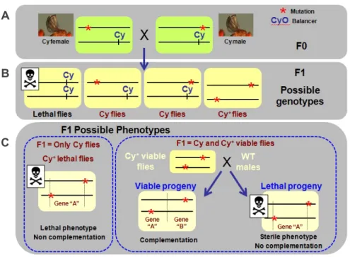
3.1. Right-arm of 2R maternal screen allowed the identification of six complementation groups……… 53
3.2. Mapping of complementation group 2………... 55
3.2.1. Complementation group 2 is allelic to the gene separation anxiety(san)……….. 55
3.2.2. sancDNA rescues the viabilitysanmutants……… 56
3.3. Mapping of complementation group 3………... 56
3.3.1. Cytological mapping of complementation group 3……….. 56
3.3.2. A genomic DNA fragment containing 2 genes rescues the sterility of complementation group 3 mutants………… 58
3.4. san and complementation group 3 alleles showed dramatic defects during syncytial nuclei divisions………... 60
4. Discussion……… 62
CHAPTER III - DIFFERENTIAL REQUIREMENTS OF A MITOTIC ACETYLTRANSFERASE IN SOMATIC AND GERM LINE CELLS………. 67-90 CHAPTER IV – dSan/dNaa50 IN VIVO N-ACETYLATES MULTIPLE TARGETS AND SHARES PARTIAL REDUNDANCY WITH OTHER N -TERMINAL ACETYLTRANSFERASES 1. Introduction……… 92
2. Materials and methods……… 94
2.1. Embryonic protein extracts……… 94
2.4. Immunofluorescence of Dmel2 Cells………... 98
2.5. Microscopy………... 99
3. Results………. 100
3.1. dSan/dNaa50 N-terminal acetyltransferase acetylates multiple different substratesin vivo……….. 100
3.2. Tissue culture cells depleted for dSan/dNaa50 showed chromosome segregation defects……… 104
3.3. Cct and dSan/dNaa50-depleted cells have similar chromosome segregation defects……… 108
3.4. Looking for mitotic substrates of dSan/dNaa50 using a N-terminal substrate consensus………... 114
3.5. Cohesin Drad1/Scc1 shares a well conserved N-terminus within higher eukaryotes……… 114
4. Discussion……….. 117
4.1. Is Ccta mitotic substrate of dSan/dNaa50?... 117
4.2. Is Drad21/Scc1 a mitotic substrate of dSan/dNaa50?... 122
4.3. Are dSan/dNaa50 knockout phenotypes mediated by internal acetylation of-tubulin?... 124
CHAPTER V – dNaa60IS REQUIRED FOR CHROMOSOME SEGREGATION DURING ANAPHASE 1. Introduction……… 128
2. Materials and methods……… 129
2.1. Bioinformatic analysis of Drosophila paralogues of dSan/dNaa50………... 129
2.2. RNA interference on culturedDrosophilaDmel2 cells…. 129 2.3. Transfection of constructs with tagged proteins………… 130
2.4. Immunofluorescence of Dmel2 Cells………... 131
2.5. Microscopy……….. 132
2.6. Creation of CG18177/dnaa60mutants……… 132
3. Results………. 134
3.1. Identification ofDrosophilaparalogues of dSan/dNaa50. 134 3.2. Identification of a new NAT required for chromosome segregation……….. 138
3.3. dnaa60is not an essential gene……….. 142
4.1. Higher complexity in the regulation of protein N
-acetylation inDrosophila melanogaster……….. 146 4.2. Potential redundancy between dSan/dNaa50 and
dNaa60 within the female germ-line……… 147 4.3. dNaa60 is possibly involved in posttranslational N
-acetylation……… 150
CHAPTER VI – GENERALDISCUSSION
1. Tissue specific functions of N-terminal acetylation?... 152
2. Regulatory functions of N-terminal acetylation…………. 154
2.1. “Net model” for N-terminal acetylation………... 155
2.2. Can different substrates require different levels of N
-terminal acetylation?... 156 2.3. Is NATs activity modulated by cofactor proteins?... 157
3. Conclusion……… 158
4. Future work……….. 158
REFERENCES……….. 161
1. Cell cycle
The cell cycle is a series of events that takes place in a
eukaryotic cell leading to its division and replication. Numerous
proteins regulate the correct progression of the cell cycle, which
ensures the accurate transmission of genetic information from a
mother cell to its daughter cells. The eukaryotic cell cycle is typically
divided on basis of chromosomal events: interphase and M phase
(mitosis) (fig. 1). Interphase is comprised by G1 phase followed by S
phase and G2 phase. The G1 and G2 phases represent “gaps” in the
cell cycle that occur between the two landmark events – DNA
synthesis (S phase) and mitosis (M phase). In these stages, the cells
grow and pass through regulatory transitions in which progression to
the next phase is controlled by a variety of signals. In G1, cells
become committed to either continue or exit from the cell cycle into a
quiescent state. The M phase is composed by two events: nuclear
division (mitosis) and cell division (cytokinesis).
Figure 1: Schematic representation of the cell cycle.The main events of the cell cycle are DNA replication (S phase), chromosome segregation and consequent nuclear division (M phase or mitosis). Between these main events exist the gap phases (G1 and G2). Cells may also enter a G0 phase that is characterized by a more or less prolonged stage of nondividing cells.
1.1. Cell cycle regulation
Cell cycle regulation involves crucial steps such as detection
and repair of genetic damage and the provision of various checkpoints
that ensure correct segregation of genetic information and prevent
uncontrolled cell division. The molecular events that control the cell
cycle are ordered and directional. Two key classes of regulatory
proteins, cyclins and cyclin-dependent kinases (Cdks) are central
players in regulating the cell cycle through its different stages. Cdks
are specific serine/threonine protein kinases that are activated at
specific points of the cell cycle. Cyclins assure activation of Cdks, as
they interact with distinct Cdks providing them with substrate
specificity. Cyclins are named from their cyclic expression over the cell
cycle, which ensures each Cdk can only be activated at specific stages
of the cell cycle. Thus, different cell cycle stages are associated with
the presence of specific cyclins and activation of specific Cdks
(Revised by Murray A.W., 2004).
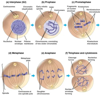
1.2. Mitosis
Mitosis results in the production of two daughter cells
containing identical genetic information and is subdivided into five
successive phases: prophase, prometaphase, metaphase, anaphase
Figure 2: Mitosis in the animal cell. (a) Interphase, where chromosomes are dispersed and not visible as distinct structures. (b) As prophase is initiated centrosomes move towards opposite sides of the cell and chromosome condensation starts to be visible. (c) Prometaphase starts when the nuclear envelope breaks down and chromosome condensation is completed. Each chromosome can be visualized as a structure composed of two sister chromatids held together at the centromeric region. During prometaphase chromosomes are captured by microtubules growing from opposite poles, a process that contributes to chromosome congression and alignment at the metaphase plate (d). (e) At anaphase onset, each chromatid pair separates and segregates to opposite poles of the cell. (f) By the end of mitosis, in telophase, chromosomes decondense and the nuclear membrane re-forms around the daughter nuclei. Cytoplasm division, or cytokinesis, occurs concomitantly as the later stages of mitosis, giving rise to two daughter cells. (Adapted from Lodish, 2003).
During prophase (fig. 2b) two major events can be easily
visualized including overall reorganization of the cytoskeleton and
condensation of chromatin into compact mitotic chromosomes.
separation and migration to opposite sides of the cell of the two
previously duplicated microtubule organizing centers (MTOCs), which
in animal cells are called centrosomes. Centrosomal maturation occurs
during G2 and plays a major role in promoting the conversion of the
highly stable MTs found in interphase to highly dynamic MTs present
in mitosis.
Prometaphase (fig. 2c) starts with nuclear envelope
break-down (NEBD). This allows MTs, nucleated by centrosomes, to invade
the nuclear space and marks the initiation of assembly of the mitotic
spindle. MTs are polymers composed of tubulin subunits that exist as
heterodimers of - and -tubulin. MTs will eventually form the mitotic
spindle. Stable interactions between MTs and chromosomes take
place at a very specific chromosomal structure called the kinetochore.
The kinetochore is a multiprotein complex located at the surface of
each sister chromatid at a region called the centromere that
corresponds to the primary constriction of condensed chromosomes.
As a result of the interaction with the spindle MTs, the chromosomes
will eventually move towards the cell center, a process known as
chromosome congression.
Metaphase (fig. 2d) occurs when all chromosomes are aligned
at the equatorial plane of the mitotic spindle so that each sister
chromatid of each chromosome is attached to MTs emanating from
opposite poles of the spindle. Chromosome segregation can only
occur when all chromosomes are correctly aligned at the spindle
equator and are under tension as a result of their attachments to the
MTs. In the next stage, which is known by anaphase (fig. 2e), sister
chromatids migrate to opposite sides of the spindle. Finally during
telophase (fig. 2f), the nuclear envelope reassembles and surrounds
each group of segregated chromatids, which then decondense giving
rise to two daughter nuclei. At the same time, the cell cytoplasm
cleavage furrow normally at the center of the cell perpendicular to the
long axis of the mitotic spindle and terminates when the two daughter
cells finally separate (reviewed by Lodish 2003, Morgan 2007).
1.3. The spindle assembly checkpoint and the metaphase to anaphase transition
The spindle assembly checkpoint (SAC) is a molecular system
that ensures the accurate segregation of mitotic chromosomes by
delaying anaphase onset until each kinetochore is properly attached to
the mitotic spindle (Kops et al., 2005; Rieder et al., 1995; Rieder et al.,
1994). The SAC is activated by kinetochores that are not yet attached
to mitotic spindle MTs and chromosome pairs that lack tension across
sister chromatids generated by spindle forces (Rieder and Maiato,
2004; Yu, 2002). The established concept is that a group of mitotic
checkpoint proteins such as budding uninhibited by benzimidazole 3
(Bub3), budding uninhibited by benzimidazole 3 related 1 (BubR1) and
mitotic arrest-deficient 2 (Mad2) bind to kinetochores that lack tension
or MT attachment leading to a delay in chromosome segregation
(Kops et al., 2005). As each pair of sister kinetochores attaches to
MTs, they become under tension leading to their stretching. This
allows checkpoint proteins to diffuse from the kinetochore silencing the
SAC. Silencing the SAC leads to activation of the Anaphase Promoting
Complex/Cyclosome (APC/C).
The APC/C regulates the metaphase to anaphase transition
through recognition and destruction of ubiquitinated proteins (fig. 3).
Ubiquitination by the APC/C is mainly controlled by activator subunits
that bind the core components at different points in the cell cycle and
promote ubiquitination of specific substrates. Of these,
cell-division-cycle 20 homologue (Cdc20) and cdc20-homologue 1(Cdh1) are the
anaphase transition allowing further degradation of securin which is an
inhibitor of the protease separase. Once securin is degraded,
separase will be active and will cleave cohesin, the complex that keeps
sister chromatids together. Also, at this stage the mitotic cyclin B is
prompted for degradation leading to the subsequent inactivation of its
binding partner Cdk1, the major mitotic cyclin dependent kinase. Cdh1
activates the APC/C at the end of mitosis to maintain cyclin destruction
(Li and Zhang, 2009).
Figure 3: Regulation of anaphase and mitotic exit by APC/C. During prometaphase, SAC proteins such as Mad2 and BubR1 are activated at kinetochores that are not (or not fully) attached with MTs. Activated Mad2 and BubR1 inhibit the capability of the APC/C to ubiquitinate securin and cyclin B and thereby prevent anaphase and mitotic exit. In metaphase, when all kinetochores are attached to MTs, APC/C ubiquitinates securin and cyclin B and thereby activates the protease separase and inactivates Cdk1. Separase then cleaves cohesin complexes which hold sister chromatids together and thereby initiates sister-chromatid separation. Cdk1 inactivation leads to the dephosphorylation of Cdk1 substrates by protein phosphatases, and thereby enables exit from mitosis. (Adapted from Peters, 2006).
1.4. Chromosome cohesion and condensation
Both cohesin and condensin complexes are essential for
Cohesin establishes sister chromatid cohesion between duplicating
DNAs in S phase. In prophase, a large structural reorganization of
chromosomes is started, with initial release of cohesin and progressive
loading of condensins, culminating with the formation of metaphase
chromosomes with well-resolved sister chromatids. Sister chromatid
resolution, is a prerequisite of the final separation of sister chromatids
that is triggered by proteolytic cleavage of cohesin at the onset of
anaphase. The dynamic behavior of cohesin and condensins must be
tightly regulated under the control of the cell cycle machinery.
1.4.1. Sister chromatid cohesion
To ensure the faithful transmission of chromosomes in mitosis,
replicated chromatids remain paired until both sister kinetochores
attach to opposite poles of the mitotic spindle, a state known as
biorientation. This pairing requires cohesin, a multiprotein complex that
ensures proper sister chromatid pairing until anaphase onset.
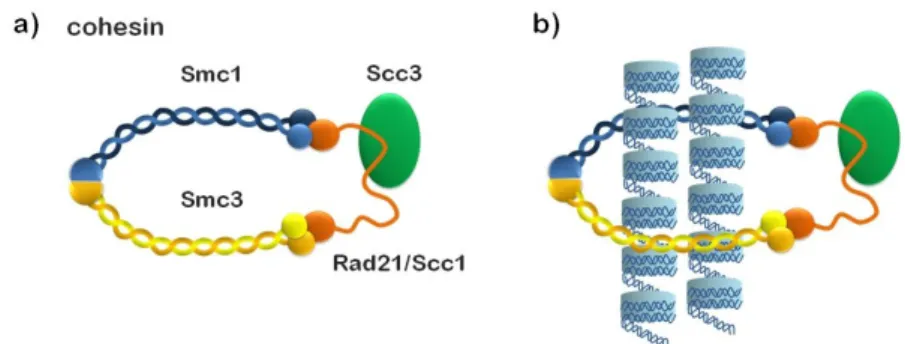
The cohesin complex contains a pair of structural maintenance
of chromosomes (SMC) subunits and ancillary non-SMC subunits.
Mitotic cohesion complexes contain a heterodimer of SMC1 and
SMC3, the non-SMC subunit Scc3 (also known as SA), and the
-kleisin subunit Mcd1/Scc1/Rad21 (fig. 4). Subunit-subunit interaction
assays have shown that cohesin forms a tripartite ring in which the
open-V structure of the SMC heterodimer is closed by simultaneous
binding of the N- and C-terminal regions of Scc1 to the head domains
of SMC3 and SMC1, respectively (fig. 4) (Haering et al., 2002). It has
been proposed that the ring-like structure of cohesin holds sister
chromatids together by embracing two DNA duplexes within the SMC
subunits coiled-coil arms (Haering et al., 2002) (fig 4). In agreement
with this model, cohesin dissociates from chromatin upon disruption of
case of circular minichromosomes, upon DNA linearization (Gruber et
al., 2003; Ivanov and Nasmyth, 2007; Uhlmann et al., 1999).
In most organisms, meiotic cohesin complexes contain the
alternative Rec8 instead of the -kleisin Drad21. There is no obvious
Rec8 orthologue inD.melanogaster. Instead, crossover suppressor on
2 of Manheim (C(2)M) encodes a distant -kleisin member that
associates with DmSMC3 and has a role in synaptonemal complex
(Heidmann et al., 2004), a regulator the overall process of homologous
recombination.
Conserved orthologues of the cohesin proteins are found in
metazoans (Darwiche et al., 1999; Losada et al., 1998; Rollins et al.,
1999; Sumara et al., 2000), suggesting cohesin is very likely the
universal mechanism of sister-chromatid cohesion.
Figure 4: (a) Composition of the cohesin complex.The head domains of cohesin’s SMC1/SMC3 dimer are bound by different ends of the Scc1/Rad21-kleisin subunit, which also recruits the HEAT repeat-containing Scc3/SA subunit to the complex.(b) The ring model. Both sister chromatids (blue cylinders) pass through the same cohesin ring structure, and the major DNA-cohesin interaction is topological.
Loading of cohesin on chromatin requires a heteromeric
complex composed of the Scc2 and Scc4 proteins. These proteins are
conserved from yeast to humans (Watrin et al., 2006). Scc2 and Scc4
are required to load cohesin onto DNA during the G1 phase of the cell
telophase in vertebrates, prior to DNA replication (Gillespie and
Hirano, 2004; Takahashi et al., 2004). Further establishment of
cohesion only occurs during or after DNA replication. Indeed,
establishment of sister chromatid cohesion is tightly coupled to
replication and some, if not all, cohesion is generated at replication
forks (Unal et al., 2007). Cohesion is established by the
acetyltransferase Eco1. This protein associates with the DNA
polymerase processivity factor PCNA and with components of the
clamp loader replication factor C (RF-C) (Kenna and Skibbens, 2003;
Moldovan et al., 2006). Deco is the name for the orthologous
acetyltransferase needed for centromeric cohesion in flies (Williams et
al., 2003). In humans, there are two paralogs of Eco1 known as
ESCO1 and ESCO2. Cohesion establishment was shown to be
dependent upon Eco1-mediated acetylation of at least two lysine
residues of SMC3 at the head region of the protein (Rolef Ben-Shahar
et al., 2008; Rowland et al., 2009; Unal et al., 2008; Zhang et al.,
2008).
There are at least three proteins which are important for
cohesin maintenance following S phase (fig. 5). These proteins include
Pds5, Sororin and Wpl1. They interact closely with cohesin and
modulate its chromatin association dynamics, although they are not
required for cohesion loading. The exact mechanism by which the
Pds5 protein modulates cohesion is not well understood. Pds5 is a
HEAT-repeat protein conserved from fungi to vertebrates. It interacts
with Eco1 and through this interaction is believed to contribute to the
dynamic association of cohesion with chromatin (Noble et al., 2006;
Tanaka et al., 2001). The phenotypes observed in Pds5 deficient cells
are somewhat variable among different organisms. PDS5 is an essential gene in budding yeast, required during G2/M phase for
In Drosophila, pds5 mutants display premature sister chromatid cohesion in brain neuroblasts (Dorsett et al., 2005). Vertebrate cells
have two Pds5-like proteins, and mild cohesion defects were observed
with decreased levels of these proteins (Losada et al., 2005). Sororin
is a vertebrate-specific component of the cohesin network (Rankin et
al., 2005; Schmitz et al., 2007). Depletion of Sororin in HeLa cells
decreased the fraction of stable chromatin-bound cohesin, causing
cohesion defects already in interphase (Schmitz et al., 2007). In
contrast to Pds5 and Sororin, Wpl1 promotes dissociation of cohesin
from chromosome arms. Drosophila mutants of the Wpl1 orthologue (wapI) prevented separation of sister chromatids in mitosis (Verni et
al., 2000). Consistently, in mammalian cells depleted of Wpl1, cohesin
remains associated with chromatids during mitosis, resulting in a
mitotic delay (Gandhi et al., 2006; Kueng et al., 2006) (fig. 5).
In metazoans dissolution of most cohesin from chromosome
arms is performed during prophase in what has been called the
“prophase pathway”. Only a small population, enriched at the
pericentromeric region remains on chromosomes until metaphase.
Cohesin dissociation from chromosome arms requires phosphorylation
of the cohesin subunit, SA, by the mitotic kinase Polo-like kinase 1
(Plk1) (Hauf et al., 2005; Sumara et al., 2002). In addition to Plk1,
Aurora B may contribute to this process by phosphorylation of other
targets. Xenopus cell-free extracts depleted of Aurora B approached metaphase with condensed sister chromatids, but the axes failed to
resolve (Losada et al., 2002; Sumara et al., 2002). Most of the cohesin
dissociated by the prophase pathway is not cleaved at anaphase, but
instead relocates to chromatin in telophase to function in the next cell
cycle. During the prophase pathway, centromeric cohesin is protected
from dissociation by Shugoshin (Sgo/Mei-S332) (Watanabe and
Sgo/Mei-S332 to mitotic chromosome segregation is modest. In contrast, HeLa
cells depleted of Sgo1, one of the two human members of this family,
causes premature sister chromatid separation during mitosis (Kitajima
et al., 2005; Salic et al., 2004; Tang et al., 2004) Sgo acts in concert
with protein phosphatase 2A (PP2A), where it is likely to counteract
Plk1-dependent phosphorylation of cohesin and thereby prevent
dissociation of cohesion from the centromeres (Kitajima et al., 2006;
McGuinness et al., 2005; Riedel et al., 2006; Salic et al., 2004; Tang et
al., 2006).
Centromeric cohesin is essential for chromosome segregation
in mitosis. For proper chromosome segregation, the sister
kinetochores must attach spindle MTs emanating from opposite poles,
and centromeric sister chromatid cohesion is essential in antagonizing
the pulling force of the spindle microtubules. Centromeric sister
chromatid cohesion is dissolved at the metaphase to anaphase
transition. Once the SAC is satisfied, the APC/C is activated and
promotes the ubiquitin dependent-destruction of the sequestering
chaperone securin, which leads to the activation of the protease
separase. Cohesin release results from site-specific cleavage of the
Scc1 subunit by separase (Uhlmann, 2001). Scc1 phosphorylation by
Figure 5: The cohesin network. The Scc2-Scc4 loading complex deposits the cohesin complex onto chromatin in the G1 phase of the cell cycle. Cohesion is established between sister chromatids concomitantly with DNA replication in an Eco1-dependent manner. Upon entry into G2/M phase cohesion is maintained by the proteins Pds5, Sororin and WpI1. Mammalian cells remove cohesin along chromosome arms during prophasevia phosphorylation of Scc1/Rad21 by Polo-like kinase. During metaphase centromeric cohesin is protected from removal by Sgo and PP2A. At the onset of anaphase, separase cleaves Scc1/Rad21 leading to the opening of the cohesin ring and the separation of sister chromatids. (Adapted from Xiong and Gerton, 2010).
1.4.2. Chromosome condensation
Chromosome condensation is essential for correct
chromosome segregation and therefore faithful transmission of genetic
information to daughter cells. One important player of the process of
chromosome compaction is the condensin complex. There are two
condensin complexes in vertebrate cells: condensin I and II. Both
complexes are pentameric composed by two SMC ATPase subunits
(SMC2 and SMC4) and three auxiliary subunits (G/G2,
CAP-D2/D3 and CAP-H/H2) (Hirano, 2005; Ono et al., 2003) (fig. 6).
Drosophila appears to have both complexes but no homologue has been identified for CAP-G2. The condensin complex is highly
conserved and is found ubiquitously among eukaryotes, while
Figure 6: Composition of the condensin complexes. Condensin’s SMC2/SMC4 dimer associates with a CAP-H-kleisin or with a CAP-H2-kleisin in condensin I or II complexes, respectively. Separate pairs of HEAT repeat-containing proteins associate with condensin I (CAP-D2 and CAP-G) or II (CAP-D3 and CAP-G2).
The two condensin complexes have different localization
patterns in the cell cycle and on chromosomes. In vertebrates,
condensin I is thought to localize to the cytoplasm during interphase
and gain access to chromosomes only at the end of prophase after
NEBD. It then dissociates again from chromosomes at the end of
telophase. Condensin II is nuclear during interphase but only
associates with chromatin until prophase, where it remains until the
end of telophase (Gerlich et al., 2006; Hirota et al., 2004; Ono et al.,
2004). Both complexes are present at centromeres and along
chromosome arms. They bind to different places along chromosome
axes and are both necessary for mitotic chromosome segregation
(Ono et al., 2004).
Recent data in yeast suggests that like cohesin, the condensin
complex might be loaded onto chromosomes by the Scc2-Scc4
complex. Budding yeast scc2 and scc4 mutants displayed
chromosome compaction defects and reduced condensin binding
(D'Ambrosio et al., 2008).
Condensin’s loading on chromosomes during early mitosis is
accompanied by the compaction of sister DNAs into largely separate
linear rod-shaped structures. Several studies showed condensin has a
role in organizing mitotic chromatids that is essential for their
successful segregation during anaphase. However the mechanism of
action and its contribution to chromosome condensation is poorly
understood. Althoughin vitrostudies suggested condensin to be a key player in chromosome condensation (Hagstrom et al., 2002; Hirano,
showed that chromosomes depleted of condensin do condense. The
first defect observed in cells depleted of condensin is associated with a
delay in the timing of prophase compaction. Elimination of both
condensin complexes in chicken DT40 cells revealed no appreciable
chromosome condensation in G2/M cells with intact nuclear envelopes
(Hudson et al., 2003). However, after NEBD, chromosomes were able
to undergo a nearly normal condensation process (Hudson et al.,
2003; Kaitna et al., 2002). Depletion of condensin II-specific subunits,
but not in those depleted of condensin I-specific subunits, leads to a
delay in chromosome condensation within prophase (Hirota et al.,
2004; Ono et al., 2004). By metaphase, depletion of condensin I- or
condensin II-specific subunits produces distinct, highly characteristic
defects in chromosome architecture, i.e., swollen or curly
chromosomes, respectively. These data are consistent with distinct
contributions of condensin I and II to mitotic chromosome structure.
Depletion of the SMC core subunits causes the severest defects
revealing cloud-like chromosomes with a very fuzzy appearance (Ono
et al., 2003). Condensin depletion was also shown to lead to the
mislocalization of a number of non-histone chromosomal proteins,
including topoisomerase II (TopoII), inner centromere protein
(INCENP) and KIF4A/chromokinesin (Hudson et al., 2008; Hudson et
al., 2003).
TopoII is an evolutionarily conserved protein, and removes
catenation between sister chromatids that remains after completion of
DNA replication (Wang, 2002). Indeed, it is thought that sister
chromatid resolution is achieved by the cooperation between
topoisomerase II and condensins (Coelho et al., 2003; Steffensen et
al., 2001). DmSMC4-depleted cultured cells failed to localize TopoII to
a clearly defined chromatid axial structure and protein extracts from
DmSMC4-depleted cells exhibited a significant decrease in
In condensin mutants, mitotic chromosomes interact normally
with spindle MTs and chromosomes congress to the metaphase plate
with rare defects (Gerlich et al., 2006; Hagstrom et al., 2002). Upon
anaphase onset, severe defects are observed in condensin mutants
and condensin depleted cells. Chromosome segregation at anaphase
almost always fails, leading to the formation of prominent chromatin
bridges and cytokinesis failure (Hagstrom et al., 2002; Hirota et al.,
2004; Hudson et al., 2003; Kaitna et al., 2002; Ono et al., 2003; Saka
et al., 1994; Savvidou et al., 2005; Steffensen et al., 2001). Premature
loss of chromosome compaction is apparently the cause of these
chromosome segregation defects in early anaphase (Gerlich et al.,
2006; Vagnarelli et al., 2006), and not due to the failure of TopoII
decatenation activity (Vagnarelli et al., 2006). On the other hand, it has
also been suggested that condensins have additional and distinct roles
during anaphase to achieve proper chromosome segregation (Wignall
et al., 2003; Yanagida, 2009). The potential roles of condensins during
anaphase remain elusive. Different studies have shown an extent of
defects observed in condensin depleted cells. The observed defects
seem however to vary among different organisms and different
conditions, thereby leaving room for different interpretations (Losada
and Hirano, 2005).
2. Posttranslational modifications
One of the most significant conclusions of the Human genome
project was the small number of annotated genes, which was
proportional increase in gene number. Indeed proteomes may be
orders of magnitude more complex than the encoding genomes would
predict. The first route of diversification of proteins is at the level of
alternative transcriptional start sites and pre-mRNA alternative splicing.
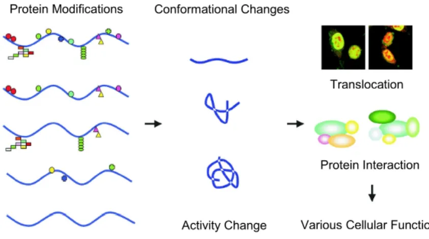
The second route of proteome expansion is by covalent
posttranslational modification (PTM) of proteins at one or more sites.
As the name implies, PTMs are covalent modifications that occur on
protein side chains or their backbones by the addition of a chemical
group either during or after assembly of the polypeptide chain (Walsh
et al., 2005). PTMs can regulate protein function, by causing changes
in protein activity, protein cellular locations and dynamic interactions
with other proteins (fig.7). Proving the relevance of these modifications
in protein processing, about 5% of the genomes of higher eukaryotic
can be dedicated to enzymes that carry out PTMs of their proteomes.
Figure 7: Schematic representation of protein modifications related to the
regulation of downstream biological processes. (Adapted from Seo and Lee, 2004).
Most common PTMs include phosphorylation (Burnett and
Kennedy, 1954), acetylation (Glozak et al., 2005), methylation (Grewal
and Rice, 2004), glycosylation (Spiro, 2002), sumoylation (Gareau and Translocation
Protein Interaction
Various Cellular Functions Activity Change
Lima, 2010), and ubiquitination (Pickart and Eddins, 2004). These
modifications regulate a variety of different cellular processes,
including cell cycle progression and its regulation.
2.1. Posttranslational modifications and the cell
2.1.1. Phosphorylation
Phosphorylation of Serine (Ser), Threonine (Thr), Tyrosine
(Tyr) and Histidine (His) residues is the best known and most
ubiquitous PTM. Because the phosphate group (-O-PO32-) carries a
large negative charge at neutral pH, phosphorylation of neutral amino
acids induces altered conformations in local protein microenvironments
(Johnson and Lewis, 2001). Phosphorylation can therefore modulate
protein function by altering protein stability, cellular localization,
substrate affinity, complex formation and activity.
The addition of a phosphate group on a substrate protein is
catalyzed by protein kinases, while hydrolyses of the phosphate group
is achieved by protein phosphatases. Eukaryotes express a large
variety of protein kinases (Manning et al., 2002) and phosphatases
(Moorhead et al., 2007; Virshup and Shenolikar, 2009), each with a
unique substrate specificity and regulation. The expansion of the
protein kinase families in higher metazoans accounts for the observed
cellular and functional diversity between these organisms. Due to its
reversible and transient nature, phosphorylation allows signal
transduction pathways to carry out diverse cellular functions. Moreover
it also allows essential events such as cell cycle and growth to occur at
precise times and locations.
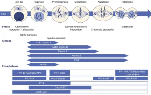
The eukaryotic cell cycle is an example of cellular
decision-making based on reversible phosphorylation and dephosphorylation of
cycle progression (Bettencourt-Dias et al., 2004). Cdks regulate the
G1, S and G2 phases of the cell cycle ensuring DNA duplication and
segregation of chromosomes into daughter cells (fig. 8). Cdks are
themselves regulated and cooperate with other protein kinases.
Figure 8: Mitotic phosphoregulation in animal cells. Upper panel shows the different mitotic stages and highlights major events known to be regulated by reversible protein phosphorylation. Middle panel indicates the activity phases of the major mitotic kinases. Lower panel shows known mitotic phosphatases with respect to the depicted mitotic events. Exact timing of activation and inactivation of most mitotic phosphatases is not known as depicted by open bars. (Adapted from Bollen et al., 2009).
Among the different kinases, Cdk1 has an essential role in
mitotic entry and progression until all chromosomes are properly
aligned along the metaphase plate (Morgan, 2007). Entry into
anaphase and subsequent mitotic exit requires inactivation of Cdk1
and other mitotic regulators (Sullivan and Morgan, 2007). This
inactivation is primarily mediated by proteosome-dependent
of mitotic phosphorylations by different phosphatases. Mitosis relies
therefore in a delicate balance between the activities of kinases and
their counteracting phosphatases.
2.1.2. Acetylation
The role of acetylation has been suggested to be analogous to
that of phosphorylation (Kouzarides, 2000). Many proteins are
posttranslationally acetylated, and at least for eukaryotic proteins,
acetylation is the most common covalent modification out of over 200
reported types. Protein acetylation is catalyzed by a variety of different
acetyltransferases. These enzymes catalyze the transfer of an acetyl
group from acetyl-coenzyme A (Acetyl-CoA) to either the -amino
group of amino-terminal residues (N-terminal acetylation, revised in
Chpater I, section 3.) or to the-amino group of lysine (Lys) residues
at various positions. The-amino group designates the position of the
central carbon atom of amino acids, whereas the -amino group of
lysine residues designates the position of a carbon atom on the side
chain.
The introduction of acetyl groups on Lys side chains potentially
converts cationic side chains into neutralized surfaces influencing
protein function or its association with other proteins. Acetylation of
-Lys residues occurs on histones, high mobility group (HMG) proteins,
transcription factors, nuclear receptors (Bannister et al., 2000; Imhof et
al., 1997; Roth et al., 2001) and-tubulin (MacRae, 1997).
Most studied acetylated proteins include N
-acetylation of Lys
side chains on the N-terminal tail of histones (Yang, 2004). Two
groups of enzymes responsible for regulating the reversible and
dynamic state of histone acetylation are histone acetyltransferases
general, leads to transcriptional activation by inducing the unpacking of
the nucleosomes from the tight 30nm chromatin fibers. This allows
manipulating the tightly packed heterochromatin to more relaxed
euchromatin state (fig. 9), which in turn allows other transcriptional
regulatory proteins to gain access to promoter elements on DNA. The
acetylation state of different promoters is maintained by specific
combinations of HATs and HDACs. Not surprisingly, histone
acetylation appears to influence other processes including cell cycle
progression, chromosome dynamics, DNA replication, recombination
and repair, silencing and apoptosis (Kouzarides, 1999).
Figure 9: Histone acetylation and transition of heterochromatin to euchromatin.
Representation of the molecular structure of acetylated and deacetylated lysine residues and resultant chromatin state. (adapted from Khan and Khan, 2010).
2.1.3. Methylation
Although there are known examples of protein side chain
methylation, histone N-terminal tail methylation draws most research
attention. Since methyl groups are relatively small, addition of these
groups to Lys and Arginine (Arg) residues does not neutralize their
charges, having little effect on histone conformation. Instead,
specific regulatory proteins (Bannister and Kouzarides, 2005).
Therefore, unlike acetylation, which generally correlates with
transcriptional activation, histone lysine methylation can signal either
activation or repression, depending on the sites of methylation
(Cuthbert et al., 2004). The -amino group of Lys can accept up to
three methyl groups and hence can be mono-, di- or trimethylated
(Martin and Zhang, 2005). For certain processes, methylation on the
same site can lead to different outcomes depending on the number of
methyl groups added.
2.1.4. Ubiquitination
Ubiquitination is likely to affect all proteins at some point in their
life cycle. Ubiquitin (Ub) a highly conserved 8.5 kDa protein that
becomes covalently attached to Lys residues of target proteins in an
inducible and reversible manner. This attachment occurs through a 3
step process involving 3 different types of enzymes, E1, E2 and E3
ligases (Pickart and Eddins, 2004). According to the number of Ubs
added, substrate proteins can be monoubiquitinated or
polyubiquitinated. Monoubiquitination is believed to serve as a
regulatory modification of a target protein in much the same way
phosphorylation regulates protein activity (Weissman, 2001). However,
the most common role associated to ubiquitination is the tagging of
proteins for degradation. The vast majority of proteins (80-90%) tagged
by Ub polypeptides are then degraded via the 26 S proteasome (Craiu
et al., 1997; Rock et al., 1994). This degradation signal usually
involves protein polyubiquitination. One example where ubiquitination
plays a central role is in cell cycle progression. The characteristic
temporal control of proteins destroyed during the cell cycle, such as
the cyclin subunits of cdks, is performed by the E1-E2-E3 ubiquitin
3. N
-terminal acetylation
Besides-amino acetylation of side chain residues, acetylation
can also occur at the -amino group of protein N-terminals. This
modification is an enzyme catalyzed reaction in which the protein
-amino group accepts an acetyl group from Acetyl-CoA. N-acetylation neutralizes positive charges that can influence protein function,
stability, interaction with other molecules, or other subsequent
modifications, such as phosphorylation.
N
-acetylation is one of the most common protein modifications
in eukaryotes. Indeed, different studies revealed N-acetylation of approximately 85% of soluble cytosolic human proteins (Arnesen et al.,
2009b; Jornvall, 1975; Persson et al., 1985) and approximately 50% in
yeast (Arnesen et al., 2009b; Lee et al., 1989).
N
-acetylation often occurs in two consecutive events
(Bradshaw et al., 1998; Polevoda and Sherman, 2002). In the first
step, the initiator methionine (iMet; fig. 10), is removed from the
nascent polypeptide by methionine aminopeptidases (MAP). This
event is not obligatory in protein biosynthesis and has been shown to
only take place if the second amino acid is small and uncharged
(Boissel et al., 1988; Sherman et al., 1985). Larger amino acids at this
position prevent removal of iMet by sterical hindrance (Lowther and
Matthews, 2000). In the second step, the acetylation of the
amino-terminus is catalyzed by N
Figure 10: Schematic representation of initial methionine removal followed by
N
-terminal acetylation.During translation of a protein, when the iMet is followed by a small and uncharged amino acid residue, it will be removed by a methionine aminopeptidase (MAP, orange). Following iMet removal, the processed amino-terminus will be acetylated at the -amine of the following amino acid residue by a NAT (green).
NATs form a subfamily within the Gcn5-related
N-acetyltransferases (GNAT) superfamily (Polevoda et al., 1999). GNATs
are one of the largest known protein superfamilies, with more than 10
000 members in all kingdoms of life (Vetting et al., 2005). Accordingly,
NATs have been found in all kingdoms: prokaryotes, archaeal, and
eukaryotes (Polevoda and Sherman, 2000; Polevoda and Sherman,
2003b). In humans, as in yeast, three NAT complexes are believed to
perform most N
-acetylation events, namely the human NatA, NatB
and NatC complexes (Arnesen et al., 2005a; Asaumi et al., 2005;
Starheim et al., 2008; Starheim et al., 2009a). Each NAT acts on
different groups of amino acid sequences, whose specificity is
determined by two or more residues at the amino-terminal positions
(table 1). However, for some proteins, acetylation does not occur even
if the appropriate amino acid sequences are present, suggesting that
additional unknown amino acid sequence patterns or other
determinants like secondary structure of the protein N-terminus may
play a role (Polevoda and Sherman, 2003b).
All eukaryotic NATs so far identified were shown to interact with
ribosomes and N-acetylation is believed to take place during protein synthesis (Gautschi et al., 2003; Polevoda et al., 2008; Starheim et al.,
2008; Starheim et al., 2009a). In vitro studies demonstrated that cotranslational N
-acetylation takes place when the nascent
polypeptide extrudes 20 to 50 amino acids from the ribosome
Type NatA NatB NatC NatD NatE Catalytic subunit Naa10p (Ard1p) Naa11p (Ard2p) Naa20p (Nat3p) Naa30p (Mak3p) Naa40p (Nat4p) Naa50p Auxiliary subunit Naa15p (Nat1p) Naa16p (Nat2p) HYPK* Naa25p (Mdm20p) Naa35p (Mak10p) Naa38p (Mak31p) -Naa15p? (Nat1p) Naa10p? (Ard1p) Substrates Ser- Ala- Thr- Val- Gly- Cys- Met-Glu- Met-Asp- Met-Asn- Met-Leu- Met-Ile- Met-Trp- Met-Phe- Ser-Gly-(H2A/H4)
Met-Leu-Table 1: Eukaryotic N-terminal acetyltransferases and there substrates.N-termini of which the iMet is followed by one of the small amino acids, Ser-, Ala-, Thr-, Val-, Gly-, Cys-, and Pro- undergo iMet cleavage performed by a methionine aminopeptidase (MAP). These N-termini, with the exception of Pro-, are often further acetylated by NatA (Arnesen et al., 2009b; Polevoda et al., 1999), or in the case of Histone H2A and H4, by NatD (Song et al., 2003). Met-Asp-, Met-Glu-, Met-Asn- and Met-Gln- are acetylated by NatB (Polevoda et al., 1999; Polevoda and Sherman, 2003b). Hydrophobic Met-Leu-, Met-Ile-, Met-Phe- and Met-Tyr- are acetylated by NatC (Kimura et al., 2000; Polevoda et al., 1999; Tercero et al., 1993). Met-Leu- N-termini have been demonstrated to be acetylated by Naa50pin vitro (Evjenth et al., 2009) *HYPK is an interactor partner of NatA in humans and may modulate NAT-activity (Arnesen et al. 2010).
In contrast to other PTMs, very little is known about the
biological relevance of N-acetylation in the context of a developing multicellular organism. Nevertheless, multiple evidences suggest it is
essential for development and is likely to play an important role during
carcinogenesis (Starheim et al., 2009b). Work in yeast and mammalian
cell culture described NATsin vivosubstrate specificities (table 1) and
3.1. The NatA acetyltransferase complex
Among the three major NAT complexes, NatA acetyltransferase
complex is the most studied one. This complex is conserved from
yeast to humans in respect to subunit composition (Arnesen et al.,
2005a) and substrate specificity (Arnesen et al., 2009b). Functional
complementation studies demonstrated that expression of the human
NatA complex (hNAA10-hNAA15) rescued the phenotypes observed in
a yeast NatA-deletion strain. Furthermore, when expressed in yeast
the hNatA complex could N-acetylate the same set of substrates as the yeast NatA complexin vivo(Arnesen et al., 2009b).
The NatA complex acetylates N-termini in which the initial iMet
was removed by MAPs. It also seems to be the most degenerate NAT
complex, including a wide range of sequences, although N-terminal
residues of Ser and alanine (Ala) are the most common ones (Arnesen
et al., 2009b; Polevoda et al., 1999) (table 1).
NatA is believed to be the major NAT complex both in yeast and
humans having a higher amount of potential substrates in comparison
to the NatB and NatC complexes (Arnesen et al., 2009b; Polevoda and
Sherman, 2003b). Based on the substrate specificity of this complex
and the N-terminal sequences of all known human proteins, it was
estimated that more than 8.000 (from a total of 20.000) unique human
proteins are hNatA substrates. Indeed, a large number of NatA
substrates were identified in vivo by analysis of the N
-terminal
acetylation levels of protein extracts from control and downregulated
NatA subunits in HeLa cells (Arnesen et al., 2009b). Surprisingly,
downregulation of the hNatA complex only led to a significant change
in the acetylation status of 6% of the identified hNatA substrates. The
authors attributed this to a residual presence of the hNatA complex
Another possibility is a partial redundancy of other NATs with the NatA
complex.
The NatA complex is composed by the catalytic subunit
Naa10p (Ard1p) and the auxiliary subunit Naa15p (Nat1p; Table 1)
(Arnesen et al., 2005a; Mullen et al., 1989). Both human subunits were
found to be associated with ribosomes, suggesting Naa10p performs
cotranslational acetylation of nascent polypeptides (Arnesen et al.,
2005a). However, a significant proportion of these subunits were also
found to be non-ribosomal (Arnesen et al., 2005a), suggesting these
proteins might have other functions independent of the NatA complex.
Naa10p protein levels are dependent on Naa15p; depletion of Naa15p
significantly diminished Naa10p protein levels on ribossomes,
suggesting Naa15p is likely to anchor Naa10p to the ribosome (Hou et
al., 2007; Polevoda et al., 2008).
Interestingly, humans contain paralogues of the hNaa10p and
hNaa15p proteins, hNaa11p and hNaa16p, respectively (Arnesen et
al., 2006b; Arnesen et al., 2009a). These were suggested to form less
abundant functional NatA complexes, allowing for four possible
combinations of the different NatA subunits (Arnesen et al., 2006b;
Arnesen et al., 2009a).
The hNatA complex was recently described to interact with the
Huntingtin (Htt) yeast two-hybrid protein K (HYPK). HYPK has
chaperone-like properties preventing Htt polyglutamine (polyQ)
aggregation. The NatA complex was essential for the proper
expression of the HYPK protein and for the modulation of Htt polyQ
aggregation. Moreover, the interaction of HYPK with the NatA complex
was shown to be required for normalin vivo andin vitroacetylation of a known NatA substrate (Arnesen et al. 2010). It is not clear in human
cells however, if HYPK influences NatA acetylation activity in general