ABSTRACT
Periodontal ligament inluence on the stress
distribution in a removable partial denture
supported by implant: a inite element analysis
Carlos Marcelo ARCHANGELO1, Eduardo Passos ROCHA2, João Antônio PEREIRA3, Manoel MARTIN JUNIOR1, Rodolfo Bruniera ANCHIETA4, Amilcar Chagas FREITAS JÚNIOR4
1-DDS, PhD, Professor, Federal Institute of Paraná – IFPR, Londrina, PR, Brazil.
2-DDS, PhD, Associate Professor, Department of Dental Materials and Prosthodontics, Faculty of Dentistry of Araçatuba, UNESP - Univ. Estadual Paulista, Araçatuba, SP, Brazil.
3-MEng, PhD, Associate Professor, Department of Mechanical Engineering, Faculty of Engineering of Ilha Solteira, UNESP - Univ. Estadual Paulista, Ilha Solteira, SP, Brazil.
4-DDS, MS, PhD student, Department of Dental Materials and Prosthodontics, Faculty of Dentistry of Araçatuba, UNESP - Univ. Estadual Paulista, Araçatuba, SP, Brazil.
Corresponding address: Prof. Adj. Dr. Carlos Marcelo Archangelo - Rua José Bonifacio, 1193, Vila Mendonça - Araçatuba - SP - Brasil - 16010-050 - Phone: 55-18-36363290 - e-mail: [email protected]
Received: October 18, 2010 - Modiication: August 7, 2011 - Accepted: September 1, 2011
O
bjective: The non-homogenous aspect of periodontal ligament (PDL) has been examinedusing inite element analysis (FEA) to better simulate PDL behavior. The aim of this study was to assess, by 2-D FEA, the inluence of non-homogenous PDL on the stress
distribution when the free-end saddle removable partial denture (RPD) is partially supported
by an osseointegrated implant. Material and Methods: Six inite element (FE) models of a
partially edentulous mandible were created to represent two types of PDL (non-homogenous and homogenous) and two types of RPD (conventional RPD, supported by tooth and
ibromucosa; and modiied RPD, supported by tooth and implant [10.00x3.75 mm]). Two
additional Fe models without RPD were used as control models. The non-homogenous PDL was modeled using beam elements to simulate the crest, horizontal, oblique and apical
ibers. The load (50 N) was applied in each cusp simultaneously. Regarding boundary conditions the border of alveolar ridge was ixed along the x axis. The FE software (Ansys 10.0) was used to compute the stress ields, and the von Mises stress criterion (svM) was
applied to analyze the results. Results: The peak of svM in non-homogenous PDL was
higher than that for the homogenous condition. The beneits of implants were enhanced
for the non-homogenous PDL condition, with drastic svM reduction on the posterior half of the alveolar ridge. The implant did not reduce the stress on the support tooth for both PDL conditions. Conclusion: The PDL modeled in the non-homogeneous form increased the
beneits of the osseointegrated implant in comparison with the homogeneous condition.
Using the non-homogenous PDL, the presence of osseointegrated implant did not reduce the stress on the supporting tooth.
Key words: Removable partial denture. Dental implant. Finite element analysis. Periodontal ligament.
INTRODUCTION
Conventional removable partial dentures (RPD) show a complex biomechanical behavior, mainly when the RPD is supported by teeth and
ibromucosa, such as in Kennedy Class I25. This
behavior has been better controlled when an
osseointegrated implant is placed and acts as an additional support, retainer, or both, improving the stability and retention of the RPD, which reduces the demand on the support structures14.
singularities according to the number and position of the remaining teeth in the arch, as well as the size of the prosthetic space and degree of bone loss 2-4,9-11,13,14,16,17,20,26,28. The usual clinical responses from
patients are represented by better RPD stability, retention and comfort when this association is done14.
Mitrani, et al.18(1993) investigated the free-end
saddle RPD combined with an implant on the distal extension wearers for a mean period of 2.5 years. Those authors showed that the implant placed on the distal extension of RPD increases patient satisfaction, reduces the marginal bone loss, and establishes the peri-implant tissues18. Nonetheless,
data about the abutment tooth behavior over time are still not reported and remain inconclusive.
Finite element (Fe) studies can provide valuable information about the stress reduction on the
support structures promoted by implants. However
this information is controversial because the
beneits of the osseointegrated implant are more
evident on the alveolar ridge and hardly conclusive for the RPD abutment tooth23.
There are some concerns about the tooth behavior in a RPD supported by implant and tooth through finite element analysis (FeA), mainly because the periodontal ligament (PDL) has not been properly modeled as a non-homogenous structure. The homogenous aspect of the PDL in FeA might negatively change the results. Atmaram and Mohammed1 (1981) reported that the homogeneous
condition of the PDL is closer to more uniform stress distribution and unable to direct the forces applied
in accordance with the set of ibers in vivo. In addition, the magnitude of stresses is shown to be less pronounced than under the non-homogeneous condition. This is particularly important in face of forces with non-axial incidence, producing terminal torqueing forces against the abutment teeth and the soft tissue19.
Other authors have pointed out that the non-homogeneous PDL was necessary in order to understand how occlusal loads are absorbed by the PDL22. Therefore, this condition is decisive
to analyze the results of RPD associated with an osseointegrated implant by FeA. Nevertheless,
the beneits of the combination between RPD and
osseointegrated implant may be analyzed from this perspective. The analysis of the stress distribution in the studies by Atmaram and Mohammed1 (1981)
and Rees and Jacobsen22 (1997) did not show the
behavior of the abutment tooth.
The objective of this study was to evaluate the
inluence of PDL condition (homogenous and
non-homogenous) on the stress distribution of a free-end saddle RPD associated with an osseointegrated implant through 2-D FeA. The hypothesis tested was
that non-homogeneous PDL increases the beneits
of implant, reducing the stress on the supporting tooth of the RPD supported by tooth and implant.
MATERIAL AND METhODS
Six mandibular models (A, B, C, D, e, and F) were modeled using graphic software (AutoCAD, Autodesk Inc., San Rafael, CA, USA). In a sagittal view, all models simulated partially edentulous hemi-arches without posterior dental support (Kennedy Class I). In addition, all models had the remaining tooth 33 and a distal extension ridge.
The characteristics of the abutment - the length of the distal extension ridge, the dimensions and characteristics of the support and protective periodontium, the mandibular bone height, the thickness of the CoCr metal structure, and the
number of artiicial teeth - were kept constant.
The difference was the PDL in models D, e, and F, which was reproduced in accordance with the in vivo
characteristic, with representation of the following
4 groups of ibers: crest, horizontal, oblique and
apical (Figure 1).
After the models had been created, the iles were
exported to ANSYS 8.0 (Swanson Analysis Systems,
Houston, PA, USA) to discriminate the regions and generate the Fe mesh. Models A, B, and C, (Figure 1) were considered homogeneous, isotropic, and linearly elastic. The mechanical properties adopted for all materials (elastic modulus and Poisson’s ratio) were established according to the literature (Table 1)1,8,15,23,24,29.
Models D, e, and MF, similar to models A, B, and C, respectively, were considered homogeneous, isotropic, and linearly elastic, except for the PDL, which was considered a non-homogeneous structure. Thus, models D, e, and F differed from models A, B and C only by their structural characteristic of the PDL, being similar in all other factors.
The element used to generate the mesh was the plane 2 element, a triangular element
deined by 6 nodes, with 2 degrees of freedom per node, and quadratic displacement behavior. The Fe mesh showed up to 80,000 nodes and up
to 40,000 elements. This coniguration allowed the appropriate reinement of the mesh in thin
structures, such as the cortical bone and the PDL, reaching the convergence norm.
As far as the creation of the non-homogeneous PDL is concern, using the methodology established by Atmaram and Mohammed1 (1981) with some modiications, the composition of the PDL in the FE
model was established under the in vivo condition5.
For this, beam elements were used to represent
4 groups of PDL ibers in a sagittal cut (crest, horizontal, oblique and apical ibers) and their
well as tensile and compression loads, the cross-sectional area and the moment of inertia for the beam elements were used to describe the PDL. Thus, the following equations were used:
Cross sectional area: A=πx(r)2 Moment of inertia: I=πx(d)4/64
where:
A=cross sectional area; I=moment of inertia;
π=3.14; d=4 μm or 4x10⁻⁶ m; r=2x10⁻⁶ m.
Grant, et al.12 (1988) determined a value of 4 µm for the PDL ibers of an RPD abutment tooth.
Thus, the values found for the cross-sectional area and the moments of inertia of each element were 1,256x10-5 mm2 and 0.785x10⁻¹² mm4, respectively.
In accordance with that established by Atmaram and Mohammed1 (1981), the number of periodontal
fibers adopted under the non-homogeneous condition was half the number of elements present
Structures E (GPa) References v References
Enamel 41 17 0.3 17
Dentin 18.6 18 0.31 18
Homogenous PDL 0.175 15 0.45 15
Non-homogenou PDL 0.35 15 0.45 15
Fibromucosa 0.68 17 0.45 17
Cortical bone 13.7 18 0.3 18
Medullar bone 1.37 18 0.3 18
Implant (Ti) 103.4 20 0.35 20
Healing abutment (Ti) 103.4 20 0.35 20
CoCr cast alloy 185 21 0.35 21
Acrylic resin 8.3 14 0.28 14
Artiicial teeth 8.3 14 0.28 14
Table 1- Mechanical properties of the materials. E – Elastic modulus. v – Poisson’s ratio. PDL – Periodontal ligament. Ti - Titanium
in the PDL under the homogeneous condition. Thus, all models with a homogeneous PDL (A, B, and C) contained 824 elements in the PDL region, and the models with a non-homogeneous PDL (D, e, and
F) contained 412 ibers for composition of the PDL.
As the boundary condition, the left and right sides of the models were fixed only in the x direction, in order to prevent lateral movement of the structures. This allowed the vertical movement of the RPD base over the fibromucosa and, consequently, the deformation of the cortical and medullar bone beneath it, as well as simulation of the bilateral mandibular condition. Only the cortical
bone at the base of the models was ixed in the x
and y directions (Figure 1).
The models were loaded with vertical forces (50
N) distributed at each cusp, fractionated into 5 point loads of 10 N each to prevent the occlusal contact characterized by only one point load (Figure 1).
The von Mises equivalent stress criterion (svM) was adopted to analyze the stress distribution. each structure was individually analyzed following
speciic areas (Figure 1), as well as to allow the data to be reined into regions of interest, as follows:
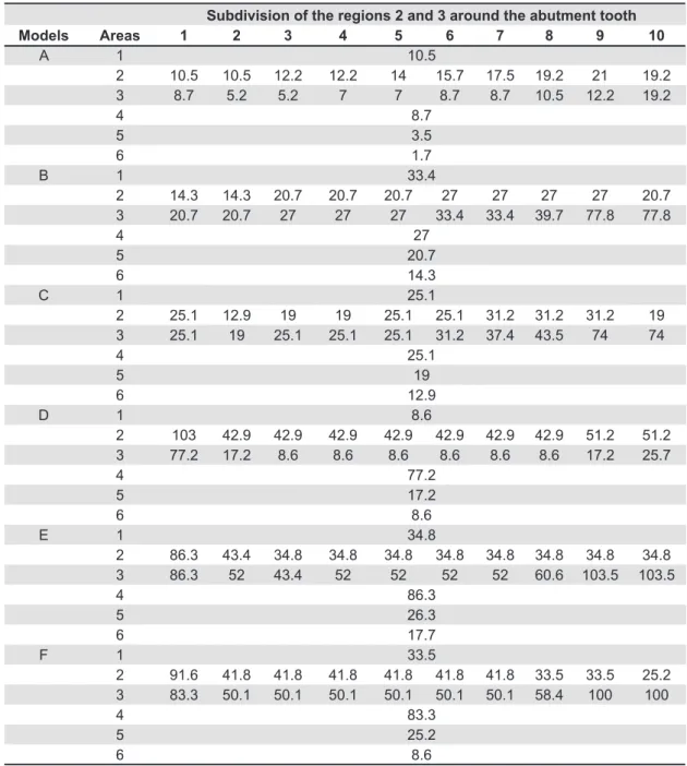
Area 1: Root apex; Area 2: Mesial side of the abutment tooth and adjacent structures; Area 3: Distal side of the abutment tooth and adjacent structures; Area 4: Distal bone crest of the abutment tooth; Area 5: Anterior half of the alveolar ridge; Area 6: Posterior half of the alveolar ridge; Area 7: Osseointegrated implant.
For more details and to allow the comparison
Subdivision of the regions 2 and 3 around the abutment tooth
Models Areas 1 2 3 4 5 6 7 8 9 10
A 1 10.5
2 10.5 10.5 12.2 12.2 14 15.7 17.5 19.2 21 19.2
3 8.7 5.2 5.2 7 7 8.7 8.7 10.5 12.2 19.2
4 8.7
5 3.5
6 1.7
B 1 33.4
2 14.3 14.3 20.7 20.7 20.7 27 27 27 27 20.7 3 20.7 20.7 27 27 27 33.4 33.4 39.7 77.8 77.8
4 27
5 20.7
6 14.3
C 1 25.1
2 25.1 12.9 19 19 25.1 25.1 31.2 31.2 31.2 19 3 25.1 19 25.1 25.1 25.1 31.2 37.4 43.5 74 74
4 25.1
5 19
6 12.9
D 1 8.6
2 103 42.9 42.9 42.9 42.9 42.9 42.9 42.9 51.2 51.2 3 77.2 17.2 8.6 8.6 8.6 8.6 8.6 8.6 17.2 25.7
4 77.2
5 17.2
6 8.6
E 1 34.8
2 86.3 43.4 34.8 34.8 34.8 34.8 34.8 34.8 34.8 34.8 3 86.3 52 43.4 52 52 52 52 60.6 103.5 103.5
4 86.3
5 26.3
6 17.7
F 1 33.5
2 91.6 41.8 41.8 41.8 41.8 41.8 41.8 33.5 33.5 25.2 3 83.3 50.1 50.1 50.1 50.1 50.1 50.1 58.4 100 100
4 83.3
5 25.2
6 8.6
with the study of Atmaram and Mohamed1 (1981),
the previously determined areas 2 and 3 were sub-divided into 10 parts.
RESULTS
Cortical bone
Fo r t h e h o m o g e n e o u s P D L , t h e s t r e s s concentration on the cortical bone around tooth 33 gradually increased in the bone crest (tooth apex direction) in areas 2 and 3 of models A, B, and C. The presence of the conventional RPD (model B) drastically increased the stress on areas 5 and 6 in comparison with MA, and the presence of the osseointegrated implant (model C) reduced the svM on the posterior half of the ridge, mainly in area 6 (Table 2).
For the non-homogeneous PDL (models D, e, and F), the svM in areas 2 and 3 of model D diminished in the bone crest (root apex direction). The presence of the conventional RPD (model e) increased the
svM in areas 1, 4, 5, and 6 compared with model D, and the presence of the osseointegrated implant (model F) drastically reduced the stresses in area 6 of the alveolar ridge (Table 2).
Medullar bone
The incorporation of the RPD (model B) drastically increased the svM on the trabecular bone for areas 5 and 6 by approximately 1235% and 834%, respectively, when compared with the
respective areas of model A (Table 3). The presence of the osseointegrated implant (model C) reduced the svM in those areas, similar to what occurred in the cortical bone.
For the non-homogeneous PDL, model e increased the svM in areas 5 and 6 (Table 3) compared with areas 5 and 6 of model B for the homogeneous PDL. The osseointegrated implant (model F) also reduced the stress levels in area 6 of the ridge (Table 3).
Fibromucosa
The high svM in areas 5 and 6 of the ibromucosa in model B, corroborate those found for the same cortical and trabecular bone areas in relation to the homogeneous PDL. The osseointegrated implant
(model C) also provided the ibromucosa with stress
relief in areas 5 and 6 of the ridge, when compared with the svM of model B (Table 4).
For the PDL under the non-homogeneous condition in areas 5 and 6, the RPD (model e) increased the svM, however, in lower ratio than those observed under the homogeneous condition, approximately 69% in both areas. The osseointegrated implant also provided model F with a reduction in svM in areas 5 and 6 of the
ibromucosa (Table 4).
Implant
T h e o s s e o i n t e g ra t ed i m p l an t s h o w e d a similar behavior for the homogeneous and non-homogeneous PDL conditions. The pitch of the internal threads of the implants was responsible for the high svM in models C and F.
DISCUSSION
The conventional RPD applies moment of force or binary forces on the abutment tooth and alveolar ridge6,7. The associations of the RPD with
an osseointegrated implant aims to increase the retention and stability, as well as provide a reduction
of the stress on the support tooth, ibromucosa and
alveolar ridge14.
In a previous FeA study with the RPD and
Areas
Models 4 5 6
A 5.7 0.9 0.9
B 7 8.6 7.8
C 7.1 8 3.7
D 95 8.3 8.3
E 59.7 14.1 14.1
F 61.3 13.7 6.9
Table 4- von Mises stress (svM), in MPa, for the ibromucosa according to speciic areas (4 to 6) in the models (A to F)
Areas
Models 1 2 3 4 5 6
A 8.3 2.8 2.8 1.4 0.7 0.7
B 17.8 5.1 5.1 3.7 9.3 6.5
C 18.1 4.4 6.4 4.4 8.3 4.4
D 11.4 5.7 1.9 3.8 1.9 1.9
E 17.4 4.4 5.8 11.6 10.2 7.3
F 17.6 4.9 6.1 11.8 11.9 4.3
implant23, this reduction of stress on the support tooth was not conirmed23. It was considered that
the homogeneous PDL condition might reduce the
stress in the alveolar ridge, but was not eficient in
reducing the stress on the abutment tooth when an osseointegrated implant was distally supporting the acrylic resin base of the RPD.
In the present study, when the homogeneous PDL condition was simulated, the stress concentration on the cortical and medullar bone remained high at the abutment tooth apex. The svM increased gradually in the bone crest – tooth apex direction in areas 2 and 3 (models A, B, and C). The greatest
beneit of the osseointegrated implant was the
reduction of the stresses on the posterior half of the alveolar ridge. The peak stress in the implant was observed on the neck closer to the cortical bone.
In agreement with another study3, the higher
svM found in the medullar bone for the homogenous PDL condition occurred at the apex of the osseointegrated implant. It is pointed out that the stress found on the cortical bone around the osseointegrated implant remained below the stress levels found at the abutment tooth apex, showing that RPD associated with an osseointegrated implant to be a feasible and safe alternative, according Mitrani, et al.18 (2003).
Another beneit of the implant (model C) was the stress reduction in the ibromucosa in the posterior
half compared with the model B. The implant provided anchorage for the acrylic resin base of the RPD, limiting its vertical intrusion movement and reducing the svM when it was compared with the model B.
Nevertheless, the use of the non-homogeneous PDL (models D, e, and F) changed the stress distribution pattern in practically all regions analyzed. The peak of stress was higher for almost all structures with the non-homogenous condition than with the homogeneous condition (models A,
B, and C). However, the stress variation around the
abutment tooth was lower compared with models D, e, and F, and its distribution was similar to that established by others authors1,21,27.
The modeling type for PDL has a signiicant
effect on the nature and magnitude of the alveolar stress1,21,27. It occurs because the modeling of
homogeneous PDL is like an assumption of a hypothetical soft interactive medium in which the stress is shared uniformly; hence, it uniformly distributes the stresses to bone. In contrast, in the
heterogeneous PDL assumption with the main ibers
of the PDL incorporated, there is no direct contact
between the individual ibers, and the stresses among the ibers are not shared equally, resulting
in the wide variation of the stresses in the adjoining structure, such as the cortical bone, reproducing more accurately the events of the PDL in function.
Similarly to Atmaram and Mohammed1 (1981),
in the present study there was an inversion in the direction of stress, since the stresses around tooth 33 gradually diminished in the direction of the bone crest to the tooth apex when the non-homogeneous PDL was simulated. Furthermore, the increase of svM on the anterior and posterior halves of the alveolar ridge for areas 5 and 6 (Table 4) were accentuated
in model B. A similar behavior was not veriied in
models D and e, as the svM increased 69.1% and 69.1% for those areas, respectively.
The results found in models A, B, and C, with the homogeneous PDL condition, might be not
represent the potentially artiicial, since the peak of
svM, as well the stress variation was not repeated in the equivalent models, D, e, and F, respectively. In addition, the stress around the abutment tooth, and in the posterior area of the alveolar ridge in models C and F, should be closer to that observed in models A and D, with no RPD, respectively. It was observed that the result closest to this hypothesis was showed
by the posterior half of the ibromucosa. Even
with the use of non-homogeneous PDL, the stress variation around the abutment tooth in models e and F was small, rejecting the hypothesis of the present study. This aspect partially supports the results pointed by Keltjens, et al.14 (1993) that the
osseointegrated implant does not reduce the stress on the abutment tooth.
The results of the present study keep valid the data from Atmaram and Mohammed1 (1981)
because the direction of demand on the abutment tooth and the behavior of the bone showed correlation with the experiment1. This aspect
reinforces the condition that the PDL used in its non-homogeneous form appropriately reproduces the PDL in function21,27.
The main beneit of the osseointegrated implant
placed in the position as performed in the present
study is reducing the stress on the ibromucosa and alveolar ridge. These beneits are more evident
when the PDL is modeled in its non-homogenous form.
CONCLUSION
The PDL modeled in the non-homogeneous
form increases the beneits of the osseointegrated
REFERENCES
1- Atmaram GH, Mohammed H. Estimation of physiologic stresses with a natural tooth considering ibrous PDL structure. J Dent Rest. 1981;60:873-7.
2- Attard NJ, Zarb GA. Long-term treatment outcomes in edentulous patients with implant-ixed prostheses: the Toronto study. Int J Prosthodont. 2004;17:417-24.
3- Barbier L, Sloten LV, Krzesinski G, Schepers e, Van Der Perre G. Finite element analysis of non-axial versus axial loading of oral implants in the mandible of the dog. J Oral Rehabil. 1998;25:847-58.
4- Battistuzzi PGFCM, Van Slooten H, Käyser AF. Management of an anterior defect with a removable partial denture supported by implants and residual teeth: a case report. Int J Oral Maxillofac Implants. 1992;7:112-5.
5- Berkovitz BKB, Moxham BJ, Newman HN. The periodontal ligament in health and disease. 2nd ed. London: ed.Mosby- Wolfe;
1995. 446 p.
6- Chou TM, Caputo AA, Moore JD, Xiao B. Photoelastic analysis and comparison of force-transmission characteristics of intracoronal attachments with clasp distal-extension removible partial dentures. J Prosthet Dent. 1989;62:313-9.
7- Chou TM, eick JD, Moore DJ, Tira De. Stereophotogrammetric analysis of abutment tooth movement in distal-extension removable partial dentures with intracoronal attachements and clasps. J Prosthet Dent. 1991;66:343-9.
8- Farah JW, Craig RG, Meroueh KA. Finite element analysis of a mandibular model. J Oral Rehabil. 1988;15:615-24.
9- Ganz SD. Combination natural tooth and implant-borne removable partial denture: a clinical report. J Prosthet Dent. 1991;66:1-5.
10- George MA. Removable partial denture design assisted by osseointegrated implants. J Calif Dent Assoc. 1992;20:64-6. 11- Gifin KM. Solving the distal extension removable partial denture base movement dilemma: a clinical report. J Prosthet Dent. 1996;76:347-9.
12- Grant DA, Stern IB, Listgarten MA. Periodontics. 6th ed. St.
Louis: Mosby; 1988. 1154 p.
13- Jang Y, emtiaz S, Tarnow DP. Single implant-supported crown used as an abutment for a removable cast partial denture: a case report. Implant Dent. 1998;7:199-204.
14- Keltjens HMAM, Käyser AF, Hertel R, Battistuzzi PGF. Distal extension removable partial dentures supported by implants and residual teeth: considerations and case reports. Int J Oral Maxillofac Implants. 1993;8:208-13.
15- Ko CC, Chu CS Chung KH, Lee MC. Effects of posts on dentin stress distribution in pulpless teeth. J Prosthet Dent. 1992;68:421-7.
16- McAndrew R. Prosthodontic rehabilitation with a swing-lock removable partial denture and a single osseointegrated implant: a clinical report. J Prosthet Dent. 2002;88:128-31.
17- Mijiritsky e and Karas S. Removable partial denture design involving teeth and implants as an alternative to unsuccesful ixed implant therapy: a case report. Implant Dent. 2004;13:218-22. 18- Mitrani R, Brudvik JS, Phillips KM. Posterior implants for distal extension removable prostheses: a retrospective study. Int J Periodontics Restorative Dent. 2003;23:353-9.
19- Ohkubo C, Kobayashi M, Suzuki Y, Hosoi T. Effect of implant support on distal-extension removable partial dentures: in vivo
assessment. Int J Oral Maxillofac Implants. 2008;23:1095-101. 20- Pellecchia M, Pellecchia R, emtiaz, S. Distal extension mandibular removable partial denture connected to an anterior ixed implant-supported prosthesis: a clinical report. J Prosthet Dent. 2000;83:607-12.
21- Qian H, Chen J, Katona TR. The inluence of the PDL principal ibers in a 3-dimensional analysis of orthodontic tooth movement. Am J Orthod Dentofacial Orthop. 2001;120:272-9.
22- Rees JS, Jacobsen PH. Elastic modulus of the periodontal ligament. Biomaterials. 1997;18:995-9.
23- Rocha eP, Luersen MA, Pellizzer eP, Del Bel Cury AA. Distal-extension removible partial denture associated with an osseointegrated implant. Study by inite element method [Abstract. No. 1948]. J Dent Res. 2003;82:254.
24- Sertgoz A, Gunever S. Finite element analysis of the effect of cantilever and implant length on stress distribution in an implant-supported ixed prosthesis. J Prosthet Dent. 1996;76:165-9. 25- Shahmiri RA, Atieh MA. Mandibular Kennedy Class I implant-tooth-borne removable partial denture: a systematic review. J Oral Rehabil. 2010;37:225-34.
26- Starr NL. The distal extension case: an alternative restorative design for implant prosthetics. Int J Periodontics Restorative Dent. 2001;21:61-7.
27- Toms SR, Eberhardt AW. A nonlinear inite element analysis of the periodontal ligament under orthodontic tooth loading. Am J Orthod Dentofacial Orthop. 2003;123:657-65.
28- Uludag B, Celik G. Fabrication of a maxillary implant-supported removable partial denture: a clinical report. J Prosthet Dent. 2006;95:19-21.