101 Radiol Bras. 2013 Mar/Abr;46(2):101–105
Temporal bone trauma and complications: computed
tomography findings
*
Trauma dos ossos temporais e suas complicações: aspectos na tomografia computadorizada
Ana Maria Doffémond Costa1, Juliana Oggioni Gaiotti1, Caroline Laurita Batista Couto1, Renata
Lopes Furletti Caldeira Diniz2, Emília Guerra Pinto Coelho Motta2, Natália Delage Gomes1
Most temporal bone fractures result from high-energy blunt head trauma, and are frequently related to other skull fractures or to polytrauma. Fractures and displacements of ossicular chain in the middle ear represent some of the main complications of temporal bone injury, and hence they will be more deeply approached in the present article. Other types of injuries include labyrinthine fractures, dural fistula, facial nerve paralysis and extension into the carotid canal. Computed tomography plays a fundamental role in the initial evaluation of polytrauma patients, as it can help to identify important structural injuries that may lead to severe complications such as sensorineural hearing loss, conductive hearing loss, dizziness and balance dysfunction, perilymphatic fistulas, facial nerve paralysis, vascular injury and others. Keywords: Temporal bone trauma; Ossicular injuries; Ossicular trauma.
A maioria das fraturas dos ossos temporais resulta de traumas cranianos bruscos, de alta energia, estando muitas vezes relacionadas a outras fraturas cranianas ou a politraumatismo. As fraturas e os deslocamentos da cadeia ossi-cular, na orelha média, representam umas das principais complicações das injúrias nos ossos temporais e, por isso, serão abordadas de maneira mais profunda neste artigo. Os outros tipos de injúrias englobam as fraturas labirínticas, fístula dural, paralisia facial e extensão da linha de fratura ao canal carotídeo. A tomografia computadorizada tem papel fundamental na avaliação inicial dos pacientes politraumatizados, pois é capaz de identificar injúrias em importantes estruturas que podem causar graves complicações, como perda auditiva de condução ou neurossensorial, tonturas e disfunções do equilíbrio, fístulas perilinfáticas, paralisia do nervo facial, lesões vasculares, entre outras.
Unitermos: Trauma do osso temporal; Injúrias ossiculares; Trauma ossicular. Abstract
Resumo
* Study developed at Unit of Radiology and Imaging Diagnosis, Hospital Mater Dei – Mater Imagem, Belo Horizonte, MG, Brazil.
1. MDs, Radiology and Imaging Diagnosis Trainees at Hospi-tal Mater Dei, Belo Horizonte, MG, Brazil.
2. MDs, Radiologists, Preceptors at Unit of Radiology and Imaging Diagnosis, Hospital Mater Dei – Mater Imagem, Belo Horizonte, MG, Brazil.
Mailing Address: Dra. Ana Maria Doffémond Costa. Rua Pla-tina, 56, ap. 302, Prado. Belo Horizonte, MG, Brazil, 30411-092. E-mail: [email protected].
Received June 30, 2012. Accepted after revision October 9, 2012.
Costa AMD, Gaiotti JO, Couto CLB, Diniz RLFC, Motta EGPC, Gomes ND. Temporal bone trauma and complications: computed tomog-raphy findings. Radiol Bras. 2013 Mar/Abr;46(2):101–105.
The relationship between types of inju-ries demonstrated by computed tomogra-phy, types of CET (severity of the injury) and prognosis has been described by sev-eral authors in the literature, all of them demonstrating approximately the same re-lationship: the more severe the CET, the more numerous and more severe are the findings at computed tomography(5).
Fractures and displacements of the os-sicular chain in the middle ear represent one of the main complications of temporal bone injuries, the latter being frequently observed in cases of severe CET, and for that reason, are more deeply approached in the present study(2,6–8).
The radiologist must be familiar with the possible trauma mechanisms and with the temporal anatomy, allowing the proper classification of fracture types in order to predict possible associated complications and to guide the appropriate treatment. Multidetector computed tomography
plays a fundamental role in the assessment of such patients. According to a study pub-lished by Morgado et al.(5), in spite of most
CET cases (82.4%) being classified as mild, tomographic changes were observed in approximately 80% of their patients. Such data highlight the importance of skull computed tomography as the method of choice in the initial assessment of such patients(1), in addition to the Glasgow
Coma Scale and information regarding the accident(5).
Multiplanar reconstructions allow de-tailed evaluation of the base of the skull, temporal anatomy as well as the extent of injuries involving specific structures(3,4,6).
Currently, in addition to representing the imaging study of choice for the diag-nosis and progdiag-nosis in cases of CET, com-puted tomography also plays an important role in the follow-up of injuries(5). INTRODUCTION
Cranioencephalic trauma (CET) is one of the leading morbimortality causes in Brazil and in the world, most commonly occurring in the young adult age group(1).
CLASSIFICATION OF TEMPORAL FRACTURES AND TRAUMA MECHANISMS
The classification of temporal bone fractures is useful in the prediction of com-plications associated with the trauma, thus providing guidance for the management and treatment of the patient(2,4,6).
It is very important that the radiologist accurately describes the affected anatomic structures, particularly in cases where the injury may cause functional compromis-ing.
The traditional classification indicates the relationship between the fracture line and the longer axis of the petrous portion of the temporal bone(3,4).
Oblique fractures, also called mixed or complex fractures, are the most common types, followed by longitudinal and trans-verse fractures(4).
As regards trauma mechanisms and main complications of each type of tempo-ral fracture, they may be summarized as follows: a) longitudinal fractures generally occur in cases of temporoparietal trauma, commonly affecting the extra labyrinthine segment, and presenting as main compli-cations ossicular lesion and hemotym-panum(3); b) on the other hand, transverse
fractures generally occur in cases of fron-tal/occipital traumas, with translabyrin-thine involvement, whose main complica-tion is facial nerve weakness(3).
Longitudinal fractures
Such fractures are characterized by a line of force running lateral to medial. In such cases, temporoparietal trauma is most frequently observed(3,4,7,9).
At axial computed tomography, a radi-olucent line parallel to the longest axis of the petrous pyramid is observed (Figure 1). The most common complications of longitudinal fractures include ossicular in-juries, tympanic membrane rupture and hemotympanum, with conductive hearing loss. Less commonly, the facial nerve may be affected(3).
Transverse fractures
Such fractures typically result from trauma either in the frontal or occipital re-gions, as well as in the craniocervical
junc-Figure 2. Transverse fracture. Axial computed to-mography image demonstrating temporal bone transverse, translabyrinthine fracture, seen as a radiolucent line perpendicular to the largest axis of the petrous pyramid.
Figure 3. Oblique fracture. Axial computed tomog-raphy image demonstrating longitudinal and trans-verse elements of the oblique fracture.
tion, with a line of force running anterior to posterior(3,4). The fracture line is
perpen-dicular to the longest axis of the petrous pyramid (Figure 2).
The sensorineural hearing loss is more common in patients with transverse frac-ture, sometimes secondarily to transection of the cochlear nerve, injuries to labyrin-thine structures or to the stapes footplate, which results in labyrinthine fistula(10,11).
Facial nerve paralysis is also more fre-quently observed in this type of fracture.
Oblique fractures
Such fractures include both elements, longitudinal and transverse (Figure 3), with frequent involvement of the otic capsule, which causes sensorineural hearing loss(3,4,9). In the event of ossicular injury, it
may also cause conductive hearing loss.
IDENTIFICATION OF THE AFFECTED STRUCTURES: INJURIES TO THE OSSICULAR CHAIN
Ossicular injuries represent a frequent complication from temporal trauma, pos-sibly leading to interruption of different segments of the ossicular chain.
In cases of patients who suffered tem-poral trauma, conductive hearing loss is the most common consequence of this type of injury, with dislocations being more fre-quently observed than ossicular frac-tures(3,7,10,12).
There are five types of dislocations as follows: incudostapedial joint dislocation; malleoincudal joint dislocation; dislocation of the incus; dislocation of the malleoincu-dal complex; stapediovestibular disloca-tion(7,8,12,13).
Fractures of the malleus, incus and stapes are uncommonly observed.
High resolution computed tomography is the method of choice to evaluate ossicu-lar trauma. Axial images allow a better evaluation of ossicular continuity. Coronal and oblique reconstructions may be used in the evaluation of the long process of the incus as well as its relationship with the malleus, as it can be seen below(3).
Incudostapedial joint dislocation
Incudostapedial dislocation is the most common post-traumatic abnormality of the ossicular chain, on account of the tenuous
suspension of the incus between the firmly attached malleus and the stapes(3,8,12).
On axial or oblique reconstructions, the interruption of such joint is seen as an en-larged space between the head of the stapes and the long process of the incus (Figure 4).
Malleoincudal jointdislocation
The malleoincudal joint is protected by the epitympanic recess. The malleus is the most firmly attached ossicle, and such an attachment is guaranteed by the tympanic membrane, by the anterior and lateral liga-ments of the malleus and by the tensor muscle and tendon of the tympanic mem-brane. Usually, in cases of trauma, the malleus remains at its position or moves slightly. On the other hand, the incus, the heaviest ossicle, is not attached to any muscle structure and its ligaments are rather weak(3,12,13).
The malleoincudal dislocation is clearer seen at axial computed tomography im-ages, which show the dislocation of the malleus head from the body/long process of the incus (Figure 5).
Reconstructions are important to high-light the position of the ossicles in those cases of significant dislocation.
Dislocation of the incus
The incus is relatively vulnerable to traumatic dislocations, on account of its weakly anchored position between the firmly attached malleus and stapes(3,12).
Af-ter severe skull trauma, the incus may suf-fer dislocation on account of its inertia(14).
Penetrating traumas through the external ear canal may also cause dislocation of the incus. The incus may remain in the epitym-panic recess, dislocate to the most inferior portion of the tympanic cavity or of the external ear canal, or even be destroyed(13).
A thorough evaluation by means of computed tomography in axial and coronal planes of the middle ear and external acoustic meatus is necessary to identify the exact position of the incus in relation to the malleus and the stapes (Figure 6).
Stapediovestibular dislocation
The annular stapedial ligament firmly attaches the stapes to the oval window, and for this reason the stapediovestibular dis-location is not common(7,14).
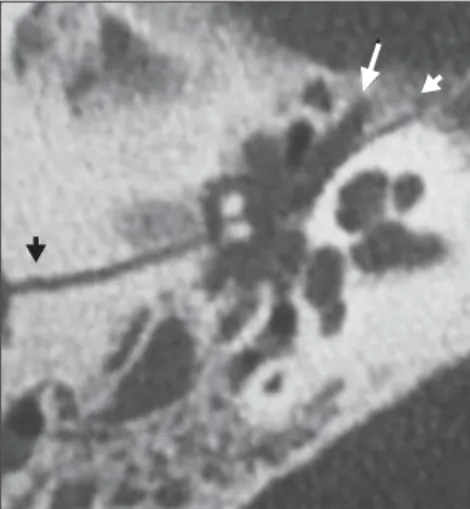
Figure 4. Incudostapedial dislocation. Axial computed tomography image of the incudostapedial joint reveals separation between the incus lenticular process and the stapes head.
Figure 5. Malleoincudal dislocation. Axial Computed tomography image demonstrates dislocation of the malleoincudal joint (A) in a comparative analysis with a normal joint (B).
A B
Figure 8. Labyrinthine fractures. Coronal computed tomography image shows transverse fracture extending into the osseous labyrinth (arrow) and with pneumovestibule (circle).
Penetrating injuries through the exter-nal ear caexter-nal (manipulation with cotton swabs) can dislocate the stapes through the oval window into the vestibulum (internal dislocation) (Figure 7).
Ossicular fractures
Among the ossicular fractures, the most relevant one fracture of the stapes arch, which occurs secondarily to its torsion(8,14).
Fracture of the stapes footplate occurs mainly in cases of transverse fractures (translabyrinthine fractures) crossing the oval window. Footplate fracture (either with or without fragments displacement) may cause perilymphatic fistula with pneumolabyrinth(14).
OTHER TYPES OF INJURIES
Labyrinthine fractures
Generally related to transverse fractures with sensorineural hearing loss(11), such
fractures are followed by pneumolaby-rinth(12,14) and perilymphatic fistulas.
Peri-lymphatic fistulas may also cause vertigo, and occur on account of injury to the otic capsule(9,15) (Figures 8 and 9).
Dural fistulas
Generally related to injuries of the teg-men tympani or of the sphenoid sinuses walls, dural fistulas evolve with otoliquor-rhea or rhinoliquorotoliquor-rhea. Such lesions should not be neglected, because of the risk for meningitis(7,12,15) (Figure 10).
Facial paralysis
The facial nerve pathway may be evalu-ated along its entire extent in the axial plane, with oblique reconstructions being
Figure 9. Labyrinthine fractures. Coronal computed tomography reveals trans-labyrinthine transverse fracture (arrow) Pneumolabyrinth (circle) and fluid inside the middle ear (perilymphatic fistula) are highlighted.
Figure 10. Dural fistulas. Coronal computed tomography image demon-strating tegmen tympani rupture (arrow).
of great relevance to analyze the integrity of the mastoid and tympanic segments. The facial nerve is affected in up to 7% of pa-tients with temporal fracture. Most injuries occur in the labyrinthine portion, in the region of the geniculate ganglion, and manifest as nerve contusion, edema and hematoma of the neural sheath and partial or complete nerve transection(7,9,15).
Immediate-onset post-traumatic paraly-sis frequently indicates presence of nerve transection or compression by a bone frag-ment (Figure 11).
Carotid canal
The petrous portion of the temporal bone includes the petrous segment of the internal carotid artery, which is located in the carotid canal, medially to the styloid process and anteriorly to the jugular fossa. The carotid canal thoroughly evaluated in the axial plane. Patients with fractures ex-tending to the carotid canal present in-creased risk for injuries to the internal ca-rotid artery. Associated complications
in-REFERENCES
1. Gasparetto EL. Tomografia computadorizada no traumatismo cranioencefálico [editorial]. Radiol Bras. 2011;44(2):vii.
2. Dal Secchi MM, Moraes JFS, Castro FB. Fratura de osso temporal em pacientes com traumatismo crânio-encefálico. Arquivos Int Otorrinolaringol. 2012;16:62–6.
3. Little SC, Kesser BW. Radiographic classification of temporal bone fractures: clinical predictabil-ity using a new system. Arch Otolaryngol Head Neck Surg. 2006;132:1300–4.
4. Saraiya PV, Aygun N. Temporal bone fractures. Emerg Radiol. 2009;16:255–65.
5. Morgado FL, Rossi LA. Correlação entre a escala de coma de Glasgow e os achados de imagem de tomografia computadorizada em pacientes víti-mas de traumatismo cranioencefálico. Radiol Bras. 2011;44:35–41.
6. Lasak JM, Van Ess MJ, Kryzer TC, et al. Penetrat-ing middle ear trauma through the external audi-tory canal. Otolaryngol Head Neck Surg. 2004; 131:P92.
7. Brodie HA, Thompson TC. Management of com-plications from 820 temporal bone fractures. Am J Otol. 1997;18:188–97.
8. Hasso AN, Ledington JA. Traumatic injuries of the temporal bone. Otolaryngol Clin North Am. 1988;21:295–316.
9. Dahiya R, Keller JD, Litofsky NS, et al. Tempo-ral bone fractures: otic capsule sparing versus otic capsule violating clinical and radiographic con-siderations. J Trauma. 1999;47:1079–83. 10. Swartz JD, Berger AS, Zwillenberg S, et al.
Os-sicular erosions in the dry ear: CT diagnosis. Ra-diology. 1987;163:763–5.
11. Swartz JD, Swartz NG, Korsvik H, et al. Comput-erized tomographic evaluation of the middle ear and mastoid for posttraumatic hearing loss. Ann Otol Rhinol Laryngol. 1985;94:263–6. 12. Cvorovic L, Jovanovic MB, Markovic M, et al.
Management of complication from temporal bone fractures. Eur Arch Otorhinolaryngol. 2012;269: 399–403.
13. Lourenco MT, Yeakley JW, Ghorayeb BY. The “Y” sign of lateral dislocation of the incus. Am J Otol. 1995;16:387–92.
14. Mafee MF, Valvassori GE, Kumar A, et al. Pneumolabyrinth: a new radiologic sign for frac-ture of the stapes footplate. Am J Otol. 1984;5: 374–5.
15. Zayas JO, Feliciano YZ, Hadley CR, et al. Tem-poral bone trauma and the role of multidetector CT in the emergency department. Radiographics. 2011;31:1741–55.
Figure 12. Carotid canal. Axial computed tomog-raphy image showing transverse fracture (arrows) crossing the carotid canal (asterisk).
clude arterial dissection, pseudoaneurysm, complete transection, occlusion and arte-riovenous fistulas(7,15) (Figure 12).
CONCLUSION
Computed tomography plays a funda-mental role in the initial evaluation of polytrauma patients, as it is capable of iden-tifying injuries to important structures which may lead to severe complications such as conductive or sensorineural hear-ing loss, dizziness and balance disorders, perilymphatic fistulas, facial nerve paraly-sis, vascular injuries, among others(3).
Investigations by means of computed tomography also allow the radiologist to classify temporal fractures and to predict such possible complications, providing guidance for treatment.
The temporal bone anatomy is rather complex, with several critical structures associated to each other(3,7,8,11). It is
impor-tant for the radiologist to be familiar with such anatomy and by team-working with other specialists, such as otolaryngologists and neurosurgeons, allowing the appropri-ate management of cases of temporal bone trauma, thus reducing the risk for severe sequels in such patients.
Figure 11. Facial paralysis. Axial computed tomog-raphy image showing longitudinal fracture (arrow heads) crossing the tympanic segment of the fa-cial nerve (arrow).