Correlation between molecular features and genetic
subtypes of Glioblastoma: critical analysis in 109 cases
Thais F GalatroI, Paula SolaI, Isabele F MorettiI, Flavio K MiuraI, Sueli M Oba-ShinjoI, Suely KN MarieI,II, Antonio MLerarioIII,IV
DOI: 10.5935/MedicalExpress.2017.05.05
I Universidade de Sao Paulo, Faculdade de Medicina FMUSP, Department of Neurology, Laboratory of Molecular and Cellular Biology, LIM15, São Paulo, SP, Brazil. II University of São Paulo, Center for Studies of Cellular and Molecular Therapy (NAP-NETCEM-NUCEL), São Paulo, SP, Brazil.
III University of Michigan, Division of Internal Medicine Metabolism, Endocrinology and Diabetes, Ann Arbor, MI, USA. IV Universidade de Sao Paulo, Faculdade de Medicina, Bioinformatics coordinator of SELA (NGS facility core), São Paulo, SP, BR.
OBJECTIVE: Glioblastoma, the most common and lethal brain tumor, is also one of the most defying forms of malignancies in terms of treatment. Integrated genomic analysis has searched deeper into the molecular architecture of GBM, revealing a new sub-classiication and promising precision in the care for patients with speciic alterations. METHOD: Here, we present the classiication of a Brazilian glioblastoma cohort into its main molecular subtypes. Using a high-throughput DNA sequencing procedure, we have classiied this cohort into proneural, classical and mesenchymal sub-types. Next, we tested the possible use of the overexpression of the EGFR and CHI3L1 genes, detected through immunohistochemistry, for the identiication of the classical and mesenchymal subtypes, respectively.
RESULTS: Our results demonstrate that genetic identiication of the glioblastoma subtypes is not possible using single targeted mutations alone, particularly in the case of the Mesenchymal subtype. We also show that it is not possible to single out the mesenchymal cases through CHI3L1 expression.
CONCLUSION: Our data indicate that the Mesenchymal subtype, the most malignant of the glioblastomas, needs further and more thorough research to be ensure adequate identiication.
KEYWORDS: glioblastoma, classiication, DNA sequence analysis, CHI3L1, EGFR.
Galatro TF, Sola P, Moretti IF, Miura FK, Oba-Shinjo SM, MarieI SKN, Lerario AM. Correlation between molecular features and genetic subtypes of Glioblastoma: critical analysis in 109 cases. MedicalExpress. 2017 Oct; 4(5):M170505
Received for Publication on July 28,2017; First review on September 6, 2017; Accepted for publication on October 3, 2017; Online on October 20, 2017
E-mail: [email protected]
■
INTRODUCTIONGlioblastoma multiforme (GBM) is the most common and most malignant form of brain tumor in adults. Glioblastomas belong to the glioma group of tumors. The World Health Organization1 classifies
gliomas according to their resemblance to their cell of origin, along with histological and molecular features. Glioblastomas, the most prevalent within the glioma group, are extremely aggressive grade IV tumors, exhibiting high mitotic rates, micro-vascular proliferation and necrosis; they also present the poorest prognosis, with a median survival of 15 months from the time of diagnosis.2 Due to their invasive nature, complete surgical
resection is very difficult to achieve. The presence of
residual tumor cells results in recurrence and malignant progression, albeit at different intervals.
Glioblastomas can be further divided into two subgroups: primary GBMs, which arise de novo, and
secondary GBMs, which result from the progression of a lower grade tumor.3 These two clinical forms of
GBM have different and extensively characterized molecular features.3 The past decade has seen the rise
of high-throughput sequencing techniques that provide in-depth knowledge of molecular alterations in tumor
cells. GBM has been one of the most molecularly profiled
tumors by several groups, including The Cancer Genome Atlas (TCGA) Networks.4-10 These studies have singled
out specific determinant mutations within the newly identified subtypes: proneural, neural, classical and
customized panel included genes such as NF1, RB1, EGFR, TP53, PTEN, IDH1 and PDGFRA, whose alterations have all been previously associated with the three main GBM subtypes: proneural, classical and mesenchymal. Using the SureDesign tool (Agilent Technologies Inc., USA), the targeted RNA capture enrichment baits were designed to include coding exon regions and 50 bp from both the 3’ end
and 5’ end of the flanking intronic sequence. The purpose
was to incorporate possible splicing site mutations in our analysis. A total of 973 regions from the 40 genes were
targeted for a final capture size of 2.79 Mega base pairs
(Mb).
A target-enrichment DNA library was constructed using the Agilent SureSelect XT Target Enrichment Kit (Agilent Technologies Inc.), following the recommended protocol. Two hundred nanograms of tumor DNA was sheared using a E220 focused-ultrasonicator (Covaris, USA) to generate DNA fragments with a mean peak around 150bp. Indexed and adaptor-ligated libraries were multiplexed and paired-end sequenced on Illumina NextSeq 500 platform (Illumina, USA). An additional library was built from peripheral blood DNA from all the patients pooled in equal amounts.
Bioinformatics analysis. Sequencing data was generated as 150-bp paired-end reads using the Illumina Next-Seq platform. Raw data was aligned to the hg38 assembly of the human genome using the BWA mapping software.12 Aligned reads were coordinate-sorted with
the bamsort tool from Biobambam2.13 Variant calling was
performed simultaneously in all the tumor samples with the reference genome–free algorithm Platypus,14 and in
the germline pool with freebayes, with the parameters properly set to call pooled data (pooled-discrete and ploidy parameter set to 132, which corresponds to the number
of alleles present in the pool). The resulting VCFs file were
annotated with SnpEff and SnpSIFT.15
Tissue microarray (TMA) construction and Immunohistochemistry. Two representative areas of each tumor were chosen by neuropathologists and marked both
on HE sections and on the original paraffin block. Each of the 0.6 mm-diameter three cores of tumor tissue was extracted
from the marked area of each donor block using an arraying machine (MTA-1, Beecher Instruments Inc., USA). The cores were inserted into a TMA recipient block in predetermined
sites. Sections of 3μm-thickness were cut from the TMA
block. A representative TMA section was initially stained with HE to assess the suitability of each core, and all other
sections were paraffin coated and stored at -20°C until use. For immunohistochemical detection, serial TMA
sections were deparaffinized, rehydrated, treated for
endogenous peroxidase blocking and subjected to antigen retrieval. Slides were immersed in 10 mM citrate buffer, pH 6.0 and incubated at 122°C for 3 min using an electric pressure cooker (BioCare Medical, USA). Specimens were in the IDH1, PDGRFA and TP53 genes; classical tumors
display mainly the EGFR mutation/amplification and the
presence of the EGFRvIII oncogenic variant; mesenchymal tumors show RB1 and NF1 mutations as their main features. The neural subtype presents overexpression of neural
markers, but display no specific genetic alterations. Patient
overall survival was also correlated with GBM molecular subtypes, with the mesenchymal subtype presenting the worst prognosis. However, the reproducibility, clinical relevance, and functional basis of these subclasses remain to be established.
In this study, we have developed a NGS-based gene panel to detect and analyze several genes commonly
mutated in GBM. We classified a series of Brazilian GBM
cases based on the somatic mutation signatures previously established for the three main subtypes: proneural, classical
and mesenchymal. We validated our genetic findings
through orthogonal methods and immunohistochemistry. In this multi-institution cohort of Brazilian patients, GBM subtype distribution and associated patient outcomes corroborate previously published results and further validates the clinical relevance of GBM subtyping.
NGS-based approaches for GBM classification are fast, reliable,
and therefore, may have value as a diagnostic tool that may add to the clinical decision-making process.
■
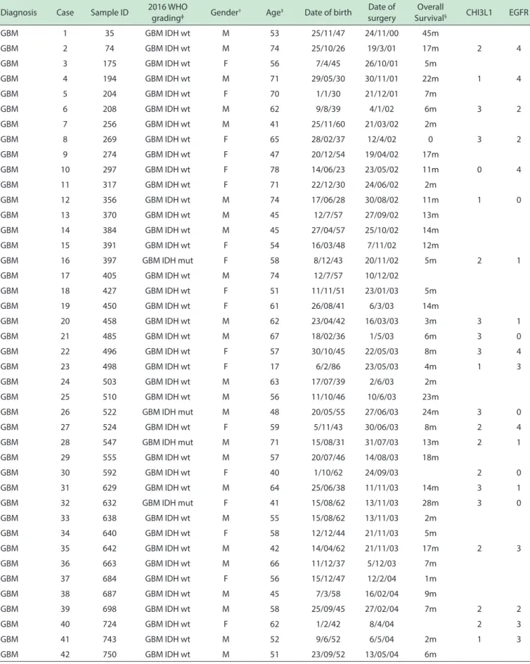
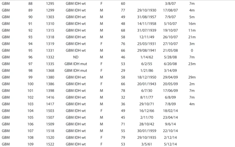
MATERIALS AND METHODSIncluded patients and ethical statement. One hundred and nine GBM samples were obtained during therapeutic surgery of patients treated by the Neurosurgery Group of the Department of Neurology at Hospital das Clínicas at the School of Medicine of the University of São Paulo, in the period of 2000 to 2014. Written informed consent was obtained from all patients according to the ethical guidelines approved by the institutional Ethics Committee. (case # 0599/10). Complete patient information, along with the current WHO grading system1
and clinical findings are presented in Supplemental Table
1, annexed to this article.
GBM sampling and diagnosis. GBM diagnosis
was confirmed by neuropathologists from the Division of
Pathological Anatomy of the same institution, according to the WHO grading system available at the time.11 Samples
were macrodissected and immediately snap-frozen in liquid nitrogen upon surgical removal. A 4µm-thick cryosection of each sample was analyzed under a light microscope after hematoxylin-eosin staining for assessment of cellular debris as well as necrotic and non-neoplastic areas; this was followed by removal from the frozen block by microdissection prior to DNA extractions.
Targeted gene panel sequencing. Based on previous
literature assessing GBM molecular sub classification,6,8,9 40
then blocked and further incubated with anti-human CHI3L1 (mouse monoclonal, clone AT4A3; Abcam, United Kingdom) and with anti-human EGFR (mouse monoclonal,
clone 31G7; Thermo Fisher Scientific, USA) at 16-20°C for
16 hours. Development of the reaction was performed with a commercial kit (Novolink; Novocastra, United Kingdom) at room temperature, using diaminobenzidine and Harris hematoxylin for nuclear staining. Optimization using positive controls suggested by the manufacturer of each antibody was performed in order to obtain optimal dilution. Two observers (TFG and IFM) evaluated staining intensity of tissue sections independently. A semi- quantitative scoring system considering both intensity of staining and the respective percentage of stained cells was applied as follows: score 0: no cells stained; score 1: 10–25% cells stained; score 2: 26–50% cells stained; score 3: 51–75% cells stained; score 4: 76–100% cells stained. Only cases with positive cell
staining with scores ≥ 2 were considered as positive. Digital photomicrographs of representative fields were captured
and processed using PICASA 3 (Google, USA).
Statistical analysis. Survival data was assessed by comparing the Kaplan-Meier survival curves using the log-rank (Mantel Cox) test. A p-value < 0.05 was considered
statistically significant. For this analysis, we included 77
cases with complete clinical follow-up. Calculations were performed using SPSS, version 23.0 (IBM, USA).
■
RESULTSMolecular classification of GBM. We used the
mutational profile to classify tumors into three major
molecular subtypes: proneural, classical, and mesenchymal.
We defined the proneural subtype by the presence of IDH1,
PDGFRA, and TP53 mutations. The classical subtype was
defined by EGFR alterations (amplification and mutations),
PTEN mutations, and the presence of the oncogenic
variant EGFRVIII. EGFR amplification results was based
on a previous publication from our group.16 Finally, for the
definition of the mesenchymal subtype, we used mutations
in NF1 and RB1. For each patient, we used DNA from blood leukocytes for germline alteration subtraction. None of the found alteration were present in these germline controls. Aside from this control, we considered in this analysis a) known pathogenic mutations, such as the IDH1 R132H, b) variants that were not present in population databases (1000kg/ExAC), c) variants with predicted pathogenicity (nonsense or frameshift, or a missense mutation with high SIFT and polyphen scores), d) variants not present in our germline pool. All the tumors which did not carry a mutation
in the genes described above and met the defined criteria were classified as “others”. Given the lack of a specific genetic profile defining the neural subtype, we were not able
to identify this subclass in our dataset. The complete list of genetic alteration can be found in Supplemental Table 2.
The targeted NGS analysis was performed on 109 GBM samples. A 20x coverage was achieved in ~97% of the targeted regions of the GBM samples. The mean coverage of the germline pool was 6063.67x.
Following the proposed criteria, we were able to classify 89 of the 109 analyzed GBM samples within the three major subtypes. Genetic alterations typical of the classical subtype were the most prevalent, accounting for
45.9% of the classified cases. Samples with alterations
inherent to the mesenchymal subtype corresponded to 19.3% of the cases; alterations particular to the proneural subtype contributed 16.5% of the cases. This information is diagrammatically shown in Figure 1. A preview of survival
rates are also shown in this figure.
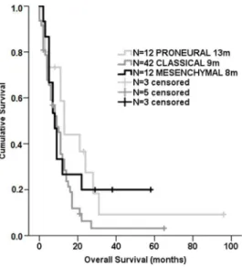
The overall survival of GBM patients according to
our molecular classification is shown in Figure 2: patients classified as mesenchymal GBM had a shorter overall
survival in comparison to other subgroups (medians of 8, 9, and 13 months for the mesenchymal, classical, and proneural, respectively; log-rank p < 0.05).
Correlation between molecular classification of GBM and immunohistochemical markers to differentiate between classical and mesenchymal subtypes.
Concomitant with the molecular classification of GBMs,
we also assessed the protein expression levels of CHI3L1 (YKL-40) and EGFR. CHI3L1 has been previously associated with the mesenchymal subtype of GBM, while EGFR overexpression is a known characteristic of the classical subtype. Immunohistochemistry staining was performed in
a subset of our molecularly classified samples, comprising a
cohort of 40 cases (8 mesenchymal, 9 proneural, 20 classical and 3 others). Figure 3 shows the results and Figure 4 depicts examples of stained tissue microarray sections. While only one of the mesenchymal samples showed negative levels of CHI3L1, cases positive for CHI3L1 (scoring above 2) comprised samples of the three subtypes (6 mesenchymal, 6 proneural and 10 classical cases were positive for CHI3L1). For EGFR, all proneural and 6 mesenchymal cases proved to be negative for this marker. Despite presenting genetic characteristics of classical subtypes, 4 of these cases did not present positivity for EGFR immunostaining. A complete listing of immunohistochemistry score can be found in Supplemental Table 2.
■
DISCUSSIONThe advent of next generation sequencing and large-scale molecular analysis over the past decade revealed that molecular alterations predict patients’ responses to treatment, overall survival and clinical outcome. A new light has been shed on the high level of GBM heterogeneity
Figure 1 - Distribution of the molecularly classiied GBM cases. Depiction of the main genetic alterations of the cases molecularly classiied in our GBM cohort. Classiication was achieved using a customized gene panel, followed by exome sequencing (refer to text for details). The color gradient at the bottom depicts patient overall survival (blue indicates long; purple short survival as shown in the color gradient at the left); gray represents cases without a complete clinical follow-up. amp: ampliication; mut: mutation.
Figure 2: Survival curve of the molecularly classiied GBM cases. Kaplan-Meier of 77 GBM cases detailing median survival (log rank test, p=0.128) times according to our molecular classiication. Patients alive at the time of the last clinical follow-up were censored from this analysis. m, months.
Figure 3: Scoring of immunohistochemical analysis for CHI3L1 and EGFR in GBM cases. Each GBM sample, previously classiied with the molecular approach, was scored for the classical (EGFR, blue) and mesenchymal (CHI3L1, pink) immunohistochemical ma-rkers. Scoring ranged from 0-4, in which score 0: no cells stained; score 1: 10–25% cells stained, score 2: 26–50% cells stained; score 3: 51–75% cells stained; score 4: 76–100% cells stained. EGFR staining was prevalent in classical samples, although classical cases did not present signiicant staining of the marker, while other mesenchymal samples presented comparable levels of EGFR. CHI3L1 was present in high levels in cases from all three GBM subtypes.
Figure 4: Representative immunohistochemical sections for the evaluation of CHI3L1 and EGFR in molecularly classiied GBM samples. Proneural (A-D), mesenchymal (E-H) and classical (I-L) GBM representative cases were stained for CHI3L1 and EGFR, whose overexpression has been associated with mesenchymal and classical subtypes, respec-tively. CHI3L1 expression was widespread among GBM samples, and not exclusive of mesenchymal cases. Both proneural (A) and classical (I) samples displayed high levels of this marker. Despite being negative in proneural cases (D), EGFR overexpression failed to diferentiate between mesenchymal and classical cases, with both groups showing positive and negative cases. The reaction was performed in parain embed-ded tissue sections with a commercial polymer kit (Novolink; Novocastra, UK), using diaminobenzidine as developer and Harris hematoxylin for nuclear counterstaining. 400x magniication for all images. Scale bar 10µm.
and mesenchymal.4–10 Their potential to aid the choice of
better treatment options is a step forward toward precision medicine.
We have performed a somatic mutation analysis in our GBM cohort utilizing a customized gene panel that comprises all the coding regions and splicing regions the most commonly mutated genes described in GBM. Our NGS panel allowed us to classify approximately ~85% of our samples according to molecular subtype using a single assay; this would have been very laborious and time-consuming using traditional methods, such as Sanger sequencing.17 The results we obtained, regarding the
percentage of each GBM subtype, as well as their association with patient overall survival, is in accordance with what has been published so far.5–9
Since the first documentation of the molecular
results indicate that the higher expression of CHI3L1 is not an event exclusive of the mesenchymal cases, because high percentages of both proneural and classical samples displayed equivalent high levels.
■
CONCLUSIONOur results indicate the need for a genetic approach to further classify GBMs, so that a higher number of patients
can profit from the precision provided by such classification
in terms of treatment options.
■
ACKNOWLEDGEMENTSThe authors sincerely thank doctors and residents from the Discipline of Neurosurgery of the Department of Neurology at Hospital das Clínicas of School of Medicine, University of São Paulo, from the Neurosurgery group at Sírio Libanês Hospital and, particularly, Drs André M Bianco and Egmond Alves, for the therapeutic and diagnostic procedures of all patients included in this study. We also thank the physicians and technicians at the Division of Pathological Anatomy of the same institutions, particularly
Dr Sergio Rosemberg, for the histological classification of
tumor samples and tissue section processing. The authors thank São Paulo Research Foundation (FAPESP), grants #2013/07704- 3, #2013/06315-3, #2013/02162-8, #2014-50137-5 and #2016/15652-1, CAPES-NUFFIC (062/15), Conselho Nacional de Pesquisa (CNPq 305730/2015-0)
and Faculdade de Medicina FMUSP for financial support.
■
AUTHOR CONTRIBUTIONAML, SKNM and SMOS conceived the study. AML performed the bioinformatics analysis. TFG and PS performed the experiments. SKNM and FKM followed-up the patients. SKMN and AML provided sfollowed-upervision. AML, SKNM and TFG wrote the manuscript. All authors contributed to the editing of the paper.
■
CONFLICT OF INTERESTThe authors declare no competing interests.
CORRELAÇÃO ENTRE AS CARACTERÍSTICAS MOLECULARES E OS SUBTIPOS GENÉTICOS DOS GLIOBLASTOMAS: ANÁLISE CRÍTICA DE 109 CASOS
OBJETIVO: O glioblastoma (GBM), o tumor cerebral mais comum e mais letal, é também um dos tipos de
tumores de mais difícil tratamento. Análises genômicas
integradas têm contribuído para um melhor entendimento to be fundamental for new discoveries that may ultimately
lead to better clinical approaches and precision medicine. For instance, Davis et al18 used the classification to compare
genetic alterations in cultured brain-tumor initiating cells. This model system is important to study treatment options and GBM biology. Natash et al19 explored the role
of oncostatin, a cytokine present in the microenvironment of GBM and its signaling pathway, which is associated with poor prognosis. They related such features to the aggressive nature in the mesenchymal GBM subtype. Chen and Xu20
have recently developed an algorithm that matches FDA approved drugs to the molecular subtype of GBMs, based on the genetic alterations of each subtype. Thus, it is important
to specifically differentiate classical from mesenchymal GBMs: classical GBMs are amenable to treatment by specific
tyrosine kinase inhibitors and adjuvant antibodies;21 in
contrast, mesenchymal GBMs have only one potentially useful drug, currently introduced as a phase 1 clinical trial
(clinical trial identifier: NCT02272270). Therefore, new
practical proposals, more suitable for major diagnostic
centers, have been looked for. The exemplified studies highlight the usefulness of GBM classification in improving
current knowledge, biological understanding and diagnosis and treatment options for this tumor.
However, and because the basis for such classification
is the genetic approach, requiring the implementation of molecular biology techniques, other more feasible methodologies have been searched for. A recent study revealed that MRI derived quantitative volumetric tumor phenotype features only moderately predict the molecular GBM subtypes, suggesting that subtypes do not generally alter the size composition of tumor areas.22 Therefore, the
more doable proteomic IHC-based approach, still based on
the reported molecular findings, has been the preferred technique for patient classification.
Both proneural and classical GBM subtypes present genetic point mutations or alterations (IDH1 R132H and EGFRvIII, respectively) that have made it possible to
develop, over recent years, specific antibodies to achieve
a cheaper and more suitable approach in major diagnostic centers.23,24 Nonetheless, the alteration of EGFRvIII was
found in only 28.4% of our GBM cohort, while classical cases corresponded to 45.9% of all cases. For those extra cases, EGFR protein overexpression still excluded 4 out of 23 samples with a classical genetic subtype.
The mesenchymal subtype presents a high level of heterogeneity of alterations within the usually mutated genes of this subtype (NF1 and RB1). CHI3L1 (also known as YKL-40) is a secreted lectin, related to the regulation of hypoxia-induced injury response and has been previously associated with the mesenchymal GBM.6,8 Previous studies
have proposed CHI3L1 as an additional marker to be used as a diagnosis tool through an immunohistochemical
da arquitetura molecular dos GBMs, revelando uma nova
subclassificação com a promessa de precisão no tratamento de pacientes com alterações específicas. Neste estudo, nós apresentamos a classificação de uma casuística brasileira
de GBMs dentro dos principais subtipos do tumor. MÉTODO: Usando sequenciamento de DNA em larga
escala, foi possível classificar os tumores em proneural, clássico e mesenquimal. Em seguida, testamos o possível uso da hiperexpressão de EGFR e CHI3L1 para a identificação dos subtipos clássico e mesenquimal, respectivamente.
RESULTADOS: Nossos resultados deixam claro que a
identificação genética dos subtipos moleculares de GBM não é possível utilizando-se apenas um único tipo de mutação,
em particular nos casos de GBMs mesenquimais. Da mesma forma, não é possível distinguir os casos mesenquimais apenas com a expressão de CHI3L1.
CONCLUSÃO: Nossos dados indicam que o subtipo mesenquimal, o mais maligno dos GBMs, necessita de uma
análise mais aprofundada para sua identificação.
PALAVRAS-CHAVE: glioblastoma, classificação,
análise de sequência de DNA, CHI3L1, EGFR
■
REFERENCES1. Louis DN, Perry A, Reifenberger G, von Deimling A, Figarella-Branger D, Cavenee WK, et al. The 2016 World Health Organization
Classification of Tumors of the Central Nervous System: a summary.
Acta Neuropathol. 2016;131(6):803–20. DOI:10.1007/s00401-016-1545-1.
2. Wen PY, Kesari S. Malignant gliomas in adults. N Engl J Med. 2008;359(5):492–507. DOI:10.1056/NEJMra0708126.
3. Ohgaki H, Kleihues P. The definition of primary and secondary
glioblastoma. Clin Cancer Res Off J Am Assoc Cancer Res. 2013;19(4):764–72. DOI:10.1158/1078-0432.CCR-12-3002. 4. Cancer Genome Atlas Research Network, Brat DJ, Verhaak RGW, Aldape
KD, Yung WKA, Salama SR, et al. Comprehensive, Integrative Genomic Analysis of Diffuse Lower-Grade Gliomas. N Engl J Med. 2015(26);372 (26):2481–98. DOI:10.1056/NEJMoa1402121.
5. Ceccarelli M, Barthel FP, Malta TM, Sabedot TS, Salama SR, Murray BA,
et al. Molecular Profiling Reveals Biologically Discrete Subsets and
Pathways of Progression in Diffuse Glioma. Cell. 2016;164(3):550–63. DOI:10.1016/j.cell.2015.12.028.
6. Verhaak RGW, Hoadley KA, Purdom E, Wang V, Qi Y, Wilkerson MD, et
al. Integrated genomic analysis identifies clinically relevant subtypes
of glioblastoma characterized by abnormalities in PDGFRA, IDH1, EGFR, and NF1. Cancer Cell. 2010;17(1):98–110. DOI:10.1016/j. ccr.2009.12.020.
7. Stieber D, Golebiewska A, Evers L, Lenkiewicz E, Brons NHC, Nicot N, et al. Glioblastomas are composed of genetically divergent clones with distinct tumourigenic potential and variable stem cell-associated phenotypes. Acta Neuropathol. 2014;127(2):203–19. DOI:10.1007/ s00401-013-1196-4.
8. Phillips HS, Kharbanda S, Chen R, Forrest WF, Soriano RH, Wu TD, et al. Molecular subclasses of high-grade glioma predict prognosis, delineate a pattern of disease progression, and resemble stages in neurogenesis. Cancer Cell. 2006;9(3):157–73. DOI:10.1016/j.ccr.2006.02.019.
9. Brennan CW, Verhaak RGW, McKenna A, Campos B, Noushmehr H, Salama SR, et al. The somatic genomic landscape of glioblastoma. Cell. 2013;155(2):462–77. DOI:10.1016/j.cell.2013.09.034.
10. Parsons DW, Jones S, Zhang X, Lin JC-H, Leary RJ, Angenendt P, et al. An integrated genomic analysis of human glioblastoma multiforme. Science. 2008;321(5897):1807–12. DOI:10.1126/science.1164382. 11. Louis DN, Ohgaki H, Wiestler OD, Cavenee WK, Burger PC, Jouvet A,
et al. The 2007 WHO classification of tumours of the central nervous
system. Acta Neuropathol. 2007 Aug;114(2):97-109. DOI:10.1007/ s00401-007-0243-4
12. Li H, Durbin R. Fast and accurate short read alignment with Burrows- Wheeler transform. Bioinforma Oxf Engl. 2009;25(14):1754–60. DOI:10.1093/bioinformatics/btp324.
13. Tischler G, Leonard S. biobambam: tools for read pair collation
based algorithms on BAM files. Source Code Biol Med. 2014;9:13.
DOI:10.1186/1751-0473-9-13.
14. Platypus, a reference genome–free algorithm that rapidly calls variants in clinical sequencing data. SciBX Sci-Bus Exch. 2014;7. DOI:10.1038/ scibx.2014.936.
15. Cingolani P, Patel VM, Coon M, Nguyen T, Land SJ, Ruden DM, et al. Using Drosophila melanogaster as a Model for Genotoxic Chemical Mutational Studies with a New Program, SnpSift. Front Genet. 2012;3:35. DOI:10.3389/fgene.2012.00035.
16. Carvalho PO, Uno M, Oba-Shinjo SM, Rosemberg S, Wakamatsu A, da Silva CC, et al. Activation of EGFR signaling from pilocytic astrocytomas to glioblastomas. Int J Biol Markers. 2014;29(2):e120-128. DOI:10.5301/JBM.5000045.
17. Le Gallo M, Lozy F, Bell DW. Next-Generation Sequencing. Adv Exp Med Biol. 2017;943:119–48. DOI:10.1007/978-3-319-43139-0_5. 18. Davis B, Shen Y, Poon CC, Luchman HA, Stechishin OD, Pontifex CS, et
al. Comparative genomic and genetic analysis of glioblastoma-derived brain tumor-initiating cells and their parent tumors. Neuro-Oncol. 2016;18(3):350– 60. DOI:10.1093/neuonc/nov143.
19. Natesh K, Bhosale D, Desai A, Chandrika G, Pujari R, Jagtap J, et al. Oncostatin-M differentially regulates mesenchymal and proneural signature genes in gliomas via STAT3 signaling. Neoplasia. 2015;17(2):225–37. DOI:10.1016/j.neo.2015.01.001.
20. Chen Y, Xu R. Drug repurposing for glioblastoma based on molecular subtypes. J Biomed Inform. 2016;64:131–8. DOI:10.1016/j. jbi.2016.09.019.
21. Padfield E, Ellis HP, Kurian KM. Current Therapeutic Advances
Targeting EGFR and EGFRvIII in Glioblastoma. Front Oncol. 2015;5:5. DOI:10.3389/fonc.2015.00005.
22. Grossmann P, Gutman DA, Dunn WD, Holder CA, Aerts HJWL. Imaging- genomics reveals driving pathways of MRI derived volumetric tumor phenotype features in Glioblastoma. BMC Cancer. 2016;16:611. DOI:10.1186/s12885-016-2659-5.
23. Kato Y. Specific monoclonal antibodies against IDH1/2 mutations as
diagnostic tools for gliomas. Brain Tumor Pathol. 2015;32(1):3–11. DOI:10.1007/s10014-014-0202-4.
24. Gupta P, Han S-Y, Holgado-Madruga M, Mitra SS, Li G, Nitta RT, et al.
Development of an EGFRvIII specific recombinant antibody. BMC
Biotechnol. 2010;10:72. DOI:10.1186/1472-6750-10-72.
25. Conroy S, Kruyt FAE, Joseph JV, Balasubramaniyan V, Bhat KP,
Wagemakers M, et al. Subclassification of Newly Diagnosed
Glioblastomas through an Immunohistochemical Approach. PLoS ONE 2014;9(12):e115687. DOI:10.1371/journal.pone.0115687.
26. Joseph JV, Conroy S, Pavlov K, Sontakke P, Tomar T, Eggens-Meijer E, et al. Hypoxia enhances migration and invasion in glioblastoma by
promoting a mesenchymal shift mediated by the HIF1α–ZEB1 axis.
Table 1 - Patient data
ANNEX
Diagnosis Case Sample ID 2016 WHO
gradingɸ Gender† Age‡ Date of birth
Date of surgery
Overall
Survival§ CHI3L1 EGFR
GBM 1 35 GBM IDH wt M 53 25/11/47 24/11/00 45m
GBM 2 74 GBM IDH wt M 74 25/10/26 19/3/01 17m 2 4
GBM 3 175 GBM IDH wt F 56 7/4/45 26/10/01 5m
GBM 4 194 GBM IDH wt M 71 29/05/30 30/11/01 22m 1 4
GBM 5 204 GBM IDH wt F 70 1/1/30 21/12/01 7m
GBM 6 208 GBM IDH wt M 62 9/8/39 4/1/02 6m 3 2
GBM 7 256 GBM IDH wt M 41 25/11/60 21/03/02 2m
GBM 8 269 GBM IDH wt F 65 28/02/37 12/4/02 0 3 2
GBM 9 274 GBM IDH wt F 47 20/12/54 19/04/02 17m
GBM 10 297 GBM IDH wt F 78 14/06/23 23/05/02 11m 0 4
GBM 11 317 GBM IDH wt F 71 22/12/30 24/06/02 2m
GBM 12 356 GBM IDH wt M 74 17/06/28 30/08/02 11m 1 0
GBM 13 370 GBM IDH wt M 45 12/7/57 27/09/02 13m
GBM 14 384 GBM IDH wt M 45 27/04/57 25/10/02 14m
GBM 15 391 GBM IDH wt F 54 16/03/48 7/11/02 12m
GBM 16 397 GBM IDH mut F 58 8/12/43 20/11/02 5m 2 1
GBM 17 405 GBM IDH wt M 74 12/7/57 10/12/02
GBM 18 427 GBM IDH wt F 51 11/11/51 23/01/03 5m
GBM 19 450 GBM IDH wt F 61 26/08/41 6/3/03 14m
GBM 20 458 GBM IDH wt M 62 23/04/42 16/03/03 3m 3 1
GBM 21 485 GBM IDH wt M 67 18/02/36 1/5/03 6m 3 0
GBM 22 496 GBM IDH wt F 57 30/10/45 22/05/03 8m 3 4
GBM 23 498 GBM IDH wt F 17 6/2/86 23/05/03 4m 1 3
GBM 24 503 GBM IDH wt M 63 17/07/39 2/6/03 2m
GBM 25 510 GBM IDH wt M 56 11/10/46 10/6/03 23m
GBM 26 522 GBM IDH mut M 48 20/05/55 27/06/03 24m 3 0
GBM 27 524 GBM IDH wt F 59 5/11/43 30/06/03 8m 2 4
GBM 28 547 GBM IDH mut M 71 15/08/31 31/07/03 13m 2 1
GBM 29 555 GBM IDH wt M 57 20/07/46 14/08/03 18m
GBM 30 592 GBM IDH wt F 40 1/10/62 24/09/03 2 0
GBM 31 629 GBM IDH wt M 64 25/06/38 11/11/03 14m 3 1
GBM 32 632 GBM IDH mut F 41 15/08/62 13/11/03 28m 3 0
GBM 33 638 GBM IDH wt M 55 15/08/62 13/11/03 2m
GBM 34 640 GBM IDH wt F 58 12/12/44 21/11/03 5m
GBM 35 642 GBM IDH wt M 42 14/04/62 21/11/03 17m 2 3
GBM 36 663 GBM IDH wt M 66 11/12/37 5/12/03 7m
GBM 37 684 GBM IDH wt F 56 15/12/47 12/2/04 1m
GBM 38 687 GBM IDH wt M 45 7/3/58 16/02/04 9m
GBM 39 698 GBM IDH wt M 58 25/09/45 27/02/04 7m 2 2
GBM 40 724 GBM IDH wt F 62 1/2/42 8/4/04 2 3
GBM 41 743 GBM IDH wt M 52 9/6/52 6/5/04 2m 1 3
GBM 43 792 GBM IDH wt M 35 21/12/69 16/07/04 5m 2 0
GBM 44 795 GBM IDH wt M 28 15/06/76 22/08/04 11m 3 4
GBM 45 852 GBM IDH wt M 60 19/08/44 22/10/04 13m 2 0
GBM 46 854 GBM IDH wt M 46 17/06/58 27/10/04 1 4
GBM 47 875 GBM IDH wt M 35 21/12/69 23/11/04 1 0
GBM 48 879 GBM IDH wt M 61 27/07/43 2/12/04 3m 3 1
GBM 49 881 GBM IDH wt M 49 25/01/55 7/12/04 4m 1 4
GBM 50 884 GBM IDH wt F 52 15/01/52 10/12/04 27m 0 4
GBM 51 885 GBM IDH wt F 86 24/04/18 10/12/04 2m
GBM 52 891 GBM IDH wt M 57 25/10/47 21/12/04 7m
GBM 53 901 GBM IDH mut M 16 7/11/88 5/1/05 2m 0 1
GBM 54 903 GBM IDH wt M 55 8/2/45 11/1/05 22m 3 1
GBM 55 925 GBM IDH wt M 40 8/9/64 25/02/05 12m
GBM 56 930 GBM IDH mut M 26 1/10/78 3/3/05 31m
GBM 57 1002 GBM IDH wt M 40 29/01/65 15/07/05 2m 3 2
GBM 58 1003 GBM IDH wt F 68 25/03/37 17/07/05 4m 2 3
GBM 59 1007 GBM IDH mut F 28 18/06/77 22/07/05 3m 1 1
GBM 60 1009 GBM IDH wt F 38 25/10/66 25/07/05 21m 1 1
GBM 61 1070 GBM IDH wt M 72 9/3/33 29/11/05 8m
GBM 62 1074 GBM IDH wt M 32 25/07/73 3/12/05 3 0
GBM 63 1084 GBM IDH wt M 54 18/04/51 13/01/06 5m 2 1
GBM 64 1091 GBM IDH wt M 55 5/9/49 3/2/06 21m
GBM 65 1103 GBM IDH wt M 54 18/04/1951 3/3/06 3 2
GBM 66 1118 GBM IDH wt F 61 10/7/59 18/04/06 2m
GBM 67 1122 GBM IDH wt M 68 26/07/37 1/5/06 5m
GBM 68 1123 GBM IDH wt M 53 29/10/53 2/5/06 11m
GBM 69 1124 GBM IDH wt M 63 19/03/43 5/5/06 4m 1 4
GBM 70 1133 GBM IDH wt M 52 28/12/53 26/05/06 9m
GBM 71 1144 GBM IDH wt M 76 15/08/29 25/06/06 2m
GBM 72 1161 GBM IDH wt M 39 13/01/67 26/07/06 13m
GBM 73 1162 GBM IDH wt F 68 20/05/38 31/07/06 15m
GBM 74 1169 GBM IDH wt F 56 13/03/50 9/8/06 2m
GBM 75 1190 GBM IDH wt F 58 16/09/48 24/10/06 9m
GBM 76 1194 GBM IDH mut M 26 1/10/78 3/11/06
GBM 77 1199 GBM IDH mut M 30 6/11/75 24/11/06 11m
GBM 78 1205 GBM IDH wt M 69 13/12/36 5/12/06 3 3
GBM 79 1212 GBM IDH mut M 31 6/11/75 5/1/07
GBM 80 1232 GBM IDH wt M 58 7/4/48 16/02/07 12m
GBM 81 1237 GBM IDH wt M 59 18/05/47 27/02/07 7m
GBM 82 1243 GBM IDH wt M 47 22/09/59 13/03/07 10m
GBM 83 1250 GBM IDH wt M 63 11/1/44 30/03/07 7m
GBM 84 1252 ND M 45 12/2/61 4/9/07 11m
GBM 85 1272 GBM IDH wt M 70 5/5/37 18/05/07 6m
GBM 86 1274 GBM IDH wt M 58 29/05/1948 25/05/07 13m
GBM 87 1282 GBM IDH wt M 56 29/01/1951 6/7/07 9m
GBM 88 1295 GBM IDH wt F 60 3/8/07 7m
GBM 89 1299 GBM IDH wt M 77 29/10/1930 17/08/07 4m
GBM 90 1303 GBM IDH wt M 49 31/08/1957 7/9/07 5m
GBM 91 1310 GBM IDH wt M 48 14/11/1958 5/10/07 16m
GBM 92 1315 GBM IDH wt M 68 31/07/1939 19/10/07 11m
GBM 93 1318 GBM IDH wt M 58 12/11/49 26/10/07 21m
GBM 94 1319 GBM IDH wt F 76 25/03/1931 27/10/07 3m
GBM 95 1331 GBM IDH wt M 66 29/08/1941 21/05/08 0
GBM 96 1332 ND M 46 1/14/62 5/28/08 7m
GBM 97 1335 GBM IDH mut F 53 6/2/55 6/20/08 23m
GBM 98 1368 GBM IDH mut F 29 1/21/86 3/14/09
GBM 99 1380 GBM IDH wt M 58 18/12/1950 29/04/09 29m
GBM 100 1386 GBM IDH wt F 66 20/01/1943 20/05/09 2m
GBM 101 1398 GBM IDH wt M 78 6/7/30 17/06/09 7m
GBM 102 1416 GBM IDH wt M 32 8/11/77 6/8/09 7m
GBM 103 1417 GBM IDH wt M 36 29/10/71 7/8/09 4m
GBM 104 1503 GBM IDH wt F 49 16/12/66 18/02/14
GBM 105 1507 GBM IDH wt M 45 2/11/70 23/04/14
GBM 106 1509 GBM IDH wt M 71 28/10/42 9/6/14
GBM 107 1518 GBM IDH wt M 55 30/01/1959 22/10/14
GBM 108 1520 GBM IDH wt F 79 29/10/1935 2/12/14
GBM 109 1522 GBM IDH wt F 53 3/5/61 5/12/14
Continuation Table 1
† M, male. F, female, ‡ Age at diagnosis, years, § m, months, ɸ Classiicaion according to the 2016 WHO grading, based on IDH1 mutational status. Wt, wild-type; mut,
mutated; ND, non-determined. last update - dec/2014
Table 2 - Classiicatory genetic alterations in GBMs
Diagnosis Case# GBM subtype
EGFR
ampliica-tion
EGFR
mutation EGFRvIII
NF1 mutation
RB1 mutation
or loss
PDGFRA mutation
IDH1 mutation
TP53 mutation
PTEN mutation
GBM 35 MESENCHYMAL loss
GBM 74 CLASSICAL +
GBM 175 MESENCHYMAL c.1630.T
GBM 194 CLASSICAL + +
GBM 204 OTHERS
GBM 208 OTHERS
GBM 256 CLASSICAL + +
GBM 269 CLASSICAL + + c.375G>T
GBM 274 CLASSICAL + c.260_322dup
GBM 297 CLASSICAL + + c.388C>T
GBM 317 OTHERS
GBM 356 CLASSICAL +
GBM 370 OTHERS
GBM 384 OTHERS
GBM 391 MESENCHYMAL c.1882delT
GBM 397 PRONEURAL c.395G>A
GBM 427 CLASSICAL +
GBM 450 CLASSICAL +
GBM 458 CLASSICAL +
GBM 485 CLASSICAL c.1874C>T
GBM 496 CLASSICAL +
GBM 498 CLASSICAL +
GBM 503 PRONEURAL c.764_766
delTCA c.51_53delAGA
GBM 510 OTHERS
GBM 522 PRONEURAL c.395G>A c.298C>T/
c.742C>T
GBM 524 CLASSICAL + c.287C>T
GBM 547 PRONEURAL + c.395G>A
GBM 555 OTHERS
GBM 592 PRONEURAL c.863A>G
GBM 629 CLASSICAL +
GBM 632 PRONEURAL c.395G>A
GBM 638 CLASSICAL +
GBM 640 OTHERS
GBM* 642 CLASSICAL c.210 +1G>A
GBM 663 OTHERS
GBM* 684 CLASSICAL
GBM 687 CLASSICAL +
GBM 698 MESENCHYMAL
IVS45--1G>C (splice site)
c.271G>T
GBM 724 CLASSICAL +
GBM 743 CLASSICAL + c.335T>C
GBM 750 CLASSICAL + + c.407G>A
GBM 792 MESENCHYMAL c.6514_
6515delGA
c.742C>T/ c.473G>A
GBM 795 CLASSICAL +
GBM 852 PRONEURAL c.1087A>T
GBM 854 CLASSICAL + +
GBM 875 MESENCHYMAL c.1882delT c.210_
211insA
GBM 879 MESENCHYMAL loss
GBM 881 CLASSICAL + + c.388C>T
GBM 884 CLASSICAL +
GBM 885 OTHERS
GBM 891 OTHERS c.524G>A
GBM 901 PRONEURAL + c.395G>A
GBM 903 MESENCHYMAL c.12_13del/
c.45_53del
GBM 925 CLASSICAL +
GBM 930 PRONEURAL + c.395G>A
GBM 1002 OTHERS
GBM 1003 CLASSICAL + +
GBM 1007 PRONEURAL c.395G>A c.206delG
GBM 1009 PRONEURAL c.733G>A
GBM 1070 MESENCHYMAL loss
GBM 1074 MESENCHYMAL c.60+1G>T c.2211+
1G>T c.184G>T c.407G>A
GBM 1084 MESENCHYMAL c.6852_
6855del
c.210_ 211insA
GBM 1091 CLASSICAL c.158_160
delTAG
GBM 1103 MESENCHYMAL + c.6852_
6855del c.465T>G
GBM 1118 CLASSICAL c.355G>T
GBM 1122 MESENCHYMAL c.499_
502del
GBM 1123 CLASSICAL + +
GBM 1124 CLASSICAL
GBM 1133 CLASSICAL + +
GBM 1144 CLASSICAL +
GBM 1161 CLASSICAL + +
GBM 1162 CLASSICAL +
GBM 1169 MESENCHYMAL loss c.355_356
insGG
GBM 1190 MESENCHYMAL
c.7190-
11_7190-8del (splice)
GBM 1194 PRONEURAL c.395G>A
GBM 1199 PRONEURAL c.395G>A c.658T>C
GBM 1205 OTHERS
GBM 1212 PRONEURAL + c.395G>A c.658T>C
GBM 1232 OTHERS
GBM 1237 OTHERS
GBM 1243 CLASSICAL + c.1043T
>C
GBM 1250 CLASSICAL +
GBM 1252 OTHERS
GBM 1272 CLASSICAL +
GBM 1274 CLASSICAL +
GBM 1282 MESENCHYMAL + c.3739_
3742del
GBM 1295 MESENCHYMAL c.323G>A loss c.210_
211insA
GBM 1299 CLASSICAL +
GBM 1303 OTHERS
GBM 1310 CLASSICAL + c.1859G
>A c.464A>G
GBM 1315 PRONEURAL
c.368--3C>T (splice)
c.97-6C>T (splice)
GBM 1318 CLASSICAL c.2240_
2254del
GBM 1319 OTHERS
GBM 1331 CLASSICAL + c.685A>T c.641dupA
GBM 1332 OTHERS
GBM 1335 PRONEURAL c.395G>A
GBM 1368 PRONEURAL c.395G>A
GBM 1380 MESENCHYMAL c.1475
delA c.822G>A
GBM 1386 CLASSICAL c.760T>A c.517C>T
GBM 1398 CLASSICAL +
GBM 1416 CLASSICAL +
GBM 1417 MESENCHYMAL c.4742_
4749del
GBM 1503 PRONEURAL loss
GBM 1507 MESENCHYMAL + c.1546C>T c.451C>T
GBM 1509 CLASSICAL c.115G>A
GBM 1518 CLASSICAL +
GBM 1520 CLASSICAL c.1280G
>T
GBM 1522 MESENCHYMAL c.1924C>T c.765dupA