413
Radiol Bras. 2017 Nov/Dez;50(6):405–415Letters to the Editor
http://dx.doi.org/10.1590/0100-3984.2016.0055
Felipe Welter Langer1, Daiane dos Santos1, Giordano Rafael Tronco Alves1, Gustavo Suertegaray1, Carlos Jesus Pereira Haygert1
1. Department of Radiology and Imaging Diagnosis, University Hospital of Santa Maria, Federal University of Santa Maria (UFSM), Santa Maria, RS, Bra-zil. Mailing address: Dr. Felipe Welter Langer. Department of Radiology and Imaging Diagnosis, University Hospital of Santa Maria, Federal University of Santa Maria. Avenida Roraima, 1000, Camobi. Santa Maria, RS, Brazil, 97105-900. E-mail: [email protected].
with high local invasion, rapid growth, and early distant metasta
-sis unless they are excised in a timely manner(2). The most com -mon locations for MPNST in neuroibromatosis patients are the extremities, head, and neck. Thoracic involvement, however, is remarkably rare, few cases having been reported(3). According
to the size and location of the intrathoracic tumor, compressive manifestations such as pain, dyspnea, dysphagia, and superior vena cava syndrome may be the presenting manifestations, as
seen in our patient, who reported dyspnea as the sole symptom related to his MPNST(3,4).
The identiication of MPNST in neuroibromatosis patients may be troublesome for several reasons. First, the existence of multiple benign neuroibromas may delay the identiication of changes in plexiform neuroibromas. In addition, because su
-pericial cutaneous neuroibromas do not undergo malignant transformation, MPNSTs often remain undetected until they
reach a moderate size or cause compressive symptoms.
Further-more, CT and magnetic resonance imaging might not be ac -curate enough to differentiate benign from malignant lesions
with any degree of reliability in the very early stages, although
advances have been made in the area of positron emission to-mography(4–6). Therefore, any suspicious lesions should gener
-ally prompt histological sampling(7).
Although the mainstay of successful treatment of an
MPNST is surgical excision after disease staging, neoadjuvant
chemotherapy may be employed in order to reduce its
dimen-sions beforehand, especially in patients with ledimen-sions surrounding
vital organs. Radiotherapy might also delay recurrence, although
it has not been shown to improve survival in MPNST patients(8).
REFERENCES
1. Hirbe AC, Gutmann DH. Neuroibromatosis type 1: a multidisciplinary approach to care. Lancet Neurol. 2014;13:834–43.
2. Porter DE, Prasad V, Foster L, et al. Survival in malignant peripheral nerve sheath tumours: a comparison between sporadic and neuroibroma -tosis type 1-associated tumours. Sarcoma. 2009;2009:756395.
3. Chao BH, Stogner-Underwood KA, Kiev J, et al. Intrathoracic malignant peripheral nerve sheath tumour in neuroibromatosis 1. Journal of Clini -cal Oncology. 2008;26:2216–8.
4. Grimer R, Judson I, Peake D, et al. Guidelines for the management of soft tissue sarcomas. Sarcoma. 2010;2010:506182.
5. Yap YS, McPherson JR, Ong CK, et al. The NF1 gene revisited – from bench to bedside. Oncotarget. 2014;5:5873–92.
6. Salamon J, Veldhoen S, Apostolova I, et al. 18F-FDG PET/CT for detec -tion of malignant peripheral nerve sheath tumours in neuroibromatosis type 1: tumour-to-liver ratio is superior to an SUVmax cut-off. Eur Radiol. 2014;24:405–12.
7. Gutmann DH, Aylsworth A, Carey JC, et al. The diagnostic evaluation and multidisciplinary management of neuroibromatosis 1 and neuroibroma -tosis 2. JAMA. 1997;278:51–7.
8. Brems H, Beert E, de Ravel T, et al. Mechanisms in the pathogenesis of malignant tumours in neuroibromatosis type 1. Lancet Oncol. 2009; 10:508–15.
Burkitt-like lymphoma of the brain mimicking an intraventricular colloid cyst
Dear Editor,
A 32-year-old male sought treatment, complaining of head
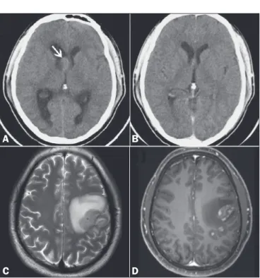
-ache. Computed tomography (CT) of the brain revealed hyper -dense intraventricular nodule to the right of the foramen of Monro,
highly suggestive of a colloid cyst (Figure 1A). The patient was using dexamethasone as pain therapy. In a CT scan of the brain obtained one month later, no nodules were observed (Figure 1B). Cervical and thoracoabdominal CT scans also showed no abnor
-malities. At two months, the patient presented with convulsions. Magnetic resonance imaging (MRI) of the brain showed a cere
-bral mass (Figures 1C and 1D). Histopathological and immuno -histochemical analysis of a biopsy sample revealed Burkitt-like
lymphoma, which is one of the non-Hodgkin lymphomas. Ancil
-lary examinations ruled out systemic disease and viral infection. Lymphomas are designated primary when they originate at and are conined to a given site(1–3). Primary central nervous sys -tem (CNS) lymphomas account for up to 6% of brain neoplasms and 1–6% of extranodal lymphomas; approximately 90% of pri -mary CNS lymphomas are non-Hodgkin lymphomas of the dif-fuse large B-cell subtype(1–6). The incidence of CNS lymphoma is higher in the presence of certain immunodeiciencies, especially human immunodeiciency virus (HIV) infection(2). Among
im-munocompetent individuals, the prevalence of CNS lymphoma is
highest (60–67%) in men 45–75 years of age. In that group, CNS lymphomas present as a single homogeneous mass (in 62%), often in the supratentorial compartment (in 83%) and notably in the deep white matter (in 57%). The corpus callosum and regions surrounding the ventricles are typically affected. Perilesional
Figure 1. A: Non-contrast-enhanced CT scan of the brain, showing well-delineated, discretely hyperdense intraventricular nodule to the right of the foramen of Monro (arrow), promoting slight dilation of the lateral ventricles (obstructive hydrocephalus). B: Follow-up CT of the brain, obtained one month later, showing no such nodule. C,D: MRI of the brain after episodes of seizures, T2-weighted sequence (C) and paramagnetic contrast-enhanced T1-weighted sequence (D), showing an intra-axial frontoparietal mass in the left cerebral hemisphere, with intense perilesional vasogenic edema and heterogeneous enhancement.
A
B
414
Radiol Bras. 2017 Nov/Dez;50(6):405–415 Letters to the Editorhttp://dx.doi.org/10.1590/0100-3984.2016.0065
Rodolfo Mendes Queiroz1, Lucas Giansante Abud1, Thiago Giansante Abud2, Cecília Hissae Miyake1, Antonio Carlos dos Santos3
1. Documenta – Hospital São Francisco, Ribeirão Preto, SP, Brazil. 2. Hospital Israelita Albert Einstein, São Paulo, SP, Brazil. 3. Hospital das Clínicas da Facul-dade de Medicina de Ribeirão Preto da UniversiFacul-dade de São Paulo (HCFMRP--USP), Ribeirão Preto, SP, Brazil. Mailing address: Dr. Rodolfo Mendes Queiroz. Documenta – Centro Avançado de Diagnóstico por Imagem. Rua Bernardino de Campos, 980, Centro. Ribeirão Preto, SP, Brazil, 14015-130. E-mail: rod_queiroz @hotmail.com.
edema is also common, being seen in 77–90% of cases(1,3–6). On CT scans, CNS lymphomas are typically hyperdense, be -cause they are hypercellular and have a high nucleus-cytoplasm ratio(1,3). On MRI, they often demonstrate a hypointense or isointense signal in T1-weighted sequences and an isointense or hyperintense signal in T2-weighted sequences. After intra
-venous administration of contrast medium, they show homoge
-neous (90%) or, in rare cases, annular enhancement. They also exhibit signs of restricted water diffusion. Perfusion-weighted imaging shows less vascularization than that seen in other ma -lignant brain tumors. On magnetic resonance spectroscopy,
CNS lymphomas show elevated lipid and choline peaks, as well
as a reduction in N-acetyl-aspartate levels(1,3–5). The deinitive
diagnosis is made by biopsy(1,2,4,6). Such lymphomas respond to
chemotherapy and radiotherapy, the surgical option being used for tumor mass reduction(1,3–5). Overall survival ranges from 15% to 80%, depending on the age of the patient, as well as on
the characteristics and stage of the disease(2,4).
The list of differential diagnoses of expansile CNS lesions in imaging studies is extensive, including glioma, acute ischemia, inlammatory processes, and infectious diseases(1,3–5,7–11). When
such lesions appear in an intraventricular location and are
hyper-dense on CT, they can be confused with colloid cysts, which are common at that site and exhibit similar density(4).
Burkitt-like lymphomas are highly malignant, with cellular characteristics intermediate between those of diffuse non-Hodg -kin large B-cell lymphoma and those of Burkitt lymphoma(12–14). Burkitt-like lymphomas are typically associated with infection— HIV or the Epstein-Barr virus. They account for 2–3% of
non-Hodgkin lymphomas in immunocompetent adults, being most common among the elderly(12–14). Burkitt-like lymphomas can
affect the brain, intestines, skin, ovaries, kidneys, liver, and bone
marrow(12). Chemotherapy is the most widely used treatment, al -though, even with treatment, survival is less than one year(13,14). The term “vanishing tumor” refers to a tumor that shows marked regression or disappears, with or without nonspeciic therapy, and can recur or progress to new forms(2,4,15,16). In the
brain, lymphomas often occur after corticosteroid therapy,
demy-elinating diseases, or inlammatory disorders(15,16).
REFERENCES
1. Mansour A, Qandeel M, Abdel-Razeq H, et al. MR imaging features of intracranial primary CNS lymphoma in immune competent patients. Cancer Imaging. 2014;14:22.
2. Bellesso M, Bizzetto R, Pereira J, et al. Primary central nervous system lymphoma. Rev Bras Hematol Hemoter. 2008;30:54–60.
3. Haldorsen IS, Espeland A, Larsson EM. Central nervous system lymphoma: characteristic indings on traditional and advanced imaging. AJNR Am J Neuroradiol. 2011;32:984–92.
4. Sasani M, Bayhan M, Sasani H, et al. Primary central nervous system lymphoma presenting as a pure third ventricular lesion: a case report. J Med Case Reports. 2011;5:213.
5. Reis F, Schwingel R, Nascimento FBP. Central nervous system lymphoma: iconographic essay. Radiol Bras. 2013;46:110–6.
6. Alabdulsalam A, Zaidi SZA, Tailor I, et al. Primary Burkitt lymphoma of the fourth ventricle in an immunocompetent young patient. Case Rep Pathol. 2014;2014:630954.
7. Castro FD, Reis F, Guerra JGG. Intraventricular mass lesions at mag -netic resonance imaging: iconographic essay – part 1. Radiol Bras. 2014; 47:176–81.
8. Castro FD, Reis F, Guerra JGG. Intraventricular mass lesions at mag -netic resonance imaging: iconographic essay – part 2. Radiol Bras. 2014; 47:245–50.
9. Destefani MH, Mello AS, Oliveira RS, et al. Chordoid glioma of the third ventricle. Radiol Bras. 2015;48:338–9.
10. Schwingel R, Duarte SBL, Oshima MM, et al. Multiple hemangio -blastomas, association with von Hippel-Lindau syndrome. Radiol Bras. 2015:48(2):xi–xiii.
11. Dultra AHA, Noro F, Melo ASA, et al. Primary intercavernous lymphoma of the central nervous system. Radiol Bras. 2015;48:337–8.
12. Simcock DE, O’Shaughnessy T, Balasanthiran A, et al. Pulmonary Burkitt’s-like lymphoma. Respir Med Extra. 2005;1:81–3.
13. Re M, Di Massimo U, Romeo R, et al. Burkitt-like lymphoma of the sphenoid sinus: case report. Acta Otorhinolaryngol Ital. 2004;24:30–2. 14. Johnson KA, Tung K, Mead G, et al. The imaging of Burkitt’s and
Burkitt-like lymphoma. Clin Radiol. 1998;53:835–41.
15. Okita Y, Narita Y, Miyakita Y, et al. Long-term follow-up of vanishing tumors in the brain: how should a lesion mimicking primary CNS lym -phoma be managed? Clin Neurol Neurosurg. 2012;114:1217–21. 16. Pohl P, Oberhuber G, Dietze O, et al. Steroid-induced complete remis
-sion in a case of primary cerebral non-Hodgkin’s lymphoma. Clin Neurol Neurosurg. 1989;91:247–50.
Giant cell tumor of the frontal sinus: a typical inding in an unlikely
location
Dear Editor,
A 32-year-old female patient was admitted to the emergency
room complaining of a knot on her forehead that had appeared
24 hours earlier. The patient underwent computed tomography (CT) of the skull, with and without intravenous administration of iodinated contrast medium. The CT scans revealed a dense, spontaneous, expansile extra-axial formation with its epicenter in the right frontal sinus, featuring an evident air-luid level and well-deined borders (Figure 1A). On T2-weighted magnetic resonance imaging (MRI) sequences, the lesion also showed an air-luid level (Figure 1B). A contrast-enhanced axial MRI scan showed peripheral enhancement (Figure 1C). The patient un
-derwent surgery for complete resection of the lesion. The patho
-logical examination demonstrated tumor-free margins, and im
-munohistochemistry showed that the lesion was characteristic of a giant cell tumor (GCT) of bone (Figure 1D).
GCT is one of the most common primary bone tumors, ac
-counting for approximately 10% of all bone tumors and 25% of
all benign bone tumors(1). It mainly affects individuals 20–40 years of age and has an insidious onset, presenting with pain and
a local increase in volume(1). It is usually located in the epiphyses
or metaphyses of the long bones, most commonly in the knees
(distal femur or proximal tibia). Although it affects less than 1% of all bone sites within the skull (mainly the temporal and sphe
-noid bones), GCT tends to be more aggressive when it occurs at
such sites(2–4).
Based on the classical radiographic aspects, GCT of bone can be deined as a lytic, expansile lesion, resulting in thin -ning or erosion of the cortical bone(5). CT is the best method to
evaluate bone destruction and to identify pathological fractures.