J Appl Oral Sci. 538
ABSTRACT
INTRODUTION
Pyogenic granuloma (PG) is a benign non-neoplastic mucocutaneous lesion, with the term “pyogenic” being used erroneously since this condition does not produce purulent secretion1 and
is not related to infection7. PG is also known as
pregnancy granuloma or pregnancy tumor when occurring in pregnant women, or as vascular epulis, benign vascular tumor and hemangiomatous granuloma4.
Jafarzadeh, et al.7
caused by minor trauma or irritation. According to Neville, et al.9 (1998), these injuries might be
to poor hygiene, trauma or local infection. Lin and Janniger8 (2004) reported an association between
dermatosis and the occurrence of PG.
PG is manifested as a sessile or pedunculated, resilient, erythematous, exophytic and painful papule or nodule with a smooth or lobulated surface that bleeds easily4,7. The gingiva is the
most commonly affected site by PGs, accounting
Pyogenic granuloma on the upper lip: an unusual
location
Eduardo Sanches GONÇALES1 DAMANTE2, Cassia Maria FISCHER RUBIRA3,
Luís Antônio de Assis TAVEIRA4
1- DDS, PhD, Assistant Professor, Department of Stomatology, Bauru School of Dentistry, University of São Paulo, Bauru, SP, Brazil. 2- DDS, PhD, Professor, Department of Stomatology, Bauru School of Dentistry, University of São Paulo, Bauru, SP, Brazil. 3- DDS, PhD in Stomatology, Bauru School of Dentistry, University of São Paulo, Bauru, SP, Brazil.
4- DDS, PhD, Associate Professor, Department of Stomatology, Bauru School of Dentistry, University of São Paulo, Bauru, SP, Brazil.
Corresponding address: Eduardo Sanches Gonçales - Faculdade de Odontologia de Bauru - USP - Departmento de Estomatologia - Al. Dr. Octavio Pinheiro Brisolla 9-75 - 17.012-901 - Bauru, SP - Brasil - Phone: 55 14 32358250 - e-mail:[email protected]
!"#$!!
P
yogenic granuloma (PG) is a benign non-neoplastic mucocutaneous lesion. It is a reactional response to constant minor trauma and might be related to hormonal changes. In the mouth, PG is manifested as a sessile or pedunculated, resilient, erythematous, exophytic and painful papule or nodule with a smooth or lobulated surface that bleeds easily. PG preferentially affects the gingiva, but may also occur on the lips, tongue, oral mucosa and palate. The most common treatment is surgical excision. This paper describes a mucocutaneous PG on the upper lip, analyzing the clinical characteristics and discussing the features that distinguish this lesion from other similar oral mucosa lesions. The diagnosis of oral lesions is complex and leads the dentist to consider distinct lesions with different diagnostic methods. This case report with a 4 year-follow-up calls the attention to the uncommon mucocutaneous labial location of PG and to the fact that surgical excision is the safest method for diagnosis and treatment of PG of the lip, even when involving the mucosa and skinKey words: Pyogenic granuloma. Pregnancy tumor. Chemically induced.
for 75% of all cases8, although occurrence of these
lesions on the lips, tongue, oral mucosa, palate9 2 has also been reported. PGs affecting
the labial mucosa are rare4.
A higher frequency of PG is observed in the second decade of life1, especially among women,
probably because of the vascular effects of female hormones8. A preference for children has been
reported by some investigators7.
Microscopically, PG is characterized by marked vascular proliferation amidst granulation tissue and ! "
#$ 10.
In view of its clinical characteristics, the differential diagnosis of PG includes peripheral giant cell granuloma, peripheral ossifying " " hemangioma, conventional granulation tissue, "$$"&$' sarcoma, angiosarcoma, non-Hodgkin’s lymphoma7
and cutaneous horn in the lower lip11 *
diagnosis depends on biopsy9.
Surgical excision is the most common www.scielo.br/jaos
J Appl Oral Sci. 539 treatment12; however, this may result in scars5,
which is why the use of more conservative treatments, such as cryosurgery6,14 and laser
surgery, is preferred3,5. The recurrence is about 3%
after simple excision13.
This is a case report of PG on the upper lip. It presents the clinical characteristics and discusses the features that distinguish this lesion from other similar lesions of the oral mucosa, with special emphasis on the diagnosis and treatment of this condition.
CASE REPORT
A 12-year-old mulatto girl presented a 3-month history of a “blister on the upper lip”. The initial aspect of the blister changed after application of thinner because the lesion bled easily. The lesion was asymptomatic, grew slowly and continued to bleed. The patient’s main complaint was her fear of going to school because of the cruel comments by
other students. Medical history revealed no other systemic or local contributory factors associated + $$ $ nodes were detected.
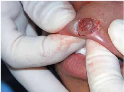
Clinical examination revealed a reddish-yellow, " $ / diameter, involving the mucocutaneous line of the upper lip on the left, which bled on touch and was resilient to palpation (Figure 1). The diagnostic hypotheses were PG, recent oral syphilis (hard chancre) or tuberculous ulcer. Complementary blood tests, complete blood count, erythrocyte sedimentation rate, VDRL (venereal disease research laboratory) test and radiography of the $ $ < infectious diseases, a fact also supported by the patient’s general good health.
The nodule was removed by excisional biopsy (Figure 2). Microscopic analysis showed an oral mucosa consisting of continuous, parakeratinized, $ $
Figure
1-Figure 2- Excisional biopsy to remove the nodule
Pyogenic granuloma on the upper lip: an unusual location
J Appl Oral Sci. 540
*
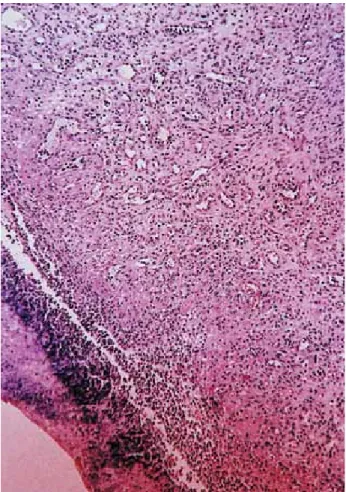
granulation tissue was rich in blood vessels, $ => $ (Figure 3). The diagnosis was PG. After 6 months ? @B E$ $
with the esthetic outcome, and 4 years (Figure @K $ discharged.
DISCUSSION
There is consensus that PG is a reactional lesion formed in response to minor trauma and/or chronic irritation, with reports of its occurrence after low-intensity traumatic injuries1,2,4,8-10.
PGs are preferentially found in the gingiva9,
whereas its occurrence in the upper lip seems to be rare5. Thus, the present case of PG affecting the
upper lip, a region rarely subjected to trauma, is unusual, especially the location of the lesion in the lip vermilion (with involvement of the mucocutaneous line and skin). This unusual location led to other diagnostic hypotheses for ulcerated lesions of the upper lip, such as hard chancre (lesion resulting from inoculation of Treponema pallidum) and labial tuberculous ulcer.
Although Jafarzadeh, et al.7 (2006) have
suggested that the differential diagnosis of PG should include a large number of lesions such as malignant ulcers, sarcoidosis, syphilis, aphthous ulcers, mycotic infection and traumatic injuries. Neville, et al.9 (1998)suggested the inclusion of
tuberculous oral lesions in the differential diagnosis since, although uncommon, these lesions may present nodular, granular or ulcerated areas.
Treponema pallidum is the etiological agent of
syphilis, a chronic, sexually transmitted disease of universal incidence that can affect various organs and systems, including the mouth, which is one of the main extragenital sites of syphilis. Oral primary syphilis manifests as a deep solitary ulcer with a red or brown base and irregular border usually located on the upper lip. The disease is followed by cervical lymphadenopathy, which is observed 1 to 3 weeks after acquisition of the infection. The condition might be confused with other solitary ulcerated lesions, especially traumatic lesions. Diagnosis of primary syphilis is made analyzing
Figure 3- Microscopic aspect showing an oral mucosa membrane. The underlying granulation tissue was rich !"#$%
Figure 4- Labial aspect 6 months (A) and 4 years (B) postoperatively
GONÇALES ES, DAMANTE JH, FISCHER RUBIRA CM, TAVEIRA LAA
J Appl Oral Sci. 541 the patient’s social and sexual lifestyle and his/her $"$ " UWXY " $ antibody and treponemal immobilization9,10,12.
Tuberculosis is also a universal disease whose understanding is of utmost importance for the Brazilian population because of the lack of basic sanitation and adequate sanitary conditions.
Mycobacterium tuberculosis, the causal agent of
tuberculosis, is contracted through sputum or air, reaching the lung and causing primary infection. * $ $< "<E Tuberculosis can also affect the mouth, especially ""$"" soft palate, uvula, gingiva and alveolar mucosa as $ " central bone lesions (tuberculous osteomyelitis). * $<" Mantoux reaction, chest x-rays and biopsy of the lesion9,10,12.
In the present case, lesion type and location led to the suspicion of syphilis. This was ruled out by investigation of the patient’s sexual life and a non-reactive VDRL test. Similarly, tuberculosis was raised as a diagnostic hypothesis, especially because of the characteristic of the lesion. This was ruled out based on chest radiography, which $ patient was in good general health.
Once syphilis and tuberculosis were ruled out in this case, only PG remained as a diagnostic hypothesis. Initially, chemical irritation resulting from the use of a paint solvent for cauterization of the initial blister (possibly a blister resulting from the fusion of herpes vesicles frequently found at this site) was believed to be the etiological agent. This hypothesis was not supported since the patient reported use of the solvent only to control bleeding, which is uncommon for a herpes lesion. When asked, the patient denied the habit of biting on her upper lip, which made the determination of a traumatic etiological agent inconclusive.
In a 12-year old female patient, an age characterized by important hormonal changes and hormonal factors that can be related to the PG’s etiology7, this lesion may be related to hormonal
etiology.
Despite the possible occurrence of scars5,
biopsy is the method for adequate diagnosis of oral lesions9 and surgical excision is the most
common treatment of PGs12. In this case, an
excisional biopsy was performed for diagnosis and treatment. The procedure was simple, essential for "$$ patient expectations. In the present case, there was a discrete scar and the patient was psychologically
CONCLUSIONS
This case reports emphasizes that the diagnosis of oral lesions is complex and leads the dentist to consider distinct lesions with different diagnostic methods. We call attention to the uncommon mucocutaneous labial location of PG and to the fact that surgical excision is the safest method for diagnosis and treatment of PG of the lip, even when involving the mucosa and skin.
REFERENCES
1- Al-Khateeb T, Ababneh K. Oral pyogenic granuloma in Jordanians: a retrospective analysis of 108 cases. J Oral Maxillofac Surg. 2003;61:1285-8.
2- Damm DD, Fantasia JE. Elevated and ulcerated nodule of lip. Pyogenic granuloma. Gen Dent. 2002;50:466-8.
3- Galeckas KJ, Uebelhoer NS. Successful treatment of pyogenic granuloma using a 1,064-nm laser followed by glycerin sclerotherapy. Dermatol Surg. 2009;35:530-4.
4- Graham RM. Pyogenic granuloma: an unusual presentation. Dent Update. 1996;23:240-1.
5- Ichimiya M, Yoshikawa Y, Hamamoto Y, Muto M. Successful treatment of pyogenic granuloma with injection of absolute ethanol. J Dermatol. 2004;31:342-4.
6- Ishida CE, Ramos-e-Silva M. Cryosurgery in oral lesions. Int J Dermatol. 1998;37:283-5.
7- Jafarzadeh H, Sanatkhani M, Mohtasham N. Oral pyogenic granuloma: a review. J Oral Sci. 2006;48:167-75.
8- Lin RL, Janniger CK. Pyogenic granuloma. Cutis. 2004;74:229-33.
9- Neville BW, Damm DD, Allen CM. Patologia oral e maxilofacial. Rio de Janeiro: Guanabara Koogan; 1998.
10- Regezi JA, Sciuba JJ. Oral pathology, clinical pathological correlations. Philadelphia: Saunders; 1989.
11- Saravana GH. Oral pyogenic granuloma: a review of 137 cases. Brit J Oral Maxillofac Surg. 2009;47:318-9.
12- Shafer WG, Hine MK, Levy BM. A textbook of oral pathology. Philadelphia: Saunders; 1983.
13- Souza LN, Martins CR, Paula AM. Cutaneous horn occurring on the lip of a child. Int J Paediatr Dent. 2003;13:365-7. 14- Wang SQ, Goldberg LH. Treatment of recurrent pyogenic granuloma with excision and frozen section for margin control. Dermatol Surg. 2008;34:1115-6.
Pyogenic granuloma on the upper lip: an unusual location