Universidade de Lisboa
Faculdade de Motricidade Humana
Morphological and functional adaptations of the
abdominal wall during pregnancy and in the
postpartum period
Tese elaborada com vista à obtenção do grau de Doutor em Motricidade Humana, na Especialidade de Biomecânica
Tese por compilação de artigos, realizada ao abrigo da alínea a) do nº2 do art.º 31 do Decreto-lei nº230/2009
Orientador:
Professor Doutor Augusto Gil Pascoal
Coorientador:
Professora Doutora Kari Bø
Júri: Presidente:
Professor Doutor Francisco José Bessone Ferreira Alves Vogais:
Doutora Kari Bø – Professora Catedrática, Departamento de Medicina do Desporto da Norwegian School of Sport Sciences, Oslo
Doutor António Prieto Veloso – Professor Catedrático, Faculdade de Motricidade Humana da Universidade de Lisboa
Doutora Maria Filomena Soares Vieira – Professora Auxiliar, Faculdade de Motricidade Humana da Universidade de Lisboa
Doutor Augusto Gil de Andrade Pascoal – Professor Auxiliar, Faculdade de Motricidade Humana da Universidade de Lisboa
Doutora Rita Alexandra Prior Falhas Santos Rocha – Professora Coordenadora, Escola Superior de Desporto de Rio Maior do Instituto Politécnico de Santarém Doutora Anneke Steensma – Investigadora Principal, Departamento de Obstetricia e Ginecologia, do Erasmus Medical Center, Roterdão (Países Baixos)
II
Reprodução da Tese
Declaração
Nome: Patrícia Gonçalves Fernandes da Mota Endereço electrónico: [email protected] Telefone: (+351) 934 479 492
Número do Cartão de Cidadão: 12208458
Título:Morphological and functional adaptations of the abdominal wall during pregnancy and postpartum period
Orientador(es): Professor Doutor Augusto Gil Pascoal (Orientador) e Professora Doutora Kari Bø (Co-Orientadora)
Ano de conclusão: 2014
Designação do ramo de conhecimento do Doutoramento: Motricidade Humana, na Especialidade de Biomecânica
Nos exemplares das teses de doutoramento entregues para a prestação de provas na Universidade e dos quais é obrigatoriamente enviado um exemplar para depósito legal na Biblioteca Nacional e pelo menos outro para a Biblioteca da FMH/UL deve constar uma das seguintes declarações:
1. É autorizada a reprodução integral desta tese/trabalho apenas para efeitos de investigação, mediante declaração escrita do interessado, que a tal se compromete.
2. É autorizada a reprodução parcial desta tese/trabalho (indicar, caso tal seja necessário, no máximo de páginas, ilustrações, gráficos, etc.) apenas para efeitos de investigação, mediante declaração escrita do interessado, que a tal se compromete.
3. De acordo com a legislação em vigor, (indicar, caso tal seja necessário, no máximo de páginas, ilustrações, gráficos, etc.) não é permitida a reprodução de qualquer parte desta tese/trabalho.
Faculdade de Motricidade Humana – Universidade de Lisboa, ____/ _____ / _____
Para as minhas filhas, Maria Miguel e Mercedes, Por fazerem querer superar-me todos os dias, Com todo o meu amor.
Acknowledgements
It would not have been possible to write my doctoral thesis without the help and support of the kind people around me, only some of whom it is possible to give particular mention here. Especially I would like to thank:
My supervisor, Professor Augusto Gil Pascoal, thank you for your confidence and trust in me. From the beginning of my PhD application your enthusiasm and knowledge have pushed me forward. I had a great freedom to plan and execute my ideas in research without any pressure. This allowed me to identify my own strengths and weaknesses, and particularly boosted my self-confidence. It was great working with you Gil, and my heartfelt thanks to you.
My co-supervisor, Professor Kari Bø. I feel privileged to have worked with one of the best known international researchers in sports medicine and women’s health. You helped me to design the project and guided me through the challenge of publishing, and I have learned so much… I believe from my heart that you are a dream supervisor for a student who wants to do research and I am lucky to be one of those who had an opportunity to work with you. Thank you for everything Kari.
Professor Ana Isabel Carita. Thanks for your statistical counseling and patience in explaining to me the different statistical approaches needed to survive in research and for all the discussions about technical questions and life in general.
My colleagues and friends at the Biomechanics and Functional Morphology Laboratory, particularly Ana Cristina Vidal, Liliana Aguiar, Silvia Cabral, Vera Moniz-Pereira and Wangdo Kim. We have shared a lot and I appreciate the friendship we have developed. It is good to have someone to share our joys and frustrations. A special thanks to Professor Filomena Vieira, for helping me with the data collection and for always being supportive and positive about my research. Filipa João for making me realize how interesting and powerful a biomechanics analysis can be, and for constantly being supportive, pushing me forward in the writing progress with your positive attitude. Professor António Veloso, for receiving me in your laboratory, and always being available to discuss biomechanical issues of my thesis.
VI
valuable. A special thanks to my dear friend Mari Gundersen and her life partner Eirik Herland, for receiving me at your place and for making me feel at home even when I had to wear a winter jacket in August.
I would like to express my sincere thank to all participating women (and their babies) for giving your time to one or more of the five studies. It was amazing to feel how you got involved with my studies and made the effort to attend one or sometimes 4 testing sessions in a period of life where time and priorities are in constant adjustments.
Tatiana Dominguez, Miguel Basto, and all the team from Centro Pré e Pós Parto (Lisboa, Portugal), for being so enthusiastic about my studies, for motivating so many pregnant women to participate on this project, and for helping me out with a room to make examinations and other logistic tasks. You saved me a lot of time and effort.
Fátima Sancho for being my partner in the reliability studies, always giving your contribution to my work, and the team from R’Equilibrius Clinic (Oeiras, Portugal) for access facilitation to the pregnant and postpartum women.
Professor Jorge Infante for the construction of the transducer cluster of markers used in one of the studies.
Gill Brook (Women’s Health Physiotherapy Team Leader, Bradford Teaching Hospitals NHS Foundation Trust, Bradford, United Kingdom) for English revision.
Dr. José Luís García (Centro Hospitalario Policlinico San Carlos, Denia, Spain) for counseling on ultrasound imaging issues and suggestions.
To all my friends and family wondering what I have been doing these years and to those who have known exactly what I have been doing by voluntarily participating in the projects, thank you for being there for me.
A particular thank to my grandmother ‘vó Rosa for always believing in me, and for being so happy with the start of this project and mostly for having me near. I miss you more than I can express, but I´m sure you would have been very pleased and proud today.
My parents for raising me up to believe that “nothing is imposible” to “always try my best”, and for helping me out in so many ways. Without you this PhD degree would have taken so much longer. You took care of Maria and Mercedes when they were too young to go to the kindergarden, picked them up when the days had too few hours, cooked dinner for the whole family, and did everything to help me and my husband to survive the hard working periods. I cannot express enough my gratitude to you.
Maria Miguel (4 years old) and Mercedes (1 year old), you are the most amazing and delightful children one can have. You help me to prioritize and to make time out of situations I would never imagine to be possible. Now there is a whole new dimension of happiness and exhaustion and I could never have dreamt you could bring so much joy to our lives everyday.
Finally my warmest thanks go to my husband Rodrigo, for constant support, endless patience and belief in me. You know how to push me to work hard and, from time to time, not to work at all. My work is so much more good-looking thanks to you. I look forward to doing more things we like to do. To travel, to try new things, to appreciate our routines, to make plans, and to enjoy life with the children.
Adaptações morfológicas e funcionais da parede
abdominal durante a gravidez e pós-parto
Resumo
A diástase dos retos abdominais (DRA) carateriza-se pela separação dos músculos
rectos abdominais, sendo que o incremento da distância inter-rectos (DIR) se inicia
durante a gravidez e prolonga-se pelo puerpério. A fiabilidade dos instrumentos de registo
desta condição é reduzida sendo escasso o conhecimento sobre a prevalência e factores
de risco que lhe estão associados. Adicionalmente existe pouca evidência sobre o efeito
na prevenção e/ou agravamento da DIR induzida pelos trabalho abdominal.
Assim, foram objetivos desta tese 1) o desenvolvimento de uma metodologia fiável de
avaliação da morfologia da parede abdominal feminina; 2) descrição da prevalência da
DIR, factores de risco e relação com dor lombo-pélvica aos 6 meses no pós-parto; 3) e
avaliar a resposta imediata da DIR ao exercício, nomeadamente no crunch abdominal e
no drawing-in.
Cento e oitenta e sete mulheres participaram nos cinco estudos apresentados na tese. Os
resultados dos três estudos metodológicos demonstraram a fiabilidade da medição da
DIR com base na ultrassonografia, nomeadamente face à palpação. Nos dois estudos
longitudinais foi avaliada a prevalência e potenciais fatores de risco da DRA a par da
associação do incremento de DIR com a incidência de dor lombo-pélvica assim como o
efeito imediato dos exercícios crunch e drawing-in.
Os resultados revelaram que aos 6 meses de pós-parto a DRA tem uma prevalência de
39% não apresentando relação significativa com a dor lombo pélvica. A resposta imediata
produzida pelo exercício drawing-in foi um aumento da DIR, enquanto o crunch induziu a
redução imediata da DIR, tanto na gravidez como no pós-parto.
Morphological and functional adaptations of the
abdominal wall during pregnancy and in the
postpartum period
Abstract
Diastasis recti abdominis (DRA) or increased inter rectus distance (IRD) is characterized by the separation of the rectus abdominis muscles. It has its onset during pregnancy and the first weeks following childbirth. The reliability of the instruments used to assess this condition is unclear. There is scant knowledge on the prevalence and risk factors for development of the condition. There is little evidence on which exercises are most effective in reduction of DRA.
The aims of the present thesis were to establish a reliable method for the assessment of the morphology of the abdominal wall, describe the natural recovery of IRD from late pregnancy till 6 months postpartum and evaluate IRD during drawing in and abdominal crunch exercises.
One hundred and eighty-seven women participated in the different studies comprising this thesis. The results of the three methodological studies showed ultrasound imaging to be a reliable method for measuring IRD. Palpation has sufficient reliability to be used in clinical practice. However, ultrasound is a more accurate and valid method. The ultrasound transducer can be held relatively stationary in a clinical setting, to evaluate IRD.
DRA is prevalent at 6 months postpartum, with a prevalence rate of 39% but is not linked with lumbo-pelvic pain.
The drawing in exercise widened the IRD in postpartum women while the abdominal crunch narrowed the IRD compared to rest both during pregnancy and in the postpartum period.
Index
Resumo IX
Abstract XI
Index XIII
List of Figures XVII
List of Tables XVIII
List of Abbreviations XX
Chapter I General Introduction 21
Preview 23
1. Background 24
1.1 Anatomy of the abdominal muscles and their aponeuroses 24 1.2Changes in the abdominal wall morphology during pregnancy 27 1.3Inter-rectus distance and diastasis recti abdominis 28 1.4 Classification and Prevalence of Diastasis Recti 28 1.5 Risk factors for diastasis recti abdominis 29 1.6 Procedures and instruments to assess the inter-rectus distance 29 1.7 The effect of exercise on diastasis rectis abdominis 30
2. Thesis goals and overview 32
3. Methodological considerations 34
3.1Ultrasound measurements 34
3.2 Procedure for transducer motion analysis 35
4. Participants 37
5. Ethics 38
Chapter II Test-retest and Intrarater Reliability of 2D Ultrasound Measurements of
Distance Between Rectus Abdominis in Women 39
Abstract 41
1. Introduction 42
2. Methods 43
2.1 Design 43
2.2 Participants 43
2.3 Instrumentation and procedures 43 2.4 Inter-rectus distance (IRD) measurement 45
2.5 Statistical Analyses 45
3. Results 47
Augusto Gil Pascoal 13/9/2014 15:04
XIV
3.2 Test-retest across days (inter-image) 47
4. Discussion 49
5. Conclusion 52
Acknowledgements 60
References 61
Chapter III Reliability of the Inter-Rectus Distance Measured by Palpation.
Comparison of Palpation and Ultrasound Measurements 65
Abstract 67
1. Introduction 68
2. Methods 69
2.1 Design 69
2.2 Participants 69
2.3 Assessors 69
2.4 Procedure 70
2.5 Ultrasound imaging 70
2.6 Statistical Analyses 71
3. Results 72
4. Discussion 73
5. Conclusion 74
References 79
Chapter IV Ultrasound Imaging Transducer Orientation and Translation during Static
Positions of Drawing-in and Abdominal Crunch Exercise 81
Abstract 83
1. Introduction 84
2. Methods 86
2.1 Participants 86
2.2 Procedures for ultrasound imaging 86 2.3 Procedures for transducer motion analysis 87
2.4 Statistical analysis 88
3. Results 89
4. Discussion 90
5. Conclusion 92
Acknowledgements 97
References 98
Chapter V Prevalence and risk factors of Diastasis Recti Abdominis from late pregnancy to 6 months postpartum, and relationship with lumbo-pelvic pain 101
1. Introduction 104
2. Methods 105
2.1 Participants 105
2.2 Instrumentation and procedures 105
2.3 Ultrasound data collection 106
2.4 Inter-rectus distance measurement and cut-off point for diastasis recti abdominis 106 2.5 Anthropometric measurements 107
2.6 Joint hypermobility 107
2.7 Lumbo-pelvic pain 107
2.8 Statistical Analyses 108
3. Results 109
4. Discussion 110
5. Conclusion 113
References 117
Chapter VI Inter-recti Distance at Rest, During Abdominal Crunch and Drawing in
Exercises during Pregnancy and Postpartum 121
Abstract 123
1. Introduction 124
2. Materials and Methods 126
2.1 Participants 126
2.2 Instrumentation and Examiner 127
2.3 Procedures 127
2.4 Inter-recti distance (IRD) measurement 129
2.5 Statistical Analyses 129
3. Results 130
4. Discussion 131
5. Conclusion 134
5.1 Key Points 134
Acknowledgements 137
References 138
Chapter VII General Discussion 143
1. Summary of main findings 145
1.1 Procedures and instruments to accurately measure IRD 147 1.2 Changes in IRD from late pregnancy till 6 month postpartum 148 1.3 Influence of two abdominal exercises on the IRD 149
XVI
Further research 152
References 153
Apendix I Published Papers 157
Apendix II Thesis related outcomes 157
Awards 161
Papers in international scientific journals 161 Papers in conference proceedings 162
Oral communications 163
Posters in conferences 163
List of Figures
Fig. I-1. Abdominal muscles and their aponeuroses. A. The rectus abdominis. B, C and D shows a common arrangement of the sheath as seen in horizontal section. 25 Fig. I-2. A. Rectus anterior ultrasound image. B. Points digitalized by the examiner on the
muscles contour (red dots). Interpolated points are using an algorithm according to a parabola-shape like curve (white points); parabola inflection point (white asterisk) suggesting the end-points for IRD measurement on the medial margin of
both RA. 35
Fig. I-3. Ultrasound transducer with a customized cluster composed by 4 non-collinear reflective markers fixed to the long axis of the transducer. 35 Fig. I-4. Two local reference coordinate systems were: 1) the pelvis reference coordinate
system, which origin was in the midpoint between the right and left anterior iliac spines; 2) transducer reference system which origin was located at the midpoint of the two virtual markers on the base of the transducer. 36
Fig. I-5. Flowchart of the participants in this study. 37
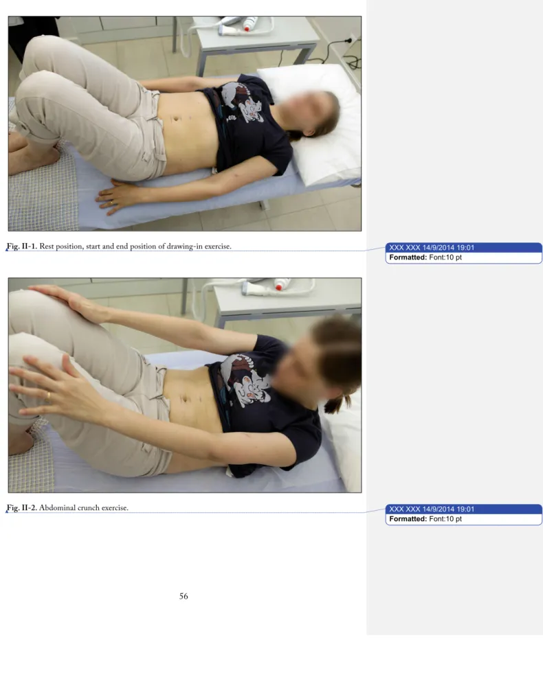
Fig. II-1. Rest position, start and end position of drawing-in exercise. 56
Fig. II-2. Abdominal crunch exercise. 56
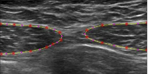
Fig. II-3. RA ultrasound image. Points digitalized by the examiner on the muscles contour
(red dots). 57
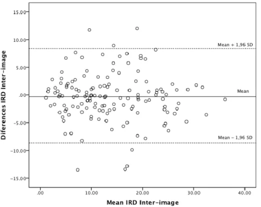
Fig. II-4. RA ultrasound image. Interpolated points using an algorithm according to a parabola-shape like curve (white points); parabola inflection point (white asterisk) suggesting the end-points for IRD measurement on the medial margin of both RA. 57 Fig. II-5. Plot of difference against mean (in mm) for measurements of the same stored
images, with mean difference and 95% limits of agreement indicated. 58 Fig. II-6. Plot of difference against mean (in mm) for the recaptured images, with mean
difference and 95% limits of agreement indicated. 59 Fig. III-1. Rest position and start position for the abdominal crunch exercise. The subject
was supine, in the crook lying position, arms resting along the body. 75 Fig. III-2. Abdominal crunch. End position for the abdominal crunch exercise. The subject was
supine, in the standard crook lying position, hands touching the knees. 75
Fig. III-3. Position of the transducer for the ultrasound imaging. 76
Fig. IV-1. Rest position and the static position of the drawing-in exercise. 94
Fig. IV-2. Static position of the abdominal crunch exercise. 94
Fig. IV-3. Ultrasound transducer with a customized cluster composed by 4 non-collinear reflective markers fixed to the long axis of the transducer. 95 Fig. IV-4. Two local reference coordinate systems were: 1) the pelvis reference coordinate
XVIII
Fig. IV-5. Raw data from transducer orientation with respect to pelvis local coordinate system (origin) on XY-plane (A). YZ-plane (B) and XZ-plane (C) during the 60 observations (20 participants x 3 trials) on each condition (abdominal crunch and
drawing in). 96
Fig. V-1. Flowchart of the participants in this study. 116
Fig. VI-1. Mean IRD and Standard Error values in mm, at the four measurement time points (M1=gestational week 35 – 41; M2= 6-8 weeks postpartum; M3=12-14 weeks postpartum; M4= 24-26 weeks postpartum), on different abdominal conditions, on each location on the linea alba. 136
List of Tables
Table II-‐1. Background Variables. 53
Table II-‐2.
Verbal instructions. 53
Table II-‐3.
Inter Rectus Distance measures during Rest, Abdominal Crunch and Drawing-‐In
exercises. * N=24 54
Table II-‐4.
Inter Rectus Distance measures during Rest, Abdominal Crunch and Drawing-‐In exercises * for the women in post-‐partum and women with different parity, and
Independent t-‐test values. 54
Table II-‐5.
Intra-‐rater reliability across repeated measurement of the same image. 55
Table II-‐6.
Intra-‐rater reliability across 2 days. 55
Table III-‐1.
Experienced physiotherapist palpation intra-‐rater reliability across 2 days of
measurements. N=40 76
Table III-‐2.
Less experienced physiotherapist palpation intra-‐rater reliability across 2 days of
measurements. N=40 77
Table III-‐3.
Palpation inter-‐rater reliability between the experienced and the less experienced
physiotherapist. N=40 77
Table III-‐4.
IRD measured by ultrasound for each palpation category. 78
Table IV-‐1.
Descriptive statistics of ultrasound transducer 3D position and translation on
each direction with respect to the pelvis during Abdominal Crunch and Drawing-‐
In (N=20). 93
Table V-‐1.
Background variables and potential risk factors for diastasis recti abdominis
(DRA) at 6 months postpartum. (P<0.05) 114
Table V-‐2.
Results of binary logistic analysis to predict possible risk factors associated with
the presence of DRA at 6 months postpartum. (P<0.05) 115
Table VI-‐1.
Background variables of the study population. Means with range, or number with
percentages. (N = 84) 135
Table VI-‐2.
Mother’s weight, body mass index (BMI), mass (kg)/height(m)2 and participation in general physical activity (regular exercise classes 2 or more times a week) at each timepoint. Means with standard deviation (SD), or number with
percentages. (N=84) 135
Table VI-‐3.
Mean Inter-‐recti distance in mm with standard deviation (SD) at rest, during
XX
List of Abbreviations
• AC = Abdominal Crunch
• AU = Above the Umbilicus
• BU = Below the Umbilicus
• CI = Confidence Intervals
• DI = Drawing-In
• DRA = Diastasis Recti Abdominis
• DP = Different Parity women
• ICC1,1 = Intra Class Correlation one way random effect model (95% confidence interval) • IRD = Inter-Rectus Distance
• MDC95 = Minimum Detectable Change at the 95% confidence level • Mean diff = Mean difference between groups and confidence intervals.
• OR = Odds Ratio
• PP = Post-Partum women
• RT = Rest
Chapter
I
General Introduction
Augusto Gil Pascoal 13/9/2014 15:04
Preview
Pregnancy and becoming a mother is one of the most exciting times in a women’s life. Besides all the hormonal and physiological changes affecting women during this period, probably the most obvious morphological alteration during pregnancy is the increasing weight and dimensions of the uterus, influencing maternal trunk musculoskeletal morphology, particularly the abdominal musculature. Many women continue or even begin to exercise during pregnancy, and postnatal women are encouraged to resume abdominal exercises shortly after delivery, to restore their abdominal figure and fitness. There is a lot of information available in numerous web pages about exercise programs for women during pregnancy and in the postpartum period, and physiotherapists and exercise instructors prescribe exercises to this population everyday. However, there is little evidence available about muscular changes and the effect and safety of different abdominal exercises during and after pregnancy.
Diastasis recti (DRA) or the increased inter–rectus distance (IRD) seems to be a common condition in women during pregnancy and postpartum. The lack of evidence for the consequences of this condition and the effect of abdominal strengthening exercises in the reduction of DRA indicates a need for identification of prevalence and risk factors of DRA. Use of responsive, reliable and valid outcome measures is mandatory for evaluation of the condition, and ultrasound imaging has recently been suggested as a useful method to assess muscular geometry and to quantify DRA. However, its reliability and validity must first be tested. The overall aim of this thesis was to establish the reliability of ultrasound measurements to assess IRD, to identify the prevalence and risk factors of DRA, and to evaluate the effect of two different abdominal contractions on IRD during pregnancy and postpartum period.
24
1. Background
1.1 Anatomy of the abdominal muscles and their
aponeuroses
The anterolateral wall of the abdomen has a laminar configuration composed by six layers including, from surface to depth, the skin, the superficial fascia, fat, the abdominal muscles, the transversalis fascia and the parietal peritoneum. The muscular layer comprise four paired muscles with fibers oriented vertically (rectus abdominis muscle), obliquely (external and internal oblique muscles) and horizontally (transversus abdominis muscles) with skeletal attachments on the thoracic cage, pelvis and the spinal column via the thoracolumbar fascia (Standring, 2008). The aponeuroses of these muscles represent sheet-like tendons that form the sheath of the rectus abdominis (rectus sheath) and also serve as the medial insertion of the oblique’s and transversus muscles, along the anterior midline of the abdomen, forming a fibrous structure that connect the right and the left side of the abdominal wall, the linea alba (Fig. I-1).
The rectus abdominis (RA) and the pyramidalis are the only vertical muscles in the abdominal wall. The rectus abdominis muscle originates from the 5th through 7th costal cartilages to insert on the symphysis pubis and crest. Superiorly, the rectus is wide, broad, and thin, becoming narrow and thick inferiorly (Kahle, Leonahardt, & Platzer, 1991). Segmentation of each rectus muscle occurs by tendinous intersections that represent attachment of the rectus muscle with the anterior layer of the rectus sheath.
consists of contributions from aponeuroses of the transversus abdominis (transversalis fascia). Inferior to the umbilicus, approximately halfway between the umbilicus and the symphysis pubis, the external abdominal aponeurosis has no contribution to the formation of the posterior rectus sheath and all three aponeuroses pass anterior to the rectus muscle. This anterior displacement of the aponeuroses creates a curved line of demarcation, in the posterior lamella of the rectus sheath, called the arcuate line, below which only the transversalis fascia separates the rectus abdominis muscle from the parietal peritoneum (Fig. I-1).
Fig. I-1. Abdominal muscles and their aponeuroses. A The rectus abdominis. B, C and D shows a common arrangement of the sheath as seen in horizontal section.
In O’Rahilly, Muller, Carpenter & Sweson (1983). “Basic Human Anatomy”. Reprinted with permission from http://www.dartmouth.edu/~humananatomy/figures/chapter_25/25-6.HTM. A technical support from Dartmouth Medical School, Hanover, United States of America.
The linea alba reaches from the xiphoid process to the pubic symphysis and is defined as the fusion of the aponeuroses of the deepest abdominal muscles (Beer et al., 2009). The linea alba consists of a three-dimensional, highly structured meshwork of collagen fibers (Axer, Keyserlingk, & Prescher, 2001) which in conjunction with both rectus sheaths are regarded as the most important structures for the stability of the abdominal wall from a
XXX XXX 14/9/2014 19:01 Formatted: Font:10 pt
26
mechanical point of view (Axer et al., 2001; Grässel, Prescher, Fitzek, Keyserlingk, & Axer, 2005; Hernández-Gascón et al., 2012). The linea alba tension is important to maintain the abdominal muscles, particularly the rectus muscles, at a certain proximity to each other (Beer et al., 2009; Rath et al., 1996) in order to optimize abdominal muscles function both as on abdominal viscera support or producing thorax/pelvis movements.
Tension on the linea alba, particularly below the umbilicus, seems to be regulated by the pyramidalis, a small paired triangular-shaped muscle, present in 80% of people , which lies between the anterior surface of the rectus abdominis and the posterior surface of the rectus sheath (Lovering & Anderson, 2008). The precise function of the pyramidalis muscles is unclear, but together both muscles are thought to assists in tensing the linea alba (Lovering & Anderson, 2008).
The linea alba compliance is highest in the longitudinal direction and smallest in the transverse direction (Beer et al., 2009; Förstemann et al., 2011) which determine the great resistance offer by the LA to rectus abdominis transversal separation. Even so, the viscoelastic properties inherent to the collagen, makes the linea alba prone to increase length when the mechanical stress is prolonged in time (Hernández-Gascón et al., 2012), namely in the case of a long-lasting increased intra-abdominal pressure, such as that resulting from pregnancy (Akram & Matzen, 2014; Benjamin, van de Water, & Peiris, 2014; Boissonnault & Blaschak, 1988; Coldron, Stokes, Newham, & Cook, 2008).
The mechanical stress on linea alba is highly associated to the action of the oblique’s and transversus abdominis muscles. The external oblique arises from the lower 8 ribs posteriorly to interdigitate with both the serratus and latissimus muscles. The direction of the fibers is approximately horizontal in the uppermost portion only to become oblique in the lowest portions. After contributing to the anterior portion of the rectus abdominis sheath, the remaining fibers insert onto the linea alba.
The internal oblique arises from the anterior two-thirds of the iliac crest and lateral half of the inguinal ligament to run essentially at right angles to those of the external oblique. The fibers run perpendicular to the external oblique muscle from the thoracolumbar fascia of
the lower back, the anterior iliac crest and the lateral half of the inguinal ligament, to insert
The transversus abdominis muscleis the innermost of the abdominal muscles, being placed immediately beneath theinternal oblique muscle from the 7th to 12th costal cartilages, iliac crest, and the lateral third of the inguinal ligament. The muscle bundles run mostly horizontally, except the lower most medial fibers, which run a more inferomedial course to their insertion on the pubic crest and pubis (Kahle et al., 1991). Their extensive aponeurosis passes horizontally in the middle line of the abdomen, and is inserted into the linea alba: the upper portion lie behind the RA muscle and blend with the posterior rectus sheath while its lower part pass in front of the RA muscle (Turatti et al., 2013).
1.2 Changes in the abdominal wall morphology during
pregnancy
The functional role of the abdominal muscles during pregnancy appears to be similar to those in the non-pregnant state (Boissonnault & Blaschak, 1988) and is suggested to be important for trunk movement, pelvic stabilization, and restraint of the abdominal contents (Standring, 2008). However, the musculoskeletal morphology of the anterolateral wall of the abdomen changes as pregnancy progresses (Foti, Davids, & Bagley, 2000; Gilleard & Brown, 1996). The weight and dimensions of the uterus and its contents increases from 40 to 1000 grams, and its capacity from 4 ml in non-pregnant state to 4000 ml at term (Cunningham, Leveno, Bloom, & Spong, 2009). The maternal inferior thoracic diameter is increased (Davies, Wolfe, Mottola, & MacKinnon, 2003; Gilleard, Crosbie, & Smith, 2002) as well as the anterior and lateral dimensions of the abdomen. These changes modify the spatial relationship between the superior and the inferior abdominal muscle attachments (Cunningham et al., 2009) increasing the length of the abdominal muscles, particularly the rectus abdominis (Fast, Weiss, Ducommun, Medina, & Butler, 1990). At 38 weeks of gestation the length of the abdominal muscles increased a mean of 115% with respect to the beginning of pregnancy (Gilleard & Brown, 1996). The increment of the anterior abdominal dimensions may alter the angle of the abdominal muscle attachment in the sagittal plane (Gilleard & Brown, 1996). Alterations in the spatial relationship of muscle attachment and the muscle’s angle of insertion may alter the muscles line of action and therefore their ability to produce torque (Gilleard et al., 2002; Gilleard & Brown, 1996).
XXX XXX 14/9/2014 18:58
Deleted:
28
1.3 Inter-rectus distance and diastasis recti abdominis
One of the muscles thought to undergo change in pregnancy is the rectus abdominis. As the fetus grows, the two muscle bellies of the rectus abdominis, connected by the linea alba, elongates and curve round as the abdominal wall expands, with most separation occurring at the umbilicus (Boissonnault & Blaschak, 1988; Fast et al., 1990; Gilleard & Brown, 1996). The augmented inter-rectus distance (IRD), often referred as diastasis rectus abdominis (DRA), is described as a change in the abdominal musculature, specifically in the linea alba and rectus abdominis sheath, with onset in the last trimester of pregnancy and whose peak of incidence occurs immediately after birth and the first weeks following childbirth (Boissonnault & Blaschak, 1988; Coldron et al., 2008; Dumas, Reid, Wolfe, & McGrath, 1995; Rett, Braga, Bernardes, & Andrade, 2009). Although, some studies suggested that an augmented IRD could reduce the abdominal integrity and functional strength, contributing to pelvic instability and back pain (Gilleard et al., 2002; Gilleard & Brown, 1996; Parker, Millar, & Dugan, 2009), no scientific evidence exists about the functional implications of an augmented inter-rectus distance or even about the effect of the exercise on prevention and/or reduction of IRD.
1.4 Classification and Prevalence of Diastasis Recti
Studies have found that DRA may affect between 30% and 70% of pregnant women (Boissonnault & Blaschak, 1988), and that it may remain separated in the immediate postpartum period in 35% to 60% of women (Bursch, 1987). However the condition has also been found in 39% of older, parous women undergoing abdominal hysterectomy (Ranney, 1990) and in 52% of urogynecological menopausal patients (Spitznagle et al., 2007). Reported prevalence of DRA or increased IRD varies and may be inaccurate due to different IRD cut-off values for the diagnosis (Beer et al., 2009; Boissonnault & Blaschak, 1988; Bursch, 1987; Chiarello et al., 2005; Gilleard & Brown, 1996; Rath et al., 1996; Spitznagle et al., 2007) and use of different measurement assessment methods.
1.5 Risk factors for diastasis recti abdominis
There is scant knowledge about the risk factors for DRA. Two studies analyzed several variables such as, age, ethnicity, body mass index, height, weight gain during pregnancy, pre-pregnancy weight, gestational age at delivery, type and duration of birth (Candido et al., 2005; Rett et al., 2009). An association of DRA during pregnancy with Caucasian ethnicity and lack of regular exercise during pregnancy was suggested (Candido et al., 2005). It is considered that women with DRA have a greater number of pregnancies and deliveries (Rett et al., 2009; Spitznagle et al., 2007), and among multiparous women, it is suggested that there is a strong association between provision of childcare and DRA during pregnancy (Candido et al., 2005). However these studies were limited by the sample size, reliability of the instruments used, and were not definitive in its ability to delineate risk factors.
1.6 Procedures and instruments to assess the inter-rectus
distance
30
Morkved, 2007) in the methods and instruments used to measure the IRD. Recently, ultrasound imaging has been suggested as a useful method to assess muscular geometry and as an indirect measure of muscle activation via changes in muscle thickness during contraction (Hodges, Pengel, Herbert, & Gandevia, 2003; Rankin, Stokes, & Newham, 2006). Coldron et al. (2008) used ultrasound to characterize RA changes during the first year postpartum and Mendes (2007) et al claimed ultrasonography to be an accurate method to measure diastasis recti when compared with surgical compass during abdominoplasty. However, at the time this study was planned, search of the literature did not reveal studies addressing the intra or inter-tester reliability of the ultrasound measurement of the IRD at rest or during abdominal muscle contraction. Across-days reliability may be of interest to physiotherapists who perform repeated assessments of abdominal muscle function over time (Hides, Miokovic, Belavý, Stanton, & Richardson, 2007). Factors such as relocation of the original imaging site, reproduction of the same transducer pressure and orientation, as well as maintenance of a relatively stationary transducer position during muscle contraction, could adversely affect reliability (Hides et al., 2007) and accurate interpretation of ultrasound imaging and lead to erroneous conclusions (Klimstra, Dowling, Durkin, & MacDonald, 2007; Whittaker, Warner, & Stokes, 2009; 2010)
1.7 The effect of exercise on diastasis rectis abdominis
and transversus abdominis muscles) which are anteriorly attach to the lateral side of each rectus abdominis muscles (I. A. F. Stokes, Gardner-Morse, & Henry, 2010) and posteriorly connect to the lumbar vertebral column. Thus, the horizontal tension produced by these deep abdominal muscles could pull the rectus abdominis muscle laterally toward the fixed sites on the vertebral column, increasing the inter-rectus distance (Pascoal, Dionisio, Cordeiro, & Mota, 2014).
32
2. Thesis goals and overview
The overall goal of the thesis was to describe the adaptations of the abdominal wall from pregnancy till 6 months postpartum. Therefore, the aim of the first part of the thesis (chapters II, III and IV) was to establish a reliable method for the assessment of the morphology of the abdominal wall, specifically the IRD. The purpose of the second part of the thesis (chapters V and VI) was to describe the modifications on the IRD from pregnancy till 6 month postpartum, and evaluate the muscle function during two abdominal exercises along the time.
The thesis manuscript was organized into seven chapters arranged sequentially. Between Chapter I, “General Introduction” and Chapter VII, “General Discussion”, five studies are presented in papers format. Therefore, three methodological studies (chapters II, III and IV) were conducted to understand how to accurately measure IRD in women during pregnancy and postpartum period, at rest and during an abdominal muscles isometric contraction.
As ultrasound imaging is a useful instrument to evaluate muscle geometry, we decided to establish the reliability of the ultrasound measurements on IRD, compare it to palpation assessment method and measure a handheld transducer motion during two common abdominal exercises.
Hence, the test-retest reliability study “Test-retest and Intrarater Reliability of 2D Ultrasound Measurements of Distance Between Rectus Abdominis in Women” presented
on chapter II of this thesis aimed: to evaluate test-retest and intrarater reliability of 2D ultrasound imaging of the IRD at rest and during abdominal crunch and drawing-in exercises.
In order to evaluate intra and inter-rater reliability of abdominal palpation, and to compare the results from abdominal palpation with 2D ultrasound imaging of the IRD, the study “Reliability of the Inter-Rectus Distance Measured by Palpation. Comparison of Palpation and Ultrasound Measurements” presented in chapter III was performed. This study
As the ultrasound transducer motion may interfere with accurate measurement of IRD, the study presented in chapter IV “Ultrasound Imaging Transducer Orientation and Displacement during Static Positions of Drawing-in and Abdominal Crunch Exercise”
aimed to measure handheld transducer motion relative to pelvis, during a clinical simulation involving the two common abdominal exercises.
The purpose of the second part of the thesis (chapters V and VI) was to describe the modifications specifically on the IRD and the prevalence of rectus diastasis from pregnancy till 6 month postpartum, and evaluate the muscle function during two abdominal exercises along the time.
Chapter V presents the longitudinal study “Prevalence and risk factors of Diastasis Recti Abdominis from late pregnancy to 6 months postpartum, and relationship with
lumbo-pelvic pain”. This study describes the prevalence of DRA at gestational week 35, and 6-8,
12-14, and 24-26 weeks postpartum; the possible risk factors related to the presence of DRA at 6 months postpartum and whether women with DRA at 6 months postpartum have more lumbopelvic pain than women without DRA.
Finally in chapter VI the longitudinal study “Inter-recti Distance at Rest, During Abdominal Crunch and Drawing in Exercises during Pregnancy and Postpartum” which compared the
34
3. Methodological considerations
Although the materials and methods used to perform the studies included in this thesis are described with detail in each of the studies (chapters II to VI) here is a brief overview of the global procedures.
3.1 Ultrasound measurements
Measurements of the inter-rectus distance were made in ultrasound images in brightness mode (B-mode) recorded by mean of an ultrasound scanner (GE Logic-e) with a 4-12 MHz, 39 mm linear transducer and a fixed frequency of 12 MHz. The ultrasound protocol and ultrasound image interpretation were previously discussed and practiced with an experienced radiologist. The same investigator (Mota, P.) did all examinations and was blinded to the subjects’ identification and to the values of the IRD measurements in all studies.
Images were exported in Digital Imaging and Communications in Medicine (DICOM) format and processed offline using a customized Matlab environment (Matlab, Image Processing Toolbox, Mathworks). To calculate the IRD, each US image was assumed as a pixel based coordinate system with origin on the left superior corner of the image Using ginput Matlab function the visible contour of both left and right RA was digitized by an
examiner as pixels coordinates, i.e. column (x) and row (y).
(1)
An algorithm was used to suggest a possible location of the medial end-point of RA muscles. The first step was to interpolate the point marked by the observer and fit them to a curve. We assumed a parabola-like-curve which equation (1) defines the correspondent shape. In this equation, the x and y values of the marked points was used as an input to determine the coefficient of the polynomial. Although we used a fourth order polynomial, we also tested the fit using third and second order polynomial and found that the fourth order polynomial had a better fit. In the second step, we computed the interpolated coordinates. From our observation, the start and end-point of the linea-alba was the point of inflexion of the interpolated curves (Fig. I-2A. Red dots and yellow line). So we determined the discrete derivative of the interpolated x-coordinate and the point at which
x
=
b
1y
4+
b
2y
3+
b
3y
2the sign changed was considered as the point of inflexion. The determined inflexion point was then presented to the user, to make the selection of the end points of the linea-alba easier (Fig. I-2B).
Fig. I-2 A. Rectus abdominis (both) ultrasound image. B. Points digitalized by the examiner on the muscles contour (red dots). Interpolated points are using an algorithm according to a parabola-shape like curve (white points); parabola inflection point (white asterisk) suggesting the end-points for IRD measurement on the medial margin of both rectus abdominis.
3.2 Procedure for transducer motion analysis
Motion capture was collected with a thirteen camera Qualisys system (model: Oqus-300plus) operating at a frequency rate of 50Hz. The capture volume provided by the cameras enabled a mean camera residual below 0.5mm.
The transducer motion was tracked by means of a customized cluster composed by 4 non-collinear reflective markers fixed to the long axis of the transducer and by 2 additional markers virtually built at the lower extremity of the transducer using a digitizing pointer.
Fig. I-3. Ultrasound transducer with a customized cluster composed by 4 non-collinear reflective markers fixed to the long axis of the transducer.
XXX XXX 14/9/2014 19:01 Formatted: Font:10 pt
36
Pelvis position was tracked by means of 4 reflective markers applied to both anterior-superior iliac spines and iliac crests.
The Visual 3D software (Visual 3D Basic RT, C-Motion, Inc., Germantown, MD) was used for tridimensional reconstruction of the pelvis and the ultrasonography transducer in the resting position (static calibration). The tridimensional reconstruction of the model enabled us the quantification of the position and rotation of the transducer relative to the pelvis segment. Two local reference coordinate systems were defined: the pelvis reference coordinate system origin was in the midpoint between the right and left anterior iliac spines and the transducer reference system origin was located at the midpoint of the two virtual markers on the base of the transducer (Fig. I-4).
Fig. I-4. Two local reference coordinate systems were: 1) the pelvis reference coordinate system, which origin was in the midpoint between the right and left anterior iliac spines; 2) transducer reference system which origin was located at the midpoint of the two virtual markers on the base of the transducer.
We have quantified the orientation of the transducer relative to the pelvis around the X-axis (cranial/caudal tilt of the transducer), Y-X-axis (medial/lateral tilt of the transducer) and Z-axis (clockwise/counterclockwise rotation of the transducer). The translational movement of the transducer along the YZ plane was calculated through the scalar distance of the midpoint between the two proximal markers placed in the transducer and the midpoint between the right and left anterior-superior iliac spines. The position of the transducer relative to the pelvis during the calibration of the subject (static motion capture) was assumed as zero degrees in all the axes of rotation and zero meters of translation regarding the linear position of the transducer.
4. Participants
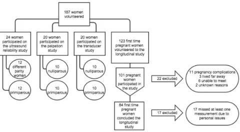
Altogether, 187 women participated in this thesis (Fig. I-5). Twenty-four (12 women in the postpartum period and 12 women with different parity (0-2 children)) and twenty (10 women in the postpartum period and 10 women with different parity (0-2 children)) healthy female volunteers were included in the test-retest study presented on chapter 2 and 3 respectively. Another twenty female volunteers (10 women in the postpartum period and 10 nulliparous women) participated in the exploratory study for the transducer motion.
In addition, one hundred and twenty-three first time pregnant women participated in the longitudinal observational studies. The participants were eligible for the study if they were first time pregnant, agreed to participate in four testing sessions (one during pregnancy at gestational week 35-41, and three in postpartum) and were able to perform the two different abdominal exercises. Exclusion criteria in the longitudinal studies were any kind of conditions affecting the ability to perform activities of daily living or any symptoms that required medical attention e.g., high-risk pregnancies, stillbirth or delivery before gestational week 37, previous spinal or abdominal surgery and neuromuscular diseases. Women were also excluded if one of the testing sessions was missed.
38
5. Ethics
Chapter
II
Test-retest and Intrarater Reliability of 2D
Ultrasound Measurements of Distance Between
Rectus Abdominis in Women
Patrícia Mota, Augusto Gil Pascoal, Fatima Sancho, Kari Bø
Abstract
Study design: Single-group test-retest reliability study.
Objectives: To evaluate test-retest intra-observer reliability of 2D ultrasound measurement of the distance between rectus abdominis, the inter-rectus distance (IRD).
Background: Diastasis recti is defined as the separation of the two rectus abdominis muscles with a reported prevalence of between 30% and 70% in women during pregnancy and in the postpartum period. The condition is difficult to measure, and ultrasound imaging has been suggested as a useful method to quantify the diastasis. However, to date no studies have investigated intra or inter-tester reliability of ultrasound to measure distance between rectus abdominis during rest and contraction.
Methods: Ultrasound images from the rectus abdominis were recorded on 24 healthy female volunteers at rest and on two conditions of abdominal contraction: Abdominal Crunch and Drawing-In exercises. The probe was positioned in two locations: below and above the umbilicus. A blinded investigator measured the IRD offline from two different ultrasound images collected on two different days (test-retest). Additionally, re-analyses of the same ultrasound images were done on two separate occasions (intra-image).
Results: Test-retest measurements of IRD demonstrated good to very good reliability with ICC values between 0.74 and 0.90. The only exception was for IRD measured 2 cm below the umbilicus on the abdominal crunch exercise, with an ICC of 0.50. For intra-tester reliability of the same images, the ICC values were all above 0.90.
Conclusion: Ultrasound imaging is a reliable method for measuring the inter rectus distance at rest and during Abdominal crunch and Drawing- in exercises.
Keywords
42
1. Introduction
Diastasis recti abdominis (DRA) has been defined as an impairment characterized by a midline separation of the two rectus abdominis (RA) muscles along the linea alba (LA).27,22
This increased inter rectus distance (IRD) has its onset during pregnancy and/or immediately after birth and the first weeks following childbirth.5,12
As the fetus grows the two muscle bellies of the RA connected by the LA, elongate and curve as the abdominal wall expands, and separation of the two muscle bellies with protrusion of the umbilicus may occur.5,14,13 Studies have found that diastasis recti may
affect between 30% and 70% of pregnant women5, and that it may remain separated in
the immediate postpartum period in 34.9%9 to 60% of women7,5,6. However the condition
has also been found in 38.7% of older, parous women undergoing abdominal hysterectomy25 and in 52% of urogynecological menopausal patients.27
Reported prevalence of diastasis recti or increased IRD may be inaccurate because of unreliable methods to measure the condition with the most common assessment method being palpation5,7,22,19, and calipers6,16. Ultrasound imaging has recently been suggested
as an useful method to assess muscular geometry and as an indirect measure of muscle activation via changes in muscle thickness.24 Coldron et al10 used ultrasound to characterize RA changes during the first year postpartum and Mendes20 et al claimed ultrasonography to be an accurate method to measure diastasis recti above and at the umbilicus when compared with surgical compass during abdominoplasty. However, search of the literature did not reveal studies addressing the intra or inter-tester reliability of the ultrasound measurement of the IRD at rest or during abdominal muscle contraction. Across-days reliability may be of interest to physiotherapists who perform repeated assessments of abdominal muscle function over time15 and factors such as relocation of the original imaging site, reproduction of the same transducer pressure and orientation, as well as maintenance of these factors during muscle contraction could adversely affect reliability.15
2. Methods
2.1 Design
This was a test–retest study evaluating the intrarater reliability of IRD measurements. For the test-retest analysis two test sessions were performed. In addition, the images collected during session 1 were analyzed a second time by the same investigator.
2.2 Participants
Twenty-four healthy female volunteers participated in this study. Twelve of the women were in the postpartum period and were recruited from a private physiotherapy clinic and the others among colleagues, friends and family. Demographic data with respect to age, body mass index (BMI) and parity are presented in Table II-1. The participants were eligible for the study if they agreed to participate in two testing sessions and were able to perform two different abdominal exercises. To ensure external validity, 12 women in postpartum period (less then 6 months) and 12 women with different parity (range 0 to 2 births), were included in the study. Pregnant women were excluded from the present study.
The study was approved by the Review Board of the Technical University of Lisbon, Faculty of Human Kinetics. Signed informed consent was obtained before participation in this study and the rights of the participants were provided in verbal and written form.
2.3 Instrumentation and procedures
44
The transducer was placed transversely along the midline of the abdomen in two locations with the center of the umbilicus as a reference: 2 cm above the umbilicus and 2 cm below the umbilicus. Initially, each measurement location was marked on the skin in order to standardize the position of the transducer. Ink marks were drawn with the subject in supine resting position with the knees bent at 90º and feet resting on the plinth, arms alongside the body (Figure II-1).
During image acquisition the bottom edge of the transducer was positioned to coincide with the correspondent skin marker and moved laterally until the medial borders of both RA muscles were visualized. The orientation of the transducer was then adjusted to optimize visualization of the image. Images were collected immediately at the end of exhalation, as determined by visual inspection of the abdomen following the recommendations of Teyhen et al.29 Additionally particular attention was paid to the
pressure imposed on the probe in order to avoid reflexive response from the participants.
Still images were obtained with subjects in the supine resting position (knees bent at 90º and feet resting on the plinth, arms alongside the body) and on two abdominal contraction conditions: abdominal crunch (Figure II-2) and drawing-in exercise (Figure II-1). One image was taken at each location under each condition. The abdominal crunch exercise was started from the resting position and the subjects were instructed to raise the head and shoulders upwards until the shoulder blades cleared the table. Subjects held this position until told to return to the starting position. The drawing-in exercise also started from the resting position, and the subjects were instructed to inhale and after exhaling draw in the abdominal musculature towards their spine. Before starting the procedure the subjects were verbally instructed about correct performance of the two exercises. The verbal instructions are provided in Table II-2. During the Drawing-In maneuver activation of the transversus abdominis muscle was confirmed by placing the transducer laterally between the iliac crest and rib cage.28 Every contraction was held for three seconds for
data collection with a resting time of 6 to 10 seconds between each repetition. After the test a convenient day for retest was scheduled with the participants.
2.4 Inter-rectus distance (IRD) measurement
Analyses of 2D ultrasound distances were conducted offline by the same investigator, using a customized code made on specific software (Matlab, Image Processing Toolbox, Mathworks Matlab, USA). Ultrasound images were assumed as a pixel based coordinate system, with the origin in the top left hand corner of the image. In this system an ‘x’ and ‘y’ coordinate could be used to locate a point in the image and distance between two or more point could be calculated. On ultrasound images the IRD is characterized by the transverse linear distance from the medial border of the rectus abdominis of one side to the corresponding position of its counterpart on the other side. Using this procedure, two points corresponding to the medial muscular insertions sites of both rectus abdominis on the linea alba, must be identified on the ultrasound images. From our observations these points are close to the inflection point of a parabola-like-curve that could be assumed for the ultrasound image of each rectus abdominis muscle contour Figure II-3 (red dots and yellow line). In order to improve the accuracy of the identification of these two points, an algorithm was developed and implemented using a customized Matlab code. Thus, the first step in the algorithm was to interpolate a set of 8-10 points manually digitalized by the examiner on the visible contour of both muscle bellies, and fit them to a parabola-like-curve. Using the coordinates of those digitized points a fourth order polynomial equation was fitted in order to determine the coefficient of the polynomial and the inflexion point of the interpolated curve. The discrete derivative of the interpolated x-coordinate and the point at which the sign changed was considered as the parabola point of inflexion Figure II-4 (white asterisks). The determined inflection point and the interpolated
parabola-like-curve were overlapped on the original ultrasound image, to guide the examiner on the identification of the medial margins of the RA and improve the accuracy of IRD measurements. Besides the software suggestion, the examiner has the final decision about the location of the medial margins of the RA muscles used on IRD measurements.
2.5 Statistical Analyses
46
ultrasound images and collected on two different days (test-retest), and across the 2 IRD measurements made on the same ultrasound image (intra-image).
The scale from Altman1 was used in the classification of the reliability values. ICC values less than or equal to 0.20 were considered poor, 0.21 to 0.40 fair, 0.41 to 0.60 moderate, 0.61 to 0.80 good and 0.81 to 1 very good.
Standard error of measurement (SEM) was used to examine the precision of measurement and it was calculated according to SEM = pooled Standard Deviation* 1−𝐼𝐼𝐼𝐼𝐼𝐼. To represent a difference in IRD beyond measurement error, the minimum detectable change (MDC) was calculated as 1.96 * SEM * 2 23. These analyses
were performed for each of the outcome variables: IRD at rest condition, abdominal crunch and drawing in exercise, 2 cm above and 2 cm below the umbilicus.
The Bland-Altman plot of difference against the mean was also used to compare the limits of agreement and mean bias between plots.4 The Standard Deviation (SD) of the differences between test and retest was calculated, and then multiplied by 1.96 to obtain the 95% random error component.2
In order to verify the differences on IRD related to the postpartum condition, the 12 postpartum women were compared to the women with different parity using an independent t-test.
3. Results
All participants returned for the second test after a mean of 3.9 days (SD = 3.9, range 1 to 16 days) and all reported that they complied with the request not to practice any of the exercises between tests. There were no dropouts. The IRD values for each measurement are shown in Table II-3. No significant differences were found in the IRD between women in postpartum and the other women with different parity (Table II-4). In general, the smallest IRD values were from abdominal crunch exercise, and the biggest were from the drawing-in exercise.
3.1 Intra-tester reliability of the ultrasound analyses
(intra-image)
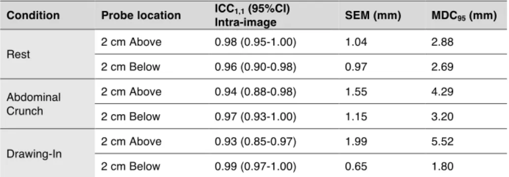
The ICC values for the IRD measured on the same image at two different occasions revealed very good reliability for every condition tested (Table II-5). The Rest condition demonstrated less variability than the measurements conducted during Abdominal Crunch and Drawing-In, but the ICC values were all above 0.90. The precision of repeated measurements of the same images was higher (revealed by lower SEMs) compared with recaptured images. The MDC values ranged from 1.80 to 5.52 mm. The Bland-Altman plot (Figure II-5) showed that the mean of differences of IRD on test-retest was closer to zero (0.052) mm, and the limits of agreement were narrower compared with the values found on different images (-1.95 mm and 2.05 mm).
3.2 Test-retest across days (inter-image)
48
4. Discussion
The present study demonstrated very good reliability for the intra-tester measurements in the same image for all the conditions tested, with ICC values above 0.90, low values of SEM (range 0.65 to 1.99) and MDC (range 1.80 to 4.29). These results are in line with the values found by Liaw et al.18 The test-retest measurements across days showed good
reliability during rest and drawing-in exercises below the umbilicus with ICC values of 0.78 and 0.74 respectively and very good reliability during rest, abdominal crunch and drawing-in exercises above the umbilicus with ICC values of 0.87, 0.83 and 0.90 respectively. The lowest ICC value of 0.50 was found below the umbilicus and during contraction, with moderate reliability for abdominal crunch. The higher values found on the SEM (range 2.28 to 4.36 mm) and on the MDC (range 6.32 to 12.08 mm) revealed lower precision of the IRD measurements.
The lower values found below the umbilicus may be explained by the influence of the amount of subcutaneous fat18 in this location. This could have interfered with the
determination of where to mark the skin, positioning of the probe and the ability to maintain a constant pressure during image acquisition. During the abdominal crunch exercise the participants had to move the upper body and this may have induced movements under the transducer. Nevertheless the ICC was moderate to good.
In general, there are several potential sources of measurement errors: the subjects, the testing, the scoring, the instrumentation and factors such as the instructions from the examiner, participant motivation, and the participants skill and motor control may affect performance in different days.17 To mitigate against some of these potential sources of
errors the position of the subject, examiner’s instructions, the transducer location and its inclination, the pressure applied to the transducer on the abdominal wall, and the room temperature were standardized.
Criteria for the diagnosis of DRA vary in the literature.5,7, 14,26,27,25,8,3 Beer et al3 suggest
50
differences were found in the IRD between women in postpartum and the other women with different parity.
In studies of postpartum women, DRA has been defined as the LA having a width greater than 2-finger breadth (1.5 cm) when measured with palpation,5 , 14, 27 or 2 cm when
measured with a dial caliper at or above the umbilicus during a partial sit-up.18 However
the inaccuracy and possible low reliability of the measurement tools used are possible limitations of previous studies.
Computed tomography (CT) and magnetic resonance imaging (MRI) are currently considered the methods of choice to examine the abdominal wall, but they are expensive and CT exposes the patient to radiation20, making it impossible to use in pregnant women.
Hence, ultrasonography has been proposed as a non-invasive technique that can be repeated several times20 during pregnancy.
The current investigation examined many aspects of reliability of the ultrasound measurements. The two RA muscles were identified in both relaxed and contracted conditions. Furthermore, repeated measurements were conducted from the same stored images as well as across images collected and measured on two different days. It would be expected that measuring the IRD repeatedly, even on different days from stored images, would be associated with higher values of ICC. This is because measuring the distance between two well defined muscles in the ultrasound images is a relatively straight forward task. Our results from the IRD and the results of Hides et al15, about the thickness
of the internal oblique and transversus abdominis muscles, support this premise, with both studies reporting very high values of ICC from repeated measures of the same image. However, accurately re-imaging the subject to obtain comparable images may require a higher level of skill. On the current study the measurements from recaptured images showed from good to very good reliability, with the only exception of moderate reliability in the abdominal crunch exercise. The lower precision shown by higher SEM and MDC values and the wide 95% limits of agreement confirm the inferior reliability of recaptured images compared to repeated measurements of the same stored image.
to treat DRA in the peripartum period. A follow-up study on the IRD in pregnancy and postpartum in different muscle contraction conditions is being conducted.
The current study is unique in the reliability tests on the IRD measurements and the use of different locations and contraction conditions to better objectively quantify the separation between the two rectus abdominis muscles. A strength of this study is the blinding of the observer to all the results of IRD measurements until the end of the process. To ensure external validity, 12 subjects in postpartum period and 12 women with different parity, were included in the study. In general the IRD was greater in postpartum women, but no significant differences were found in the IRD between the two groups. Consistent with our findings, Liaw et al18 also noted that the medial margins of the RA appear to be indistinct
where the fascial borders become less clear in postpartum women. We used a customized Matlab code to implement a method of ultrasound images segmentation based on explicit shape representation defined by a known point distribution model.11 In fact, a semi-automated ultrasound image segmentation method was used in order to help the examiner to identify the medial margins of both RA muscles and improve the accuracy of IRD measurement. However the examiner has always the final decision. We believe that in the near future this Matlab code could be implemented in the software embedded in the ultrasound scanners, helping clinicians to accurately measure the IRD or other muscular morphometric parameters (e.g. muscle cross sectional area).
The limitations of this study include the use of only one rater with limited experience in ultrasound imaging and inclusion of only healthy subjects with no musculoskeletal or neurological symptoms. It may be more difficult to reliably measure subjects with symptoms that can interfere in the performance of the exercises across the days or in the last gestational weeks where wider IRD may require a broader view of the abdominal wall to be able to see both RA muscles on the same image. Because the main goal of this study was to evaluate test-retest and intrarater reliability of the IRD in different contraction conditions, we excluded pregnant women from this study, because the IRD is constantly changing with the progress of pregnancy and movement/position of the baby.21 This may
52