Volume 2013, Article ID 737358,15pages http://dx.doi.org/10.1155/2013/737358
Research Article
Comparative Anatomy of the Hind Limb Vessels of the Bearded
Capuchins (
Sapajus libidinosus) with Apes, Baboons, and Cebus
capucinus: With Comments on the Vessels’ Role in Bipedalism
Roqueline A. G. M. F. Aversi-Ferreira,
1,2,3,4Tainá de Abreu,
2,5Gabriel A. Pfrimer,
5Sylla F. Silva,
2Janine M. Ziermann,
1Frederico O. Carneiro-e-Silva,
5Carlos Tomaz,
4Maria Clotilde H. Tavares,
4Rafael S. Maior,
4and Tales A. Aversi-Ferreira
1,21Department of Anatomy, Howard University College of Medicine, 520 W Street NW, Numa Adams Building,
Washington, DC 20059, USA
2Laboratory of Anthropology, Biochemistry, Neuroscience and Primates’ Behavior (LABINECOP), Federal University of Tocantins,
Avenida NS 15 ALC NO 14 109 Norte, 77001-090 Palmas, TO, Brazil
3Graduate School of Animal Biology, Institute of Biology, University of Brasilia, Darcy Ribeiro Campus, 70910-900 Bras´ılia, DF, Brazil 4Department of Physiology, Laboratory of Neuroscience and Behavior, University of Brasilia, Darcy Ribeiro Campus,
70910-900 Bras´ılia, DF, Brazil
5Graduate School of Veterinary, Federal University of Uberlˆandia, Rua Cear´a S/N Bloco 2D Campus Umuarama,
38400-902 Uberlˆandia, MG, Brazil
Correspondence should be addressed to Tales A. Aversi-Ferreira; [email protected] Received 22 October 2013; Accepted 18 November 2013
Academic Editor: Hisao Nishijo
Copyright © 2013 Roqueline A. G. M. F. Aversi-Ferreira et al. This is an open access article distributed under the Creative Commons Attribution License, which permits unrestricted use, distribution, and reproduction in any medium, provided the original work is properly cited.
Capuchin monkeys are known to exhibit sporadic bipedalism while performing specific tasks, such as cracking nuts. The bipedal posture and locomotion cause an increase in the metabolic cost and therefore increased blood supply to lower limbs is necessary. Here, we present a detailed anatomical description of the capuchin arteries and veins of the pelvic limb of Sapajus libidinosus in comparison with other primates. The arterial pattern of the bearded capuchin hind limb is more similar to other quadrupedal Cebus species. Similarities were also found to the pattern observed in the quadruped Papio, which is probably due to a comparable pelvis and the presence of the tail. Sapajus’ traits show fewer similarities when compared to great apes and modern humans. Moreover, the bearded capuchin showed unique patterns for the femoral and the short saphenous veins. Although this species switches easily from quadrupedal to bipedal postures, our results indicate that the bearded capuchin has no specific or differential features that support extended bipedal posture and locomotion. Thus, the explanation for the behavioral differences found among capuchin genera probably includes other aspects of their physiology.
1. Introduction
There are numerous studies about Sapajus behavior,
cogni-tion, and bipedalism [1–5], but the anatomical descriptions
of these species are scarce in scientific literature and the
majority of those works concentrate on thoracic limbs [6–
13]. Few descriptions are related to the nervous system of the
hind limbs [14, 15]. It is not surprising that the anatomical
descriptions available of the lower limbs that support the data
about its bipedalism focusing mainly on muscles and bones, although these structures are supplied by the arterial system. Indeed, some studies have focused on the kinematics
of capuchin bipedalism [16] but few have addressed the
metabolic cost of bipedal gait on hind limbs. Although this
behavior requires higher energetic cost on hind limbs [17]
and depends on an adequate arterial supply to muscles, only a handful of articles have emphasized the anatomical features
described the femoral artery of the apes and also mentioned Cebus with regard to the saphenous arteries. Manners-Smith
[19] investigated the hind limb arteries of primates and
com-pared two specimens of Cebus capucinus to other primates of different families, that is, Simiidae, Cercopithecidae, Hapali-dae, and Prosimiae. Among New World primates, however, this work focused mostly on the genus Ateles. There were only few superficial descriptions of Cebus capucinus’ vessels and no pictures were provided for this species. Furthermore,
neither of the authors mentioned above [18,19] described the
arteries supplying the muscles.
Indeed, the anatomical aspects of hind limb vessels could have considerable importance to keep the muscles working to support the weight when the animal is on a bipedal gait, because the more ramified arterial system could increase the
area for the distribution of blood [20,21]. Applying this
ratio-nale to different primate species, the comparative anatomical description of vessel plays important role to provide data about the physiology and behavior behind bipedalism.
Therefore, the main objective of this study was to generate modern and accurate data about the anatomical pattern of the vessels of Sapajus libidinosus (bearded capuchin) hind limbs and compare our findings with the anatomy of others species available in the scientific literature, namely, Cebus capucinus
[19, 22], apes [23], baboons [24, 25], and modern humans
[26].
We analyzed the vessels of the hind limbs of bearded capuchins, considering aspects as origin, distribution, and irrigation. We are discussing our results in comparison with other species and present our formatted data in a table. More-over, we made an effort to identify associations of our data that would support the physiological anatomy of the bipedal position.
2. Material and Methods
2.1. Samples. Nine adult bearded capuchin specimens (three females, six males) were used in this study. Their weights were between one and four kilograms. No animal was killed for the purposes of this study; five of them suffered accidental deaths in their natural habitats and were deposited in the anatomical collection of the Anthropology, Biochemistry, Neuroscience and Behavior of Primates Laboratory (LABINECOP) from the Federal University of Tocantins, Tocantins State, Brazil. The other four belonged to the Brazilian Institute of Envi-ronment and Renewable Natural Resources (IBAMA) archive and were donated for studies in the 1970s. This work was approved by the Institutional Ethical Committee from the Federal University of Goi´as, Goi´as State, Brazil (CoEP-UFG 81/2008, authorization from the IBAMA number 15275). 2.2. Preparation of the Animals for Dissection. All procedures involving the animals were done in accordance with the guidelines of the Brazilian Society of Animal Experimenta-tion (COBEA). After the trichotomy with a razor blade, the animals received perfusion, by the femoral artery, with 10% of formaldehyde for fixation. The animals were conserved in 10% of formaldehyde, in covered opaque cubes.
2.3. Nomenclature. We used the human nomenclature for the vessels whenever possible. In the cases where no human nomenclature was available we followed the terminology of
[23].
3. Results
3.1. Arterial Description (Table1). In the bearded capuchins investigated, the external iliac artery runs inferior and later-ally in the pelvis lying on the ilium-psoas muscle. It emits
a medial-inferior branch, the obturator artery (Figure1(a)),
immediately before crossing the aponeurosis of the external oblique muscle of the abdomen. We did not find a true inguinal ligament in any of the specimens studied here. The obturator artery emits the medial circumflex femoral artery, which crosses the aponeurosis of the external oblique
abdomen muscle medially to iliac vein (Figures1(a)and1(b)).
In one specimen, however, the medial circumflex femoral artery originates directly from external iliac artery but only at one side.
The medial circumflex femoral artery crosses inferiorly to the aponeurosis of the external oblique abdomen muscle, runs obliquely from the anterior to the posterior aspect of the thigh, and communicates branches to the gracilis and adduc-tor muscles, one single branch to the femur head and emits
the external pudenda artery (Figure1(b)). The medial
cir-cumflex femoral artery is usually a branch from the obturator artery.
In the bearded capuchin the femoral artery (Figures1(a),
1(b),2(a), and2(b)) emits first a lateral trunk that gives rise to
the lateral circumflex femoral artery and the superficial
epi-gastric artery (Figure2(b)). The superficial epigastric artery
turns laterally and superiorly and emits, superior-laterally, the superficial circumflex iliac artery. The lateral circumflex femoral artery emits two muscular branches to the vastus lateral and vastus intermedius muscles, a descendent branch, and a branch that runs inferiorly and laterally towards the femur’s head. We have identified the branches of the lateral circumflex femoral artery as the superficial epigastric artery (“ascendant branch”), and the lateral circumflex femoral artery (“descendent and transversal branches”).
In our specimens we observed that the first medial branch of the femoral artery is the profunda femoris artery that runs
medial and posterior in the thigh (Figures2(b)and2(c)). It
gives rise to muscular branches to the adductor muscles and quadriceps muscle in the medial side of the thigh, and one or two perforating branches that turn laterally. The profunda femoris artery terminates in muscular branches caudal in the lateral compartment of the thigh to the vastus lateralis muscle. The femoral artery continues in the thigh medially under the sartorius muscle through the adductor canal and inferiorly to the lower margin of the vastus medialis muscle, clearly giving rise to branches to the adductor muscles, the sartorius muscle and the vastus medialis muscle. In the distal quarter of the thigh the popliteal artery originates posteriorly and continues superficially as the saphenous artery (Figures
1(a)and2(d)). In the bearded capuchins described here, the
(a)
(a)
(b)
(b)
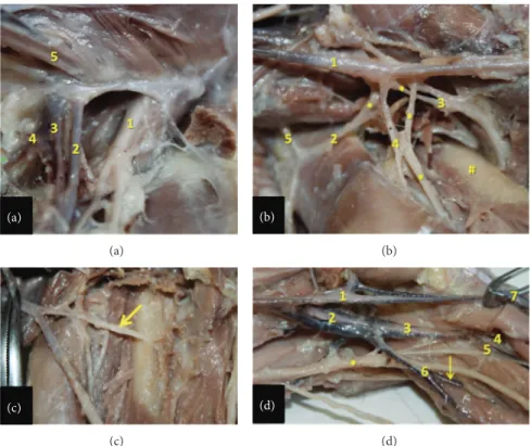
Figure 1: Ventral view of arteries in left hind limb of the bearded capuchin. Cranial to the right (a): View of the left pelvis and thigh. 1 indicates the abdominal aorta artery; 2 the left common iliac artery; 3 the internal iliac artery; 4 the external iliac artery; 5 the obturator artery; 6 the
branch to the genital organ; 7 the medial circumflex femoral artery; 8 the external pudenda artery; 9 the femoral artery; the∗ indicates the
respective veins associated with arteries; the arrows indicate the saphenous vein (0.7X). (b) View of the left inguinal region. 1 indicates the femoral artery; 2 the medial circumflex femoral artery; 3 the muscular branch to the gracilis muscle; 4 the artery to the femur head; 5 the
external pudenda artery. The arrow indicates the muscular branch to the semimembranosus muscle and the∗ indicates part of the saphenous
vein (2.4X). (a) (a) (b) (b) (c) (c) (d) (d)
Figure 2: Detailed view of the inguinal, thigh, and popliteal regions in the bearded capuchin. (a) Left inguinal region. 1 indicates the femoral nerve; 2 the femoral artery; 3 the femoral vein; 4 the medial circumflex femoral artery; 5 the spermatic cord (3X). (b) Right thigh. The arteries are indicated by numbers, where 1 is the femoral artery; 2 the lateral circumflex femoral; 3 the profunda femoris artery; 4 the branch of the
lateral circumflex femoral artery and 5 the inferior epigastric. The∗ indicates veins associated with arteries; the # indicates the femur (2.4X).
(c) Right thigh. The arrow indicates the profunda femoris artery (2X). (d) Left popliteal region. 1 indicates the femoral artery; 2 the popliteal artery; 3 the common tibial artery; 4 the anterior tibial artery; 5 the posterior tibial artery; 6 the fibular artery; 7 the saphenous artery. The
(a) (a) (b) (b) (c) (c)
Figure 3: View of the lower leg of the bearded capuchin. Superior to the top. (a) Right leg. 1 indicates the anterior tibial artery and 2 the
posterior tibial artery, both emerging in the anterior compartment of the leg. The # indicates the tibia and the∗ the fibula (1.4X). (b) Right
leg. The fascia of leg was kept and the arteries were not separated from the veins (0.8X). (c) View of the medial aspect of the left leg (1X). (b)-(c) The numbers indicate the branches from the saphenous artery. 1 indicates the anterior ramus; 2 the medium ramus; 3, the posterior
ramus. The∗ indicates the tibia.
(a)
(a)
(b)
(b)
Figure 4: View of the posterior (a) and lower (b) leg of the bearded capuchin. The number 1 indicates the fibular artery that runs in the proximal leg together with the saphenous vein. (a) View of the posterior aspect of the left leg and part of the thigh (0.8X). (b) Lateral view of the lower leg and foot and part of the right leg (0.8X).
that gives off two inferior genicular arteries and later splits anteriorly into the common tibial artery and posteriorly into
the fibular artery (Figure2(d)).
The common tibial artery in the bearded capuchin
(Fig-ure3(a)) divides into an anterior branch, the anterior tibial
artery, and in the posterior and inferior branch, that is, the posterior tibial artery. The anterior tibial artery passes bet-ween the tibia and the fibula to turn proximately towards the anterior leg compartment and terminates into muscular branches supplying anterior superficial muscles of the leg
(Figure3(a)). The posterior tibial artery runs medially to the
soleus muscle, lies at the posterior aspect of the interosseous membrane and penetrates in the middle third of the leg of this membrane to supply the deep anterior muscles of the leg
in the anterior leg compartment (Figure3(a)). The popliteal
artery was considered here as the posterior tibial artery also due to position and function; the posterior and anterior tibial arteries do not reach the foot.
The fibular artery observed in the bearded capuchins
(Figure2(d)) is a branch from the popliteal artery. It gives
rise to branches to the triceps sural muscles, continues super-ficially between the heads of the gastrocnemius muscle, and
lies parallel to the short saphenous vein (Figure4). Both, the
fibular artery and the saphenous vein, run laterally to the gastrocnemius tendon along its lateral margin, passing pos-teriorly and inferiorly to the lateral malleolus into the foot’s dorsal area. From there the arteries distribute laterally and superficially as a calcaneal branch.
The saphenous artery (Figure 3(b)) is superficial, emits
the superior genicular arteries, and divides into the anterior and posterior branches in the medial proximal third of the leg. The anterior branch generates a medium branch in the
proximal third of the leg (Figures3(b)and3(c)). Initially, all
branches from saphenous artery run under the skin of the leg
(Figure3(b)). In the foot, they split into the dorsal and the
plantar arteries. The anterior ramus of the saphenous artery
(Figure3(c)) runs, after giving rise to the medium ramus,
inferiorly and penetrates into the distal third of the leg under-neath the retinaculum of the anterior leg muscles. It passes then obliquely to the lateral aspect into the retinaculum
(a)
(a)
(b)
(b)
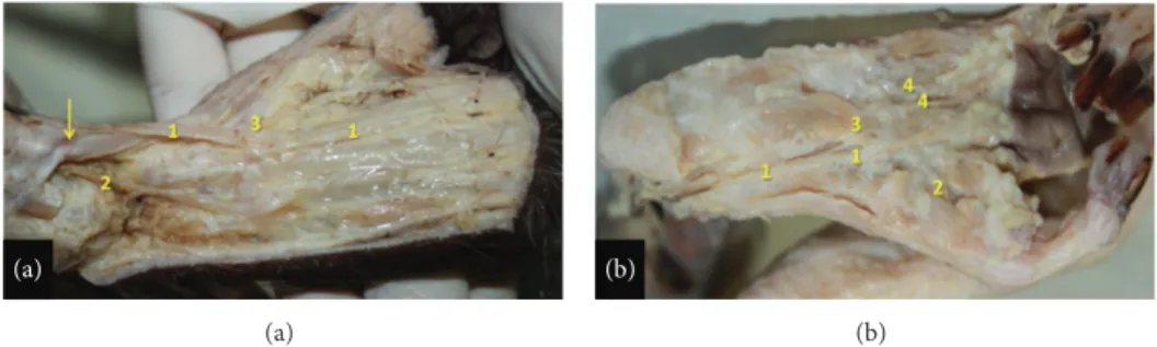
Figure 5: Right foot of the bearded capuchin. (a) Anterior and medium ramus of the saphenous artery on the dorsal foot. The number 1 indicates the dorsalis pedis medial artery; 2 the dorsalis pedis lateral artery; 3 the 1st dorsal metatarsal branch. The arrow indicates the retinaculum of the anterior leg muscles (2X). (b) Plantar view. The number 1 indicates the medial plantar artery; number 2 the hallux plantar artery; number 3 the lateral plantar artery; number 4 the 1st plantar metatarsal artery (2X).
behind the tendon of the anterior tibial muscle. From there it emerges laterally on the foot, where finally it emits the
dorsalis pedis lateral artery (Figures3(c)and4(a)).
The dorsalis pedis lateral artery distributes blood to soft tissues and skin and emits the 3rd and 4th dorsal metatarsal branches. The medium ramus from the anterior branch of the
saphenous artery (Figure3(c)) runs inferiorly, passes
anteri-orly to the medial malleolus, superficially to the retinaculum
(Figure4(a)), and emerges on the medial portion of the foot
emitting the dorsalis pedis medial artery (Figure5(a)) that
gives off the 2nd dorsal metatarsal, a perforating branch and the first dorsal metatarsal artery. The posterior ramus of the
saphenous artery (Figure 3(b)) runs inferior and medially
along the medial margin of the medial head of the gas-trocnemius muscle, continues medially to the tendon of the triceps sural muscle, and penetrates the foot inferiorly
(Fig-ure5(a)) and medially becoming the medial plantar artery
(Figure5(b)). The medial plantar artery is associated with the
plantar nerve in the sole (Figure5(b)). It splits into a branch
to the hallux (the first plantar metatarsal branch), a branch to the second interosseous space, and a communicating branch and gives off the lateral plantar artery approximately in the
proximal third of sole. The lateral plantar artery (Figure5(b))
supplies the third and fourth interosseous spaces.
3.2. Vein Description. The veins in the hind limb form a superficial and a deep group in the bearded capuchins. The deep veins are “venae comitantes,” that is, companion veins
that follow like duplicates the arteries (Figures 1(a), 1(b),
2(d), and3(b)). A highlighted superficial vein in the bearded
capuchin is the short saphenous vein (Figure4) that
origi-nates dorsal and laterally in the foot follows laterally and turns around the lateral malleolus. It ascends superficially between the heads of the gastrocnemius muscle together with the fibu-lar artery, is superficial and posterior in the knee (popliteal fossa) and thigh, and continues accompanying the muscular branch of the medial circumflex femoral artery in the
proxi-mal third of thigh (Figures1(a)and1(b)).
The short saphenous vein continues to merge with the external iliac vein in the pelvis. The other superficial veins are a group formed by companion veins of the saphenous artery’s branches that drain to the femoral vein, which represents the
long saphenous. We found three superficial veins: two along each branch of the saphenous artery and a third one along the medium ramus of the anterior ramus of the saphenous artery. The femoral vein receives the tributaries from veins accompanying the saphenous arteries. In the medial, aspect of the thigh of the bearded capuchin the femoral vein ascends passing into the femoral sheath, medial and deep to the femoral artery and lateral and anterior to the medial
circumflex femoral artery and vein (Figure2(a)). The external
iliac and internal veins join to form the common iliac vein
that drain to the inferior cava vena (Figure2(a)).
4. Discussion
4.1. Comparison with Other Primates: Arteries (Table 1). Interestingly, few variations were found among the specimens of bearded capuchin observed in the present study. The main variation, the origin of the medial circumflex artery in one of nine cases, only allowed a general description of the pattern of vessels in the bearded capuchin. We also found that the inguinal ligament in general extends between the spina iliaca anterior superior of the ilium and the tuberculum pubicum of the pubis and separates a space for muscles and vessels between abdomen and leg. In contrast to the description
of Manners-Smith [19] in Cebus we could not find a true
inguinal ligament in any of the specimens of bearded capuchin studied here.
The origin of the obturator artery in monkeys and apes is quite diverse: it was found to originate from the external iliac
artery in Cebus capucinus and in Pan [19]; from the internal
iliac artery in Papio and in ca. 50% of Pan [23,24]; directly
from the femoral artery or from the profunda femoris artery
in 25% of Papio [25]; and from the inferior epigastric artery
in Gorilla, Pongo, and in ca. 50% of Pan [23]. According to
Gibbs [23], the obturator artery originates from the femoral
artery as a variation in humans, while it originates from the
profunda femoris, internal iliac [26], and inferior epigastric
artery in 20 to 30% of all cases. The origin of obturator artery found here in bearded capuchin investigated here is similar to Cebus [19] and Pan [24]. However, we found in one specimen that the medial circumflex femoral artery originates on one side directly from external iliac artery.
T a ble 1: C o m pa ra ti ve an at o m y o f the hind lim b ar ter ies o f b ea rd ed ca p u chin (t his w o rk), Ce bu s ca pu ci n us ,b aboo n s, ape s, an d Ho m o. O o rigina tes fr o m E: emi ts. B ea rded ca p uc hin s Ce bu s Ba boo n s A p es Ho m o OE OE OE O E O E Ob tu ra to r Ex te rn al il ia c Me d ia l cir cumflex fe mor al mu sc u la r br an ch es Ex te rn al il ia c — Int er n al il ia c — Ex te rn al il ia c (Pa n [ 24 ]), in te rn al iliac (5 0% o f Pa n [ 23 ]), inf er io r ep igast ri c (Go ri lla ,Po n go ,a n d 50 % o f Pa n [ 23 ]) — Or igina tes fr o m fe mor al as var ia ti on [ 23 ], p ro funda fe mo ri s, in te rn al iliac [ 26 ], an d inf er io r ep igast ri c fr o m 20 to 30 % Ili ac b ra nches to foss a il ia c and an asto mos es w it h il io lu mb ar ar te ry [ 26 ] Me d ia l cir cumflex fe mor al Ob tu ra to r ar te ry or ext er n al il ia c (o n e ca se obs er ve d ) Mu sc u la r br an ch es , fe m u r h ea d br an ch , ext er n al p udenda Ex te rn al il ia c Mu sc u la r br an ch es Ob tu ra to r — Ex te rn al il ia c (Hy lo ba te s [ 23 ]a n d Pa n [ 24 ]) ,prof u n d a fe mor is (5 0 % o f Pa n [ 23 ]), fe mo ral (Go ri lla an d Po n go [ 23 ]) — Prof u n d a fe mor is [ 23 , 24 , 26 ]f em o ra la s va ri at io n [ 23 , 26 ] M u sc u lar br an ch es [ 26 ] Ex te rn al p udenda Me d ia l cir cumflex fe mor al — ——F em o ra l — Fe m o ra l( Pa n , unila te rall y in Go ri lla ; [ 23 ]) —F em o ra l[ 23 , 26 ] Anasto mos e w it h br an ch es of in te rn al p udenda ar ter y [ 26 ] Fe m o ra l Ex te rn al il ia c La te ra l cir cumflex fe mor al, inf er io r ep igast ric, prof u n d a fe mor is , po p li teal , sa p h en o u s, mu sc u la r br an ch es Ex te rn al il ia c La te ra l cir cumflex fe mor al, prof u n d a fe mor is , sa p h en o u s, po p li teal Ex te rn al il ia c Sa p h eno u s, po p li teal , prof u n d a fe mor is , supre m a ge nic ula r Ex te rn al il ia c [ 23 ] M edial cir cumflex fe mor al (Go ri lla ,Po n go , as a va ri at io n in Ho m o [ 23 ]), des cenden t ge nic ula r (Pa n , Po n go , Ho m o [ 23 ]), sa p h eno u s (all ap es [ 23 ]), deep cir cumflex iliac (Af rica n ap es ;[ 23 ]), m u sc ula r br an ch es [ 23 ], su p er fi cial cir cumflex iliac (gr ea t ap es an d Ho m o [ 23 ]), inf er io r ep igast ri c (Pa n , SW an d al la p es an d Ho m o;[ 23 ]), ext er n al p udenda (Ho m o, Pa n , Go ri lla [ 23 ]), p ro funda femo ri s (a ll ap es and Ho m o, ex cep t Go ri lla, G), p o p li te al (al la p es an d Ho m o, G) Ex te rn al il ia c [ 23 , 26 ] M edial cir cumflex fe mor al (a s a va ri at io n), d es cenden t ge nic ula r, m us cu la r b ra n ch es, su p er ficial cir cumflex iliac, inf er io r ep igast ri c, ext er n al p udenda, prof u n d a fe m o ri s, po p li teal [ 23 , 26 ]
Ta b le 1: C o n ti n u ed . B ea rded ca p uc hin s Ce bu s Ba boo n s A p es Ho m o OE OE OE O E O E Sup er fi ci al ep igast ric Fe m o ra l Sup er fi ci al cir cumflex il ia c —— Ex te rn al il ia c — L at eral cir cumflex fe mor al (Go ri lla [ 23 ]), fe mor al (5 0 % Po n go [ 23 ]), ext er n al iliac (Pa n [ 23 ]) —F em o ra l[ 23 , 26 ]— La te ra l cir cumflex fe mor al Fe m o ra l Mu sc u la r br an ch es , des cenden t br an ch , fe m u r h ea d br an ch Fe m o ra l A scenda n t, des cen-den t, and tra n sv er sal Prof u n d a fe mor is Ra m u s des cenden s Prof u n d a fe mor is (a ll ap es, ex cep t 50% Go ri lla [ 23 ]), fe mo ral (Go ri lla to as cenda n t an d d es cenden t ra m u s [ 23 ]) Thr ee b ra n ch es in Pa n an d A si an ap es ,fi ve br an ch es in Po n go [ 23 ] F emo ra l, ext er n al il iac, [ 24 ]p ro fu n d af em o ri s [ 23 , 24 , 26 ] Thr ee b ra n ch es [ 23 , 26 ] Sup er fi ci al cir cumflex il ia c In fe ri or ep igast ric Mu sc u la r br an ch es —— In fe ri or ep igast ric — In fe ri o r ep igast ric (Pa n [ 24 ]), fe mo ral [ 23 ] — In fe ri o r ep igast ric [ 24 ], fe mor al [ 23 ] — Prof u n d a fe mor is Fe m o ra l Mu sc u la r br an ch es , pe rf o ra n s (1 or 2) Fe m o ra l Thr ee pe rf o ra n s br an ch es F emo ral P er fo ra n s F emo ral [ 23 ] M u sc u lar br an ch es , tw o p er fo ra n s b ra n ch es in 6 6.6% o f Po n go ,Pa n , an d Hy lo ba te s,t h re e p er fo ra n s b ra nches in Go ri lla [ 23 ] Fe m o ra l[ 23 , 26 ] Thr ee p er fo ra n s br an ch es [ 23 , 26 ], la tera la nd me di al cir cumflex femo ral ar te ri es an d m u sc u la r br an ch es [ 26 ] Po p li te al F em o ra l In fe ri or ge nic ula r, co mmo n ti b ial , fib u la r Fe m o ra l An ter io r ti b ial , fib u la r, su p er io r ge nic ula r, rudimen- tar po st er io r ti b ial , media genic ula r Fe m o ra l An ter io r an d p o st er io r ti b ial ar te ri es Fe m o ra l[ 23 ] An te ri o r and p o ste ri o r ti b ial ar te rie s in all ape s, ex cep t Pa n th at di vides in to a p o ste ri o r ti b ia l an d a co m m on br an ch for the an te ri or ti bi al an d fi bu la r ar te ry , su p er io r ge nic ula r br an ch es in al la p es an d inf er io r genic ula r br an ch es in Pa n an d Hy lo ba te s [ 23 ] Fe m o ra l[ 23 , 26 ] An ter io r an d p o st er io r tib ial ar te ri es, su p er io r ge nic ula r an d inf er io r ge n ic u lar br an ch es , sur al ar ter ies [ 23 , 26 ], middle ge nic ula r ar te ri es ,m u sc u la r br an ch es ,c ut an eo u s br an ch es [ 26 ] Sup er ior ge nic ula r Sa phe n ous — P o pl ite al — P o pl ite al — P o pl ite al [ 23 ]— P o p li te al [ 23 , 26 ]— In fe ri or ge nic ula r P o p li te al — ——P o p li te al — Po p li te al in Pa n an d Hy lo ba te s [ 23 ] —P o p li te al [ 23 , 26 ]—
Ta b le 1: C o n ti n u ed . B ea rded ca p uc hin s Ce bu s Ba boo n s A p es Ho m o OE OE OE O E O E An ter io r ti b ial Co m m o n ti b ial Mu sc u la r br an ch es Po p li te al — Po p li te al — Po p li te al [ 23 ] M u sc u lar br an ch es in al l ap es, ex cep t Pa n are te rmina tin g as d o rs al ar te ry of th e fo o t [ 23 ] Po p li te al [ 23 , 24 , 26 ] Do rs al ar te ry o f th e fo ot [ 23 , 26 ], p o st er io r tib ial re cu rr en t, an te ri o r ti b ial re cu rr en t, m u sc u lar br an ch es , p er fo ra ting b ra nches, an te ri or m ed ia l ma ll eo la r, an ter io r la te ral m alleo la r [ 26 ] P o st er io r ti b ial Co m m o n ti b ial Mu sc u la r br an ch es Sa p h eno u s (f ro m po p li teal to rudi-men ta ris p o st er io r ti b ial ) Me d ia l an d la te ra l pl an ta r Po p li te al — Po p li te al [ 23 , 24 ] C al ca n ea li n gr eat ap es [ 23 ], me di al ,a nd la tera l pl an ta r [ 23 , 24 ] Po p li te al [ 23 , 24 , 26 ] C alca n ea l, medial , an d la te ra lpl an ta r [ 23 , 26 ], cir cumflex fib u la r, n u tr ien t ar ter y of th e ti b ia ,m u sc u la r b ra n ch es, p er fo ra ti n g br an ch es , co mm unica tin g br an ch ,m ed ia l ma ll eo la r [ 26 ] Fib u la r P o p li te al Mu sc u la r br an ch es , calca n eal Po p li te al — Po p li te al — P o st er io r tib ial in Go ri lla an d Hy lo ba te s, an te ri or ti bi al in Po n go ,p o p li te al in Pa n [ 23 ] La te ra lc al ca n ea l, p er fo ra tin g b ra nc h in all ape s [ 23 ] P o st er io r tib ial [ 23 , 24 , 26 ] L at eral calca n eal , p er fo ra tin g b ra nc h in all apes [ 23 , 26 ], m u sc u lar br an ch es , nu tr ie nt to fi b u la , p er fo ra ting b ra nches, co mm unica tin g br an ch [ 26 ] Sa p h eno u s F emo ral Sup er ior ge nic ula r, an te ri or an d p o st er io r br an ch es Fe m o ra l Supre m a ge nic ula r, an te ri or , an d p o st er io r br an ch es Fe m o ra l An ter io r an d p o st er io r br an ch es Fe m o ra l Do rs al is ped is in gr ea t ap es [ 23 ] D es cendin g genic ula r [ 23 ] — An ter io r br an ch of sa p h en o u s Sa p h eno u s M edi um br an ch , do rs alis p edis lateral ,3r d an d 4 th do rs al m eta ta rs al Sa p h eno u s — Sa p h eno u s — — — — —
Ta b le 1: C o n ti n u ed . B ea rded ca p uc hin s Ce bu s Ba boo n s A p es Ho m o OE OE OE O E O E P o st er io r br an ch of sa p h en o u s Sa p h eno u s Me d ia l pl an ta r Sa p h eno u s Me d ia l an d la te ra l pl an ta r Sa p h eno u s Me d ia l an d la te ra l pl an ta r —— — — M edi um br an ch of th e an ter io r br an ch of sa p h en o u s (do rs alis ped is ) An ter io r br an ch of sa p h en o u s Do rs al is p edis medi al ,1 st do rs al m eta ta rs al , 2nd d o rs al m eta ta rs al An ter io r br an ch of sa p h en o u s 1st d o rs al m eta ta rs al An ter io r br an ch of sa p h en o u s Do rs al P o st er io r tib ial in Pa n ,s ap heno us in gr ea t ape s [ 23 ] —A n te ri o r ti b ia l[ 23 , 26 ] Ta rs al ,a rc u at e, fi rs t d o rs al m eta ta rs al , cu ta n eo u s [ 26 ] Me d ia l pl an ta r P o st er io r br an ch of sa p h en o u s T o hall ux, to sec o n d in te ro ss eo us sp ace ,a co mm uni-ca tin g br an ch , la te ral pl an ta r P o st er io r br an ch of sa p h en o u s — P o st er io r br an ch of sa p h en o u s — Po st er io r ti b ia li n al l ap es [ 23 ] F ro m fir st to thir d in te ro ss eo us sp aces in Pa n ,f ro ms ec o n dt o thir d o nes in Po n go [ 23 ] Po st er io r ti b ia l[ 23 ] M u sc u lar br an ch es , co mm unica tin g br an ch [ 26 ] La te ra l pl an ta r Me d ia l pl an ta r P o st er io r br an ch of sa p h en o u s — P o st er io r br an ch of sa p h en o u s — P o st er io r tib ial in gr ea t ape s [ 23 ] C o mm unica tin g b ra nc h [ 23 ] Po st er io r ti b ia l[ 23 ]M u sc u la r b ra n ch [ 26 ]
Manners-Smith [19] described that the external iliac arteries gives off a common trunk to the inferior epigastric, obturator, and medial circumflex femoral artery in C. capuci-nus. Also, the latter emits branches to the adductor muscles and its deep portion passes between the pectineus and psoas major muscle. The medial circumflex femoral artery
originates from the profunda femoris artery in Homo [23,24,
26] and in 50% of the Pan [23], from the external iliac artery
in 50% of Pan and Hylobates, and from the obturator artery in Papio. An origin directly from the femoral artery occurs in Gorilla and Pongo [23] but is also described as a variation in Homo [23,26]. The origin of the medial circumflex femoral artery observed here in bearded capuchin is similar to Papio
[24]. Nevertheless, the medial circumflex femoral artery is
variable and may originate from external iliac, internal iliac,
or obturator arteries in primates [24].
The external pudenda artery originates directly from the
femoral artery in Homo [26] and Pan and unilaterally in
Gorilla [23] Papio [24] and in 40% of the Papio anubis
specimens [25]. Within the bearded capuchin we observed
high variability in the origin of the external pudenda artery with every specimen having a different one, but it originates mainly from medial circumflex femoral artery. The origin of the external pudenda artery is different in all species reported in previous study.
In the C. capucinus specimens studied by
Manners-Smith [19] the lateral circumflex femoral artery arises like an
independent branch from the femoral artery and divides into ascendant, descendent, and transversal ramus. The inferior epigastric artery is described as originating from the medial circumflex femoral artery or from a trunk of the external iliac artery. Nonetheless, the superficial epigastric artery was not mentioned. The lateral circumflex femoral artery is a branch from the profunda femoris in all apes (except half of the Gorilla) and divides directly from femoral artery into
ramus ascendant and descendent in Gorilla [23]. This is also
described by Swindler and Wood [24], for Pan, Papio and
Homo [26] although its origin in Homo may stem from the femoral and external iliac artery. In Papio anubis the lateral circumflex femoral artery originates from the femoral (35%) or the profunda femoris artery (65%) alone or in a common
trunk with the medial circumflex femoral artery [25]. Gibbs
[23] described that the lateral circumflex femoral artery
might arise directly from femoral artery in Homo.
In Gorilla the superficial epigastric artery is a branch from the lateral circumflex femoral artery in Pan it is a branch from the external iliac artery, and it originates from the femoral
artery in Homo and 50% of the Pongo [23,26]. According
to Swindler and Wood [24], the superficial epigastric artery
originates directly from the external iliac artery and gives off the superficial iliac artery in Papio, Pan and Homo. In Papio Anubis, however, it originates always from femoral, either alone or in a trunk together with the external pudenda artery
and/or the superficial circumflex iliac artery [25].
The description of the lateral circumflex femoral artery in the bearded capuchin in the present work is similar to that
in Manners-Smith [19] and in some cases of Homo [24] and
Gorilla [23]. Since Manners-Smith’s account is relatively unclear regarding its branches, we have identified them as
the superficial epigastric artery (“ascendant branch”), and the lateral circumflex femoral artery (“descendent and transver-sal branches”). The origin of the superficial epigastric artery
is similar to Homo [23,26] and 50% of the Pongo [23]. The
superficial circumflex iliac artery was found to originate from
the femoral artery in Homo and great apes [23,26], from the
inferior epigastric artery in Homo, Pan, and Papio [24,26],
but its origin was not described by Manners-Smith [19] in
Cebus. Therefore, according to our data, the origin of the superficial circumflex iliac artery in bearded capuchins is similar to Papio and some of the apes and Homo.
According to Manners-Smith [19] the profunda femoris
artery arises from the femoral artery, just below the lateral circumflex femoral, and gives off a branch to the adductor group and three perforating branches. The profunda femoris originates from the femoral artery and supplies the adductor
muscles in all apes [23], Papio [24], and Homo [23,24,26].
It supplies the quadriceps in Homo, Pan, and Pongo and the hamstrings in Homo and Pan. The profunda femoris does not give rise to perforating branches in one third of Pongo, but emits two perforating branches in Pan and two-third of Pongo
and three in Homo and Gorilla [23]. Nevertheless, it usually
gives off four perforating branches in Homo, Pan, and Papio,
according to Swindler and Wood [24]. Unfortunately, Dyl and
Topol [25] did not describe the perforating branches of the
profunda femoris in Papio anubis. The origin of the profunda femoris observed in our bearded capuchins is identical to the
one in Cebus of Manners-Smith [19], Homo, and apes, but the
number of perforating branches is more similar in Pan and
in the majority of Pongo, according to Gibbs [23]; whereas in
Cebus [19], the number of perforating branches is the same as in Homo and Gorilla.
Manners-Smith [19] describes in Cebus that the femoral
artery runs in the adductor canal whence it emits branches to the adductor muscles and in the lower end of the canal
divides into saphenous and popliteal arteries. Brown [18]
(citing Meckel) stated that the femoral artery divides into two branches, a superficial muscular and a deep one. The latter supplies the anterior and posterior leg and probably repre-sents the great saphenous artery, which divides into anterior small vessel that descends to dorsum foot and the posterior
ones that descend to the sole. Gibbs [23] did not describe
the trajectory of the femoral artery but mentioned that it has muscular branches t: the adductors in Homo, African apes, and Hylobates; to the vastus medialis muscle in Homo and African apes; and to the sartorius muscle in Homo. Swindler
and Wood [24] did not mention the trajectory or division
of the femoral artery in Homo, Pan, and Papio; but Dyl and
Topol [25] found its division into popliteal and saphenous
arteries in Papio anubis. The position and division of the fem-oral artery seem to be similar in all studied primates, as well as in the bearded capuchin observed here, except in Homo
where the saphenous artery appears like a variation [19] or as
a small branch from the descending genicular artery [23].
In Cebus [19], the popliteal artery gives off an anterior
tibial and a rudimentary posterior tibial artery and continues as fibular artery. It gives rise to the superior and medial genic-ular arteries. The popliteal artery lies deep in the popliteal fossa only in Homo. In all apes and Homo, it originates from
the femoral artery, except in Pan where it divides into poste-rior tibial artery and a common branch for the anteposte-rior tibial
and fibular arteries [23,24]. In Homo, the popliteal artery is
a continuation of the femoral artery. It emits the superior,
middle, and inferior genicular arteries [23] and divides at
the level of the proximal end of the crural interosseous space into tibial anterior and posterior arteries, in 90% of the cases
[26]. Nevertheless, Swindler and Wood [24] described that
the femoral artery gives off the descending genicular artery before it passes to popliteal fossa to become popliteal artery and that the saphenous artery is a thin and insignificant end branch of the descending genicular artery placed in the medial side of knee and leg. In all apes and Homo, the popliteal artery gives rise to the superior, medial, (except in Hylobates) and inferior genicular arteries (except in Gorilla
and Pongo) [23].
With respect to the branches of the popliteal artery, the pattern observed in the bearded capuchins is different from other primates described so far, but it is identical to the
description by Manners-Smith for Cebus [19]. Despite that,
the nomenclature employed in the present work is different; that is, the “rudimentary posterior tibial artery” of
Manners-Smith [19] is described here as posterior tibial artery and,
since the “superior genicular artery” branches off inferiorly to the other genicular branch of saphenous artery, it was identified here, in bearded capuchin, as the inferior genicular artery. Furthermore, the main difference of the popliteal artery branches in bearded capuchins to other primates lies in its division into common tibial and fibular arteries, whereas the majority of the apes (except to Pan) present a division into
anterior and posterior tibial arteries. Gibbs [23] described a
division of the popliteal artery into anterior tibial and fibular arteries as a variation in Homo that is somewhat similar to the one observed here in the bearded capuchin.
Manners-Smith [19] did not describe in detail the
distri-bution of anterior tibial artery and his “rudimentary posterior tibial artery” for Cebus. In Homo, Pan, and Pongo, the anterior tibial artery passes between tibia and fibula; it emits the fibular artery in Pan and Pongo and reaches the foot in Homo and Pan but not in Pongo and Gorilla, where it may be replaced distally by the saphenous artery. This replacement
seems to appear in great apes [23], similarly to what has
been observed here in bearded capuchins. The fibular artery
arises from posterior tibial artery in Homo [23,24,26]. The
posterior tibial artery is the terminal branch of popliteal artery in all apes, except in Pan; it branches off into medial and lateral plantar arteries in Pongo and Gorilla but not in Hylobates, Pan, and Homo [23,24,26].
In Papio, Manners-Smith [19] described that the popliteal
artery divides into anterior tibial and fibular arteries, but
Swindler and Wood [24] mentioned a large branch from
popliteal artery that they called the posterior tibial artery due to its position and function. Indeed, in the bearded capuchin investigated here, a branch from popliteal artery was consid-ered as the posterior tibial artery also due to position and function, in keeping with Swindler and Wood regarding the true posterior tibial artery. In the bearded capuchin, posterior and anterior tibial arteries do not reach the foot, similarly to the description in Papio where these vessels end near to ankle.
The trajectory of the fibular artery was not descripted
by Manners-Smith [19] for Cebus. The fibular artery is a
branch from the posterior tibial artery in Homo, Gorilla, and Hylobates, from the anterior tibial artery in Pongo and from
the popliteal artery in Pan [23,26]. Interestingly, according to
Swindler and Wood [24], the fibular artery is a large branch
from the posterior tibial artery in Pan, which is in
disagree-ment with Gibbs [23], but this could also be just a variation
from different studied specimens. The fibular artery gives rise to perforating and lateral calcaneal rami in all apes and Homo
[23]. The origin of the fibular artery in the bearded capuchin
and Cebus [19] is similar to Pan [23], but the distribution is
similar to all apes and Papio.
Manners-Smith [19] mentioned the saphenous artery in
Cebus, which gives rise to the suprema genicular artery and splits into the anterior (dorsal) and posterior (plantar) branch. The dorsal branch was called posterior tibial artery (see above). The plantar artery divides into superficial and deep branches. This description is similar to our observation in the bearded capuchin regarding the region of the distri-bution, although different names and position were ascribed. With respect to the position, there seems to be a discrepancy
in the Cebus descriptions of Manners-Smith [19]: the dorsal
division was first identified as “tibialis posterior” (page 120), and later the plantar division was also identified as “tibialis posterior” (page 121). The latter case seems to be correct as the dorsal division is associated with the posterior ramus of the saphenous.
The superficial part of the anterior division of saphenous artery subdivides into inner and outer branches according
Manners-Smith [19]. The inner branch is continuous as first
dorsal metatarsal artery and communicates with arteries of the sole. The outer branch joins with the perforating branch of the fibular artery to form an arch. The deep part also forms an arch that emits the dorsal metatarsal arteries in Cebus. The posterior (plantar) division of the saphenous artery enters into the plantar region and divides into the lateral plantar and medial arteries. The saphenous artery runs medially to the knee in great apes and Homo and anastomoses with the medial inferior genicular artery, but in great apes it runs together with the saphenous nerve to the foot as dorsalis pedis artery and penetrates the first interosseus space to form the
plantar arch [23].
Swindler and Wood [24] described also two branches
from saphenous artery, the anterior and posterior, both sup-plying the distal leg and foot in Papio. In Pan there is a large saphenous artery to supply the dorsal foot and the sole is sup-plied by the posterior tibial artery. According to the authors cited here, the medium ramus from the anterior branch of the saphenous artery seems to be the dorsalis pedis artery. In this work, however, to bearded capuchin, the name “dorsalis pedis” was only used when this artery was located in the foot. In the bearded capuchin, we did not observe the perforating branch from the fibular artery and its communication with the anterior branch of the saphenous. Except for that, the rest of the description is similar to Cebus from Manners-Smith
[19].
In the bearded capuchin the dorsalis pedis lateralis is a branch from the anterior ramus of the saphenous artery and
the dorsalis pedis medialis is a branch from the ramus medialis of the anterior ramus of the saphenous artery
(present work). In Cebus according to Manners-Smith [19],
the dorsalis pedis lateralis is a branch from the deep division of the saphenous artery. The dorsalis pedis medialis, on the other hand, is a division from the inner branch of the anterior division of the saphenous artery and therefore was identified as first dorsal metatarsal artery. The dorsalis pedis artery is the final branch of the anterior tibial artery in Homo
[26], posterior tibial artery in Pan, the continuation of the
saphenous in great apes, and completes the plantar arch in Homo, Pan, and Pongo [23]. Therefore, our description of the dorsalis pedis artery in the bearded capuchin is more similar to Cebus and Papio.
In Homo and Pan the medial plantar arteries supply the first to third interosseous spaces and in Pongo the second and third ones. The lateral plantar artery is absent in Hylobates. It crosses the sole obliquely and emits a communicating branch to the dorsalis pedis artery to complete the plantar arch in
great apes and Homo [23]. The origin of the plantar arteries
in the bearded capuchins is similar to Cebus and Papio. In general, the pattern of arteries of the bearded capuchin
hind limbs (Figure 6) is more similar to Cebus described
by Manners-Smith [19] (Table 1). However, we observed
specific differences in relation to origins, distribution, and names (see above discussion). The most likely explanation
is that Manners-Smith [19] analyzed capuchin from Central
America (Cebus genus) whereas we investigated the bearded capuchin from South America, which has been recently segregated as independent genus (Sapajus). On the other
hand, as discussed by Swindler and Wood [24], problems
in the interpretation were generated by the differences in nomenclature as for example, the posterior tibial artery to the posterior branch of the saphenous. Finally, other small differences could be explained by natural variation among specimens.
The arterial pattern observed for the hind limbs vessels in the bearded capuchin is somewhat similar to model observed in Papio. The similarities are probably due to a comparable pelvis and the presence of a tail. The characters become more
different in great apes and Homo (Figure 7). Despite that,
the anatomical descriptions in nonhuman primates are scarce and most of them are not thorough, rendering complete comparisons impossible. Future studies could focus on com-parative analysis among bearded capuchin and other New World monkeys to supply data for taxonomy, phylogeny and evolution of apes and monkeys.
4.2. Comparison with Other Primates: Veins. According to
Gibbs [23] in Gorilla the short saphenous vein splits into two
branches, both of which merge with the popliteal vein in the popliteal space. It is a lateral vein in the human leg and one
of two lateral veins in Pongo’s leg [23]. The short saphenous
vein begins on the lateral foot in Papio, runs in the lateral side
of the thigh, and drains into the popliteal vein [24]. Different
from the observations in baboons, apes, and Homo, the short saphenous vein in the bearded capuchins continues to merge with the external iliac vein in the pelvis. The other superficial
veins in the bearded capuchins are the group formed by com-panion veins of the saphenous artery’s branches that drain to the femoral vein, which represents the long saphenous. Gibbs
[23] described one medial superficial vein in Homo, Pongo,
and Pan and two in Gorilla as the long saphenous. Bearded capuchins on the other hand present three superficial veins: two along each branch of the saphenous artery and a third one along the medium ramus of the anterior ramus of the saphenous artery.
4.3. Comments on Bipedalism. The accurate studies consid-ering the diameter of the vessels and number of branches of arteries to muscles and to compare them among the various primates was no yet made and could present many difficulties to be performed; therefore, the anatomical data about arterial distribution can give the more objective information to discuss deeply all subjects under the bipedalism evolution. In fact, it has been recently argued that the adoption of incre-mental terrestriality by arboreal primate species may have
been crucial to the development of tool use in primates [27].
Terrestrialism does not equate to bipedalism in terms of the conditions for complex manipulation, although as noted before, bipedalism does substantially spare the upper limbs from the locomotory function. Arguably, terrestriality with-out bipedalism could still be advantageous for the arboreal species since less effort is necessary to maintain balance on the ground and consequently one or both forelimbs could be freed for the handling of objects.
Capuchins are quadrupeds in strict sense [28] but switch
easily from quadrupedal to bipedal postures and thus may be an important anthropological model for the evolution of
human bipedalism [2]. Interestingly, the intermittent
bipedal-ism observed in the bearded capuchin is a strenuous or at least
difficult activity [17], specially when coupled with other
activ-ities, such as nut-cracking [29]. More morphological studies
are required to provide important insights into this behavior as well as to associate behavior and morphology of the pri-mate musculoskeletal system, to improve the understanding of the bipedalism evolution in the order primates. Indeed, most of the morphological studies in order to understand bipedalism in primates are focused on the musculoskeletal apparatus. However, the vascular supplement to muscles could play an important role in the aspect as they supply the muscles needed for the bipedal gait.
Indeed, the aspect to be considered is the relation between volume of muscle, that is, the tissue to be supplied, and the surface covered by vessels that supply these muscles. The surface-to-volume ratio plays an important role in the metabolism in general and to muscles physiology and this might depend on the volume of body or an organ. In fact, the capuchins must have higher relative superficial area than other larger primates, and it could explain the high muscles capacity to support the weight of animal when in bipedal gait; however, this knowledge is not enough to generate a conclusion on the special behavior about the Sapajus bipedal-ism rather than others primates with same proportions. Interestingly, occasional bipedal gait has been observed in several primate species; few of them adopt this posture while
(a) (b) (c)
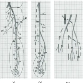
Figure 6: Schematic representation of the arterial pattern of the hind limb of the bearded capuchin. (a) General scheme. (b) Details of the circle in (a), that is, leg’s scheme. (c) Details of the circle in (b), that is, foot scheme. The dashed line indicates a position behind the plane and continuous line represents the plane level. The numbers indicate the names of arteries. 1 the aorta; 2 the common iliac; 3 the internal iliac; from 4 to 8 indicate the pelvic arteries not named in this work; 9 the external iliac; 10 the iliolumbar artery; 11 and 12 the obturator artery and its muscular branch; 13 the medial circumflex artery; 14 the external pudenda artery; 15 the femoral artery; 16 the inferior epigastric artery; 17 the superficial circumflex iliac artery; 18 the lateral circumflex femoral artery; 19 the profunda femoris artery; 20 the perforating branch; 21 the popliteal artery; 22 the fibular artery; 23 the common tibial artery; 24 the anterior tibial artery; 25 the posterior tibial artery; 26 the posterior saphenous branch; 27 the anterior saphenous branch; 28 the medium saphenous branch; 29 the medial plantar artery; 30 the first plantar metatarsal branch; 31 the lateral plantar artery; 32 the medial dorsal artery; 33 the first dorsal metatarsal artery; 34 the lateral dorsal artery. Human (a) Baboon (b) Bearded capuchin (c)
Figure 7: Schematic representation of the arterial pattern of the hind limb of the modern human and baboons, based on Swindler and Wood
handling tools. This is the case even for closely related species, for example, Cebus albifrons, C. olivaceus, C. capucinus, and Sapajus libidinosus have all been shown to engage in bipedal locomotion, but this behavior has only been observed in association with tool use in the Sapajus.
Probably, as small-bodied primates, all capuchin species would benefit from the higher surface-to-volume ratio in hind limb vessels to stay in a bipedal gait. Since only a few such species present prolonged bipedal posture during tool use, higher surface-to-volume may be a necessary but not sufficient condition for occasional bipedalism in capuchins. Moreover, the results presented here indicate that the bearded capuchin has no specific anatomical circulatory apparatus for this behavior, because it was not possible to analyze whether the Sapajus libidinosus has more arterial ramification than others studied primates or to verify the diameter of the arter-ies comparatively. Indeed, there are no data available in liter-ature that would specifically relate to this question.
5. Conclusions
Compared to all primates included in the present study, the arterial pattern observed in bearded capuchins is closest to the description of Cebus capucinus made by Manners-Smith
[19]. There are however a few differences regarding the origin,
trajectory, and branching of the tibial artery, which may
support the recent Cebus/Sapajus separation [5]. Bearded
capuchins also show a high degree of similarity to baboons, which is probably due to the presence of tails in both genera. As expected from the phylogeny, the degree of similarity to apes and humans is lower. Nevertheless, the pattern of superficial veins observed in bearded capuchins is not similar to any of the primates in comparison, namely, the short saphenous does not drain to popliteal but to extern iliac vein. The pattern of the deep veins, on the other hand, shows very few discrepancies among all studied primates. As a whole, the hind limb vessels observed in the bearded capuchin do not seem to have any fundamental influence on the intermittent bipedalism displayed by these primates in relation to other primates with similar proportions. Future studies should focus on comparative anatomical analysis among capuchin genera and other New World monkeys, apes, and monkeys to responds fundamental questions as the interesting behavior associated to Sapajus.
Acknowledgments
Roqueline A. G. M. F. Aversi-Ferreira was recipient of a doc-toral fellowship from CAPES/Brazil and Tales A. Aversi-Fer-reira received a research fellowship from National Council of Technology and Development (CNPq/Brazil).
References
[1] E. B. Ottoni and P. Izar, “Capuchin monkey tool use: overview and implications,” Evolutionary Anthropology, vol. 17, no. 4, pp. 171–178, 2008.
[2] B. Demes, “Three-dimensional kinematics of capuchin monkey bipedalism,” American Journal of Physical Anthropology, vol. 145, no. 1, pp. 147–155, 2011.
[3] E. Visalberghi, D. M. Fragaszy, P. Izar, E. B. Ottoni, P. C. Lee, and A. C. D. A. Moura, “Terrestriality and tool use,” Science, vol. 308, no. 5724, pp. 951–952, 2005.
[4] J. W. Lynch Alfaro, J. P. Boubli, L. E. Olson et al., “Explosive Pleistocene range expansion leads to widespread Amazonian sympatry between robust and gracile capuchin monkeys,”
Journal of Biogeography, vol. 39, no. 2, pp. 272–288, 2012.
[5] J. W. L. Alfaro, J. D. S. E. Silva Jr., and A. B. Rylands, “How differ-ent are robust and gracile capuchin monkeys? An argumdiffer-ent for the use of sapajus and cebus,” American Journal of Primatology, vol. 74, no. 4, pp. 273–286, 2012.
[6] T. A. Aversi-Ferreira, R. A. E. G. M. F. Aversi-Ferreira, Z. Silva, L. F. Gouvˆea-E-Silva, and N. Penha-Silva, “Anatomical study of the forearm deep muscles in Cebus apella (Linnaeus, 1766),”
Acta Scientiarum, vol. 27, no. 3, pp. 297–301, 2005.
[7] T. A. Aversi-Ferreira, M. S. L. Silva, J. P. Paula, L. F. G. Silva, and N. Penha-Silva, “Anatomia comparativa dos nervos do brac¸o de Cebus apella. Descric¸˜ao do m´usculo dorso epitroclear,” Acta
Scientiarum Biological Sciences, vol. 27, no. 3, pp. 291–296, 2005.
[8] T. A. Aversi-Ferreira, L. G. Vieira, R. M. Pires, Z. Silva, and N. Penha-Silva, “Estudo anatˆomico dos m´usculos flexores superfi-ciais do antebrac¸o no macaco Cebus apella,” Bioscience Journal, vol. 22, no. 1, pp. 139–144, 2006.
[9] T. A. Aversi-Ferreira, J. Pereira-de-Paula, M. D. S. Lima-e-Silva, and Z. Silva, “Anatomy of the arteries of the arm of Cebus
libidi-nosus (Rylands et al., 2000) monkeys,” Acta Scientiarum, vol. 29,
no. 3, pp. 247–254, 2007.
[10] T. A. Aversi-Ferreira, J. Pereira-de-Paula, Y. C. L. do Prado, M. S. Lima-e-Silva, and J. R. da Mata, “Anatomy of the shoulder and arm muscles of cebus libidinosus,” Brazilian Journal for
Morphological Sciences, vol. 24, no. 2, pp. 63–74, 2007.
[11] T. A. Aversi-Ferreira, P. J. Paula, M. S. L. Silva, Y. C. L. Prado, and Z. Silva, “Estudo anatˆomico das art´erias do ombro de Cebus
libidinosus (Rylands, 2000, Primates-Cebidae),” Ciˆencia Animal Brasileira, vol. 8, no. 2, pp. 273–284, 2007.
[12] T. A. Aversi-Ferreira, R. Diogo, J. M. Potau, G. Bello, J. F. Pastor, and M. A. Aziz, “Comparative anatomical study of the forearm extensor muscles of Cebus libidinosus (Rylands et al., 2000; primates, cebidae), modern humans, and other primates, with comments on primate evolution, phylogeny, and manipulatory behavior,” Anatomical Record, vol. 293, no. 12, pp. 2056–2070, 2010.
[13] R. A. G. M. F. Aversi-Ferreira, K. A. Marin, F. O. Carneiro e Silva, and T. A. Aversi-Ferreira, “Comparative anatomy of the thigh nerves of Cebus libidinosus (Rylands et al., 2000),” Pesquisa
Veterinaria Brasileira, vol. 31, no. 3, pp. 261–266, 2011.
[14] T. A. Aversi-Ferreira, R. S. Maior, F. O. Carneiro-e-Silva et al., “Comparative anatomical analyses of the forearm muscles of
Cebus libidinosus (Rylands et al. 2000): manipulatory behavior
and tool use,” PLoS ONE, vol. 6, no. 7, Article ID e22165, 2011. [15] T. Abreu, G. A. Pfrimer, R. A. G. M. F. Aversi-Ferreira, L. D.
Brand˜ao, H. Nishijo, and T. A. Aversi-Ferreira, “Comparative anatomical study of the leg’s nerves of Cebus (barbed capuchins) with baboons, chimpanzees and modern humans,” Pesquisa
Veterin´aria Brasileira, vol. 32, 1, pp. 113–117, 2012.
[16] M. Duarte, J. Hanna, E. Sanches, Q. Liu, and D. Fragaszy, “Kine-matics of bipedal locomotion while carrying a load in the arms in bearded capuchin monkeys (Sapajus libidinosus),” Journal of
[17] B. Demes and M. C. O’Neill, “Ground reaction forces and center of mass mechanics of bipedal capuchin monkeys: implications for the evolution of human bipedalism,” American Journal of
Physical Anthropology, vol. 150, no. 1, pp. 76–86, 2013.
[18] J. M. Brown, “The femoral artery in apes,” Journal of Anatomy
and Physiology, vol. 15, pp. 525–535, 1881.
[19] T. Manners-Smith, “The limb arteries of primates,” Journal of
Anatomy and Physiology, vol. 46, pp. 95–172, 1912.
[20] J. R. Mata, F. R. Mata, M. C. Souza, H. Nishijo, and T. A. Aversi-Ferreira, “Arrangement and prevalence of branches in the external carotid artery in humans,” Italian Journal of Anatomy
and Embryology, vol. 117, no. 2, pp. 65–74, 2012.
[21] J. C. Penereiro, “Some considerations about Galileo regarding the physics likeness, theory of strength materials and bending theory,” Caderno Brasileiro De Ensino De F´ısica, vol. 27, pp. 288– 312, 2010.
[22] T. Manners-Smith, “The limb arteries of primates,” Journal of
Anatomy and Physiology, vol. 45, pp. 23–64, 1910.
[23] S. Gibbs, Comparative soft tissue morphology of the extant
Homi-noidea, including man [Ph.D. thesis], The University of
Liver-pool, Inglaterra, England, 1999.
[24] D. R. Swindler and C. D. Wood, An Atlas of Primates Gross
Anat-omy: Baboon, Chimpanzee and Man, University of Washington
Press, Seattle, Wash, USA, 1973.
[25] Ł. Dyl and M. Topol, “The femoral artery and its branches in the baboon Papio anubis,” Folia Morphologica, vol. 66, no. 4, pp. 291–295, 2007.
[26] S. Standring, “Pelvis girdle and lower limb,” in Gray’s Anatomy:
The Anatomical basis of Clinical Practice, Churchill Livingstone,
London, UK, 2008.
[27] E. J. M. Meulman, C. M. Sanz, E. Visalberghi, and C. P. van Schaik, “The role of terrestriality in promoting primate technol-ogy,” Evolutionary Anthropology, vol. 21, no. 2, pp. 58–68, 2012. [28] D. M. Fragaszy, E. Visalberghi, and L. M. Fedigan, The Complete
Capuchin: The Biology of the Genus Cebus, Cambridge
Univer-sity Press, Cambridge, Mass, USA, 2004.
[29] Q. Liu, K. Simpson, P. Izar, E. Ottoni, E. Visalberghi, and D. Fragaszy, “Kinematics and energetics of nut-cracking in wild capuchin monkeys (Cebus libidinosus) in Piau´ı, Brazil,”
Ameri-can Journal of Physical Anthropology, vol. 138, no. 2, pp. 210–220,
Submit your manuscripts at
http://www.hindawi.com
Hindawi Publishing Corporation
http://www.hindawi.com Volume 2014
Anatomy
Research International
Peptides
Hindawi Publishing Corporation
http://www.hindawi.com Volume 2014
Hindawi Publishing Corporation http://www.hindawi.com
International Journal of
Volume 2014
Zoology
Hindawi Publishing Corporation
http://www.hindawi.com Volume 2014
Molecular Biology International
Genomics
International Journal of Hindawi Publishing Corporation
http://www.hindawi.com Volume 2014
The Scientific
World Journal
Hindawi Publishing Corporation
http://www.hindawi.com Volume 2014
Hindawi Publishing Corporation
http://www.hindawi.com Volume 2014
Bioinformatics
Advances inMarine Biology
Journal ofHindawi Publishing Corporation
http://www.hindawi.com Volume 2014
Hindawi Publishing Corporation
http://www.hindawi.com Volume 2014
Signal Transduction
Journal ofHindawi Publishing Corporation
http://www.hindawi.com Volume 2014
BioMed
Research International
Evolutionary Biology
International Journal of Hindawi Publishing Corporation
http://www.hindawi.com Volume 2014
Hindawi Publishing Corporation
http://www.hindawi.com Volume 2014 Biochemistry Research International
Archaea
Hindawi Publishing Corporation
http://www.hindawi.com Volume 2014
Hindawi Publishing Corporation
http://www.hindawi.com Volume 2014
Genetics
Research International
Hindawi Publishing Corporation
http://www.hindawi.com Volume 2014 Advances in
Virology
Hindawi Publishing Corporation http://www.hindawi.com
Nucleic Acids
Journal ofVolume 2014
Stem Cells
International
Hindawi Publishing Corporation
http://www.hindawi.com Volume 2014
Hindawi Publishing Corporation
http://www.hindawi.com Volume 2014
Enzyme
Research
Hindawi Publishing Corporation
http://www.hindawi.com Volume 2014
International Journal of