i
Antimicrobial silicones for use as dialysis
catheters
Ana Rita Noutel Lorga Gomes
Dissertation
Integrated Masters in Bioengineering
Molecular Biotechnology branch
Supervisor: Dr. Inês C. Gonçalves (i3s)
Co-supervisor: Dr. Artur M. Pinto (i3s/FEUP-LEPABE)
June 2017
ii
iii
This thesis was conducted at:
i3s – Instituto de Investigação e Inovação para a Saúde; Universidade do Porto
LEPABE/FEUP - Laboratório de Engenharia de Processos, Ambiente, Biotecnologia e Energia; Universidade do Porto
The work described in this thesis was financially supported by:
Project POCI-01-0145-FEDER-007274 (Institute for Research and Innovation in Health Sciences); Project POCI-01-0145-FEDER-006939 - Laboratory for Process Engineering, Environment, Biotechnology and Energy – LEPABE and NORTE‐01‐0145‐FEDER‐000005 – LEPABE-2-ECO-INNOVATION, funded by FEDER funds through COMPETE2020 - Programa Operacional Competitividade e Internacionalização (POCI) and Programa Operacional Regional do Norte (NORTE2020), and by national funds through FCT - Fundação para a Ciência e a Tecnologia; and Project PTDC/CTM-Bio/4033/2014 (NewCat - New biomaterials to prevent infection associated with dialysis catheters), funded by FEDER funds through COMPETE2020 - Programa Operacional Competitividade e Internacionalização (POCI) – and by national funds through FCT - Fundação para a Ciência e a Tecnologia.
v
Acknowledgements
Gostava de começar por agradecer a todos os meus amigos e colegas que partilharam comigo estes 5 anos, principalmente às “resistentes”, que estiveram sempre ao meu lado nos melhores e nos piores momentos.
Um enorme obrigada à minha orientadora, Dr. Inês Gonçalves, por toda a paciência, apoio e sobretudo disponibilidade. Só espero ter sido tão boa aluna quanto a Inês foi orientadora. Agradeço também ao meu co-orientador, Dr. Artur Pinto, por toda a ajuda e conselhos que me deu.
Muito obrigada ao André Maia, coordenador técnico da Biosciences Screening Unit do i3s, que tornou uma boa parte deste trabalho possível, mostrando sempre disponibilidade para me ajudar a melhorar tudo até ao ínfimo pormenor.
Agradeço a todo o grupo Bioengineered Surfaces que acompanhou o meu trabalho desde o início e me deu sugestões e incentivos para melhorar. Obrigada Cristina, Fabíola, Cláudia, Paula, Catarina, Mariana e Hélia.
À equipa do NewCat deixo um agradecimento especial. Ao Prof. Fernão Magalhães por me ajudar a tomar decisões, à Natacha pela companhia e apoio, à Patrícia por me tirar todas as dúvidas desde o início, à Andreia pelas grandes ajudas que me deu, e finalmente à Inês, a minha companheira dos trabalhos difíceis (e dos não tão difíceis também), um obrigada gigante.
Finalmente, gostava de agradecer ao Ricardo por toda a compreensão e toda a força que me dá, aos meus pais por terem estado incondicionalmente do meu lado não só nestes últimos tempos, mas sempre, e à minha irmã Marta que teve que me aturar todos os dias e que me inspira a dar o meu melhor.
vii
Abstract
Catheter-related infection is a severe issue, being one of the most prevalent types of infection in the hospital setting. Peritoneal dialysis catheters are no exception to this matter. These catheters are usually made of silicone, a polymer which is widely used in the biomedical field. Silicone, however, is hydrophobic and tends to attract bacteria to the surface. Some surface modification strategies are used to avoid bacterial colonization of silicone, but few have been used commercially in the production of catheters.
Graphene-based materials (GBMs) are considered antimicrobial, and can readily act and kill bacteria when immobilized in coatings. However, most of the work that combines GBMs and silicone is directed towards improving its physical characteristics, but not its microbicidal properties.
This work aims to investigate the antibacterial properties of GBM-coated silicone. Ideally these results would culminate in a more antibacterial catheter. Medical grade silicone and graphene nanoplatelets (GNPs) with 5 µm of diameter (GNP-M5) and its respective oxidized form (GNP-M5ox) were used for this purpose. The effect of GNP exposure and oxidation in the antibacterial activity of the coating was investigated. For this purpose, two distinct coating strategies – dip and spray coating – were tested to achieve the exposure of the GNPs at the surface. The GNPs were immobilized either with silicone as a binder, or coated with no binder.
The oxidation of GNP-M5 to GNP-M5ox was successfully performed. The presence of a binder (silicone) in coatings was found to be necessary to immobilize the GNPs on the surface and therefore only these samples were fully characterized. In general, spray coating exposed higher amounts of GNP at the surface and more homogeneously than dip coating, but the latter provided a better adhesion of the GNPs to the surface, despite not inducing changes in surface wettability. Spray coating with GNP-M5 produces a super hydrophobic surface while spraying with GNP-M5ox renders the surface more hydrophilic. Antibacterial testing of dip coating samples suggests that low amounts of GNP do not influence the number of adherent bacteria, but GNPs in the highest concentration tested present more bacteria attachment than control silicone samples. Nevertheless, while SR and SR/GNP-M5 coated samples have a percentage of dead bacteria between 40% and 50%, the samples coated with SR/GNP-M5ox increased bacterial death to 80%. Spray coating samples induced higher bacterial adhesion and less percentage of dead bacteria than dip coating samples. Nevertheless, coating with GNP-M5ox still resulted in higher percentage of dead bacteria, which was around 75% for the best condition.
viii
As such it was possible to conclude that the coatings with oxidized GNP induce more bacterial death, independently of the coating strategy. However, while for dip coating bacterial death is higher for increasing concentrations because more GNP is exposed, for spray coating this is not seen, possibly because GNP are more masked within the coating.
Globally, this work demonstrates the potential use of GNP coatings in silicone for application in peritoneal dialysis catheters with improved antimicrobial properties.
ix
Table of contents
Acknowledgements... v Abstract ... vii Table of contents ... ix List of figures... xiList of tables ... xiii
Abbreviations and Symbols ... xiv
CHAPTER I: Motivation and Aim... 1
1. Motivation and Aim ... 1
2. Structure of the Dissertation ... 2
CHAPTER II: Literature Review ... 3
1. Silicone: preparation, biomedical use and modification ... 3
1.1. Structure and preparation of silicone ... 3
1.2. Biomedical application of silicone ... 6
1.3. Silicone elastomer molding ... 7
1.4. Surface modification of silicone elastomers ... 9
1.4.1. Physical modification... 9
1.4.2. Chemical modification: Covalently attached coatings... 9
1.4.3. Chemical modification: Non-covalent coatings ... 10
2. Modification of silicone elastomers with graphene-based materials ... 11
2.1. Graphene-based materials (GBMs) ... 11
2.2. Silicone with GBMs ... 11
3. Graphene and its derivatives as antimicrobial materials ... 14
3.1. GBM antimicrobial properties ... 14
3.3. Antimicrobial activity of GBMs in coatings ... 15
3.3.1. Coatings with GBMs ... 15
3.3.2. GBMs coatings on silicone ... 16
4. Testing antimicrobial properties of biomaterial surfaces ... 17
4.1. Catheter-related infection ... 17
4.2. Types of antimicrobial materials ... 17
4.3. Antimicrobial surface testing ... 18
4.3.1. Testing on bacteria in suspension ... 18
4.3.2. Testing on bacteria adhered on surfaces ... 21
CHAPTER III: Materials and Methods ... 23
1. Materials Production ... 23
1.1. Graphene Nanoplatelets (GNP) ... 23
1.1.1.Graphene Nanoplatelets ... 23
1.1.2.Oxidation of Graphene Nanoplatelets ... 23
1.2. Silicone films ... 24
x
1.3.1.GNP coating with no binder ... 24
1.3.2.GNP coating with silicone as a binder ... 25
2. Materials Characterization ... 26
2.1. GNP characterization ... 26
2.1.1. X-ray Photoelectron Spectroscopy (XPS)... 26
2.1.2. Scanning electron microscopy (SEM) ... 27
2.2. GNP-coated silicone characterization ... 27
2.2.1. Optical microscopy ... 27
2.2.2. Rubbing test ... 27
2.2.3. Washing test ... 27
2.2.4. Scanning electron microscopy (SEM) ... 27
2.2.5. Water contact angle ... 27
3. Antibacterial Properties Testing ... 28
3.1. Bacteria and Growth Conditions ... 28
3.2. Antibacterial assessment of materials... 28
3.2.1.Testing in adherent bacteria ... 30
3.2.2.Testing in bacteria in suspension ... 30
4. Statistical analysis ... 31
4.1. Contact angles ... 31
4.2. Antibacterial assays ... 31
CHAPTER IV: Results and Discussion ... 33
1. Graphene nanoplatelets characterization ... 33
2. GNP-coated silicone ... 36
2.1. Silicone rubber films ... 36
2.2. GNP coating with no binder ... 36
2.2.1. Dip coating ... 36
2.2.2 Spray coating ... 37
3.1. GNP coating with binder and antimicrobial activity ... 38
3.1.1. Dip coating ... 38
3.2.2. Spray coating ... 46
CHAPTER V: Conclusion and Future Work ... 55
1. Conclusion ... 55
2. Future Work ... 56
References ... 57
xi
List of figures
Figure 1. Silicone elastomer processing by (A) extrusion, (B) compression molding, (C)
transfer molding, (D) injection molding.32,33 ...8
Figure 2. Potential routes of infection in catheters. ... 17
Figure 3. Different operating mechanisms of antibacterial surfaces. ... 18
Figure 4. Schematics of the contact method used on: (A) ISO 22196 and (B) ASTM E2180 standards. ... 19
Figure 5. Silicone film production. ... 24
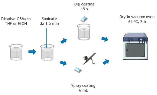
Figure 6. GNP/solvent dispersion application and material production. ... 25
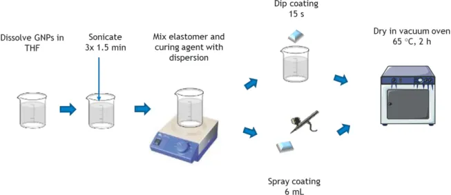
Figure 7. SR/GNP dispersion application and material production. ... 26
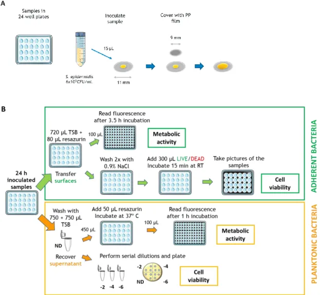
Figure 8. Schematics of the antibacterial assay. (A) Surface inoculation and PP film placement for forcing contact with the inoculum; (B) Testing after 24 h incubation. ... 29
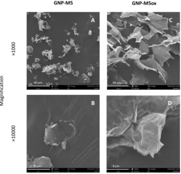
Figure 9. SEM images of the GNP-M5 powder and lyophilized GNP-M5ox. Pictures taken at the magnification of ×1000 and ×10000 (Scale bar = 80 µm and 8 µm, respectively). ... 34
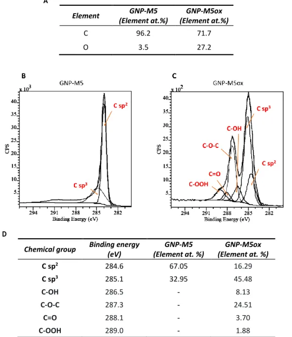
Figure 10. XPS analysis of GNP-M5 and GNP-M5ox. (A) Atomic percentage of carbon and oxygen obtained by analysis of the survey; (B) C1s high-resolution spectrum of GNP-M5; (C) C1s high-resolution spectrum of GNP-M5ox; (D) Contents of chemical groups resulting of C1s spectra fitting. ... 35
Figure 11. Silicone rubber film. (A) Stereomicroscopy image; (B) SEM image of front view; (C) SEM image of side view. SEM pictures taken at the magnification of ×300. (Scale bar= 200 µm). ... 36
Figure 12. Stereomicroscopy images of (A) silicone base film and (B) 1 mg/mL GNP-M5 in THF dip coating sample (Scale bar=5 mm). ... 36
Figure 13. Stereomicroscopy images of (A) silicone base film and (B) 1 mg/mL GNP-M5 in THF spray coating sample (Scale bar=5 mm). ... 37
Figure 14. Optical microscopy images of (A) silicone base film and (B) 1 mg/mL GNP-M5 in EtOH spray coating sample (Scale bar=1 mm). ... 37
Figure 15. Stereomicroscopy images of SR/GNP dip coating samples (Scale bar = 5mm). .. 38
Figure 16. Optical microscopy images of SR/GNP dip coating samples. Pictures taken at the magnification of ×100 and ×400 (Scale bar=1 mm and 200 µm, respectively). ... 39
Figure 17. SEM images of SR/GNP dip coating samples. Pictures taken at the magnifications of ×300, ×1000 and ×3000 (Scale bar= 200 µm, 80 µm and 20 µm, respectively). ... 41
xii
Figure 18. Water contact angle for SR/GNP dip coating samples. Statistical analysis performed by Kruskal Wallis test and statistically significant differences are indicated with * (p ≤ 0.05). ... 42 Figure 19. S.epidermidis adherent to SR/GNP dip coating samples after 24 h incubation. (A) Metabolic activity of bacteria in the surface after 3,5 hours incubation with resazurin; (B) Total adhered bacteria per mm2; (C) Percentage of live (green), dying
(orange) and dead (red) bacteria; (D) Representative images of the LIVE/DEAD staining (Scale bar=50 µm); (E) Images of bacteria adhered to SR and GNP aggregates (Scale bar=10 µm). Statistical analysis of the metabolic activity assay and total adhered bacteria performed with Kruskal-Wallis test and ordinary One-way ANOVA, respectively. Statistically significant differences are indicated with * (p ≤ 0.05)... 44 Figure 20. Planktonic S.epidermidis of SR/GNP dip coating samples after 24 h incubation. (A) Metabolic activity of bacteria in suspension after 1 hour incubation with resazurin and (B) colony forming units of the viable bacteria collected in supernatant. Statistical analysis of metabolic activity was performed with Kruskal-Wallis test and CFUs with ordinary One-way ANOVA. Statistically significant differences are indicated with * (p ≤ 0.05). ... 45 Figure 21. Stereomicroscopy images of SR/GNP spray coating samples (Scale bar = 5mm). ... 46 Figure 22. Optical microscopy images of SR/GNP spray coating samples. Pictures taken at the magnification of ×100 and ×400 (Scale bar=100 µm and 20 µm, respectively). ... 47 Figure 23. SEM images of SR/GNP spray coating samples. Pictures taken at the magnifications of ×300, ×1000 and ×3000 (Scale bar= 200 µm, 80 µm and 20 µm, respectively). ... 49 Figure 24. Water contact angle for SR/GNP spray coating samples. Statistical analysis performed by Kruskal Wallis test and statistically significant differences are indicated with * (p ≤ 0.05). ... 50 Figure 25. S.epidermidis adherent to SR/GNP spray coating samples after 24 h incubation. (A) Metabolic activity of bacteria in the surface after 3,5 hours incubation with resazurin; (B) Total adhered bacteria per mm2; (C) Percentage of live (green),
dying (orange) and dead (red) bacteria. (D) Representative images of the LIVE/DEAD staining (Scale bar=50 µm); (E) Images of bacteria adhered to SR and GNP aggregates. (Scale bar=10 µm) Statistical analysis of metabolic activity assay and total adhered bacteria performed with Kruskal-Wallis test and one-way ANOVA, respectively. Statistically significant differences are indicated with * (p ≤ 0.05). ... 52 Figure 26. Planktonic S.epidermidis of SR/GNP spray coating samples after 24 h incubation. (A) Metabolic activity of bacteria in suspension after 1 hour incubation with resazurin and (B) colony forming units of the viable bacteria collected in supernatant. Statistical analysis of metabolic activity was performed with Kruskal-Wallis test and CFUs with ordinary One-way ANOVA. Statistically significant differences are indicated with * (p ≤ 0.05). ... 53 Figure 27. Summary of obtained results. ... 54
xiii
List of tables
Table 1 – Summary of silicone synthesis reactions.3 ...3
Table 2 - Composition and polymer linking for each form of silicone. ...5
Table 3 - Biomedical applications of different forms of silicone. ...6
Table 4 - Silicone elastomer and GBMs composites: preparation and properties... 13
Table 5 – Abbreviatures used for referring to silicone binder materials. ... 26
Table 6 - Rubbing test for the dip coatings with no binder. Score of 0 is a clean rubber and 5 is a rubber presenting high GNP detachment. ... 37
Table 7 – Rubbing test for the spray coatings with no binder. Score of 0 is a clean rubber and 5 is a rubber presenting high GNP detachment. ... 38
Table 8 - Rubbing test for the SR/GNP dip coating samples. Score of 0 is a clean rubber and 5 is a rubber presenting high GNP detachment. ... 40
Table 9 – Rubbing test for the SR/GNP spray coating samples. Score of 0 is a clean rubber and 5 is a rubber presenting high GNP detachment. ... 48
xiv
Abbreviations and Symbols
CFUs Colony forming units CVD Carbon vapor deposition FLG Few-layer graphene
G Graphene
GBMs Graphene-based materials GNPs Graphene nanoplatelets GNP/EtOH GNP dispersion in ethanol
GNP/THF GNP dispersion in tetrahydrofuran GNP-M5 Non-oxidized graphene nanoplatelets GNP-M5ox Oxidized graphene nanoplatelets
GO Graphene oxide
GtO Graphite oxide
MHM Modified Hummers method
OD Optical density
PBS Phosphate-buffered saline PDMS Polydimethylsiloxane
PI Propidium iodide
PP Polypropylene
RFUs Relative fluorescence units rGO Reduced graphene oxide ROS Reactive oxygen species
RTV Room temperature vulcanization SEM Scanning Electron Microscopy
SR Silicone film coated with silicone in THF SRf Silicone rubber film
SR/GNP Silicone film coated with GNP and silicone in THF TCPET Tissue culture polyethyleneterephtalate
THF Tetrahydrofuran TSA Trypticase soy agar TSB Trypticase soy broth
1
CHAPTER I: Motivation and Aim
1. Motivation and Aim
Silicone is a synthetic polymer and the most used material for peritoneal dialysis catheters in the form of silicone elastomer. This happens because, in general, silicone is an inert material which combines absence of adverse biological reactions with good elastomeric properties. However, silicone catheters have elevated risk of infection as silicone is hydrophobic, making it likely to concentrate bacteria on the surface, facilitating bacterial adherence. Infection related to silicone catheter use in peritoneal dialysis is a significant issue, as it is the most common complication. For this reason, the quest for an infection resistant biomaterial is urgent.
To avoid infection, surface modification of polymers like silicone can be performed to enhance the antimicrobial properties of the original material. From surface oxidation to surface modification with other materials, numerous strategies have been attempted but few have been successful when the modified silicone catheter is tested in the hospital environment.
Graphene has received a great deal of attention due to its excellent mechanical, optical and electrical properties. Recently, graphene and graphene-based materials (GBMs) have been used in other applications like drug delivery and functionalization of other nanomaterials to convey antimicrobial properties. However, GBMs by themselves are generally considered intrinsically antimicrobial. Moreover, their biocompatibility has also been characterized and for low concentrations the toxicity is negligible. For these reasons, GBMs appear as an attractive choice for the development of a more effective antibacterial silicone.
This work focused on exploiting the antimicrobial activity of GBMs, namely graphene nanoplatelets (GNP), as a coating for a potential silicone catheter. GNP with 5 µm in diameter (GNP-M5) and its oxidized form (GNP-M5ox) were used to investigate the influence of the oxidation in the antibacterial properties. Each form of each GNP was produced, characterized and tested for antimicrobial properties. Two different coating strategies, dip and spray coating, were tested in terms of GNP exposure. The influence of the exposure of GNP was also subject of surface antibacterial activity evaluation.
2
2. Structure of the Dissertation
The present dissertation is organized into four chapters. Chapter I includes the motivation and aim of the dissertation.
Chapter II – Literature Review begins with summarizing aspects related to silicone as a polymer in research and medical field, including its general properties, manufacturing and the modification of the surface of silicone. This is followed by a section dedicated to graphene and graphene-based materials (GBMs) as antimicrobial agents and a review of the literature on silicone modified with GBMs for various purposes. This chapter closes with a section describing techniques to evaluate antimicrobial properties of surfaces.
Chapter III – Materials and Methods covers the materials and the procedures used to perform the work present in this dissertation. The production of oxidized graphene nanoplatelets (GNP) is described in detail. The production of two different approaches for applying GNP dispersion coatings on silicone is also described: using no binder or using silicone as a binder. The dispersions were applied dip coating or spray coating. The presence, distribution and orientation of the GNP on the surface of the samples was assessed. Samples which presented GNP as free, non-bound material on the surface were discarded for further characterization. Samples with good GNP immobilization were used to perform antibacterial testing. Surfaces and supernatants were tested in terms of the viability and metabolic activity of the bacteria. Chapter IV – Results and Discussion compiles the results obtained with the tests mentioned in Chapter III, and the discussion of the latter. The chapter is divided concerning each type of material used as a binder of the GNPs. Regarding the silicone binder surfaces, the antibacterial test results are presented and discussed.
The last chapter, Chapter V – Conclusions summarizes the main conclusions of the work, and reflects on future work.
3
CHAPTER II: Literature Review
1. Silicone: preparation, biomedical use and
modification
1.1. Structure and preparation of silicone
Silicones are a group of synthetic polymers whose backbone is constituted of silicon (Si) and oxygen (O) bonds. This basic unit of the polymer is known as siloxane. The element silicon of the backbone also forms bonds with two organic groups, namely methyl, vinyl and phenyl groups.1
Because of the inherent flexibility of the O-Si-O bond, silicone polymer-based materials have good flexibility and softness, even when at very low or high temperatures.1
Depending on the manufacturing process, silicone can possess a high tensile area, good resilience and up to 1250% of elongation.2
The synthesis of silicone polymers usually comprises four steps: i) silica reduction to silicon, ii) chlorosilane synthesis, iii) chlorosilane hydrolysis, and iv) polymerization and polycondensation.3 These steps and the associated chemical reactions are depicted in
Table 1.
4
i) Silica reduction to silicon. As silica (SiO2) is the natural source of silicon, it needs
to be reduced to the elemental silicon (Si) by the carbothermal method.4 This process
requires great amounts of heat developed by a strong electric current.5
ii) Chlorosilane synthesis. Silicon reacts with a chlorinated organic compound at an
elevated temperature.4
iii) Chlorosilane hydrolysis. Hydrolyzing the chlorosilane leads to the production of
a mixture of cyclic and linear oligosiloxanes, and hydrochloric acid.4 These oligomers
possess a chain that is too short for most applications, and therefore need to be polymerized or condensed into a chain of sufficient length.3
iv) Polymerization and polycondensation. Cyclic organosilanes must be ring-opened
and polymerized with help of an acid or basic catalyzer. Linear organosilanes can be combined when catalyzed by acids and bases by condensation of silanol terminals.3
Silicone polymers can form a three-dimensional network by a crosslinking reaction, which leads to the formation of bonds between polymer chains.3 This process is also known
as curing or vulcanization. The crosslinking reaction is used to produce silicone gels, elastomers, and resins from silicone polymers.6 Three types of crosslinking are usually
performed: using radicals, by condensation, or by addition.
Peroxide radicals are used to crosslink elastomers like silicone and low-density polyethylene which cannot be crosslinked with common curing agents.7 The temperature
used is high, the crosslinking time is short, and the process results in high consistency glossy silicone rubbers.8 However, the volatile residues must be removed post curing to
avoid depolymerization.3
There are two types of crosslinking by condensation. In the first type of condensation, the polymer starts crosslinking with contact with moisture, commonly from humidity in the air.9 These materials are named one-part RTV (room temperature
vulcanization). The second type of condensation reaction relies on the mixing of two components. An organotin salt is used as catalyst and alcohol is released as a by-product.3
Because mixing of two components, a polymer and an silane, is necessary, these are called two-part systems.
Crosslinking by addition is achieved by adding Si-H groups where vinyl groups are present. The addition is catalyzed by Pt or Rh complexes.10 Platinum cure systems are
quickly cured by heat but can be cured at room-temperature. This type of crosslinking also requires the mixture of two parts, and is therefore categorized as a two-part system.3
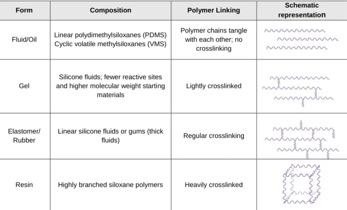
Depending on the functional group attached to the silicon atom and the degree of crosslinking of the polymer chains, silicones may be categorized into fluids (also known as oils) – the only form of silicone which does not crosslink – gels, elastomers (also known as rubbers) or resins (Table 2). 11
5
Table 2 - Composition and polymer linking for each form of silicone.
Furthermore, in most silicone elastomers, fillers are added to reinforce crosslinking thus enhancing the mechanical strength of the material. The most satisfactory silicone elastomer reinforcement is achieved by using silica fillers.8,12 Another common type of
filler is carbon black, although it is often used in low-resistivity silicones due to a lower resistivity and thermal stability relatively to silica fillers.13
Additionally, carbon nanotubes and graphene-based materials are also being studied as an alternative to commercial fillers.14 Albeit their similar chemical properties, they
possess different morphologies. Multiwalled carbon nanotubes are difficult to disperse in silicone rubber because of their cylindrical shape, which renders the use of 2D-graphene-based materials a more attractive alternative. The production of silicone and GBM composites will be discussed in section 2.
Form Composition Polymer Linking Schematic
representation
Fluid/Oil Linear polydimethylsiloxanes (PDMS) Cyclic volatile methylsiloxanes (VMS)
Polymer chains tangle with each other; no
crosslinking
Gel
Silicone fluids; fewer reactive sites and higher molecular weight starting
materials
Lightly crosslinked
Elastomer/ Rubber
Linear silicone fluids or gums (thick
fluids) Regular crosslinking
6
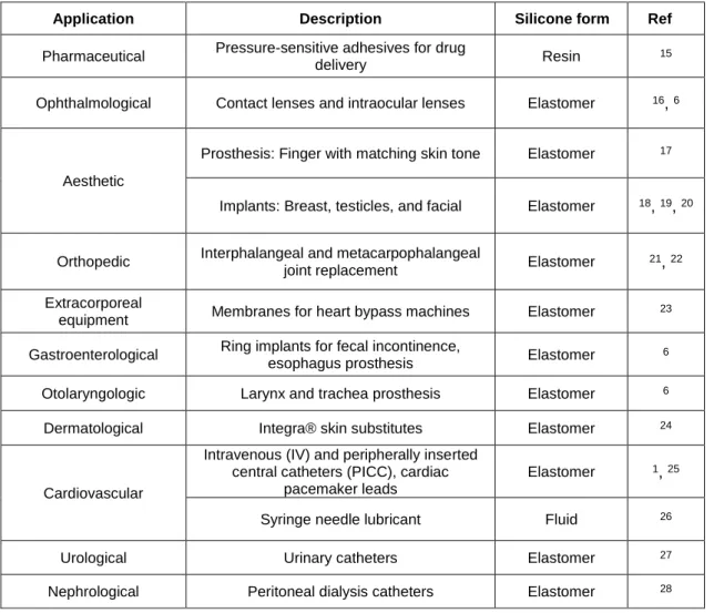
1.2. Biomedical application of silicone
Although hydrophobic, silicone is generally both biocompatible and biodurable, hence the approval of silicone-based gels, elastomers, fluids, and adhesives for medical use by several international agencies.3 This is related to the inertness of the material, and
this implies a low interaction with cells and chemicals if inside the body.6
Moreover, silicone is also permeable to gases like CO2 and Oxygen,2 which is
advantageous for some applications such as contact lenses. In fact, silicones are considered rather unique materials and attractive for medical use, mainly due to the combination of absence of adverse biological reactions and good elastomeric properties.2
Silicones used in medical practice appear in various forms like resin, elastomer, or fluid, which makes various applications possible (Table 3).
Table 3 - Biomedical applications of different forms of silicone.
Application Description Silicone form Ref
Pharmaceutical Pressure-sensitive adhesives for drug
delivery Resin 15
Ophthalmological Contact lenses and intraocular lenses Elastomer 16, 6
Aesthetic
Prosthesis: Finger with matching skin tone Elastomer 17
Implants: Breast, testicles, and facial Elastomer 18, 19, 20
Orthopedic Interphalangeal and metacarpophalangeal
joint replacement Elastomer 21, 22 Extracorporeal
equipment Membranes for heart bypass machines Elastomer 23
Gastroenterological Ring implants for fecal incontinence,
esophagus prosthesis Elastomer 6
Otolaryngologic Larynx and trachea prosthesis Elastomer 6
Dermatological Integra® skin substitutes Elastomer 24
Cardiovascular
Intravenous (IV) and peripherally inserted central catheters (PICC), cardiac
pacemaker leads
Elastomer 1, 25
Syringe needle lubricant Fluid 26
Urological Urinary catheters Elastomer 27
7
The most common silicone used in medical applications is polydimethylsiloxane (PDMS) where the organic groups bound to silicon are two methyl groups.3 Commercial
medical-grade PDMS exists in the forms of fluid, gel and elastomer.6
One of the main uses of silicone is in catheter production. A catheter is defined as a tubular device designed for insertion into vessels or cavities, to permit the withdrawal or the injection of fluids or other substances.8
Silicone has been a main material used for production of various types of central venous access IV catheters (CVC),1,29 peripherally inserted central venous catheters
(PICC),1 peritoneal dialysis catheters,30 and urinary catheters.27 Because of its resistance to
chemicals and flexibility, silicone has been considered the standard material for long-term access.30 However, for short term use some non-tunneled and non-cuffed silicone acute
dialysis catheters are available.28
Silicone as an elastomer appears as the most common form used in biomedical applications. For this reason, this thesis will focus mainly on silicone elastomer use.
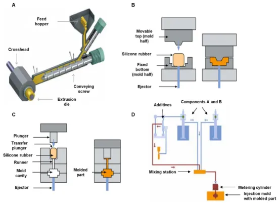
1.3. Silicone elastomer molding
As reviewed in section 1.1., silicone elastomers are obtained by crosslinking of low molecular weight, low viscosity silicone polymers.31 However, for certain applications,
silicone elastomers undergo a molding process before curing, which is used to produce solid elastomers with a pre-determined shape. The elastomers which undergo molding processes are cured by peroxide curing or addition curing, and both processes may be accelerated by heat.32
There are four main processing methods used with silicone elastomers: extrusion, compression molding, injection molding, and transfer molding (Figure 1). The extrusion process is used with solid silicone elastomer and the molding process is performed either with solid or liquid silicone rubber.32
8
Figure 1. Silicone elastomer processing by (A) extrusion, (B) compression molding, (C) transfer molding, (D) injection molding.32,33
Extrusion of silicone is a process in which the material is squeezed through a die with the help of a conveying screw and is subsequently cured by heat. This process can be used for producing tubes or cables.32
Compression and transfer molding are both press molding processes. In compression molding, a preform (rough piece of uncured elastomer) is placed on one half of a heated mold.33 When the mold is closed, the compressed rubber spreads into the entire cavity. In
transfer molding, the uncured rubber is placed in a chamber on top of the mold and placed in a press. The pressure forces the rubber to flow through an open end on the pot and spreading into the heated mold. This is useful when avoiding air trapping is crucial.33
In injection molding, the liquid or solid silicone elastomer components are pumped into a mixer which homogenizes uncured rubber before being forced through a nozzle into a heated, closed mold. This automated process is characterized by its accuracy and the production of high quality parts.32
9
1.4. Surface modification of silicone elastomers
1.4.1. Physical modification
The surface of silicone can be modified by physical techniques which generate high energy species or deposit atomic clusters at the surface.34 Some examples are plasma
treatments, UV-irradiation, laser treatments, corona treatments, and ion-beam implantation.
Plasma treatments have been used to increase surface wettability, although some hydrophobicity is recovered as cracks in the treated layer of the surface normally occur.35
Different gases such as O2, N2, NH3 and Ar are used, and different excitation frequencies
produce different results.36
UV-irradiation in ambient setting leads to the formation of ozone (UV-light in combination with oxygen) and the development of a silicon oxide layer on the surface.37
Silicon oxide films are resistant to oxygen and water, rendering them attractive to use as coatings.38
Laser technology also uses UV frequencies, but with the aim of producing local transformations or patterned surfaces.39
Corona treatments use electrically induced ionized air to bombard the surface of polymers.40 It is used to induce oxidation and to improve the overall adhesion properties of
silicone.37
Ion-beam implantation on silicone has been considered a breakthrough method in overcoming the usual surface properties of silicone, by improving hydrophilicity and making the surface able to resist biodeposition in long-term use medical devices.41 In fact
the work of Yoshihaki, et al42 showed that silicone implantation with O
2+ ions improved
antithrombogenicity.
1.4.2. Chemical modification: Covalently attached coatings
Chemical modification of silicone surface includes modifications by chemical reaction by wet treatment, and covalent bonding of macromolecular chains to the surface, also known as grafting,34 which produces covalently-attached coatings. Out of the two
types, grafting is the most commonly used to modify the surface of silicone.25 Grafting is a
process based on the activation of the silicone surface by a physical modification technique, such as plasma or ozone treatments. After this step, other molecules can be introduced on the surface with the formation of covalent bonds.
Radiation grafting and photografting have been used to introduce chemically reactive groups onto the surface of hydrophobic, inert polymers like silicone.43 Radiation
grafting in particular has been used for various compounds with different applications such as N-vinylpyrrolidone for increasing hemocompatibility,44 acrylamide for anti-inflammatory
10
delivery,45 or n-vinylimidazole for antimicrobial properties.46 This technique has also been
used to produce a patented heparin-grafted silicone surface with anticoagulant properties.47
Plasma-induced grafting has been used with PEGMA to avoid bacterial adhesion.48
The grafting of an allyl glycidyl ether (AGE) polymer brush by this method has also been applied to attach antimicrobial peptides (AMPs) on the surface of silicone to achieve antimicrobial properties.49
Laser induced graft polymerization has been used with pulsed lasers to create concentrated radical areas to effectively graft HEMA on the surface of PDMS to improve the hydrophilicity of the polymer.25
Ozonization, or ozone-induced grafting is a technique which has been used by Xu, et al50 for grafting 2-methacryloyloxyethyl phosphorylcholine (MPC) improving
hemocompatibility by avoiding platelet adhesion.
1.4.3. Chemical modification: Non-covalent coatings
The production of a thin polymer film on planar surfaces by techniques such as dip coating, spray coating and spin coating has received a great amount of attention recently, owing to the observation of unique properties of thin films compared to bulk materials.51
However, silicone, due to its relative inertness, is incompatible with most adhesives and coatings, excluding the ones already composed of silicone adhesive and coatings .52
Coating with quaternary ammonium salts (QAS), used to produce antimicrobial surfaces in polymers like silicone,53 can be achieved by spin coating of a solution
containing QAS in methanol blended with silicone and the catalyst.54 The use of silver
alloys in thin film coatings with liquid silicone rubber for conveying antimicrobial properties to catheters has also been described, although no significant decrease of infections was observed.55 Dopamine has also been used to produce an antimicrobial
coating based on a polydopamine solution (PDA) and silver nanoparticles (AgNP) on the luminal and external surfaces of silicone catheters,56 without the need of activating the
surface or using silicone in the coating solution. The preparation of the coating in alternate layers of PDA and AgNP ensured a controlled release of the latter.
11
2. Modification of silicone elastomers with
graphene-based materials
2.1. Graphene-based materials (GBMs)
Graphene (G) can be defined as a two-dimensional, single-atom plane of carbon.57
Graphene can be synthetized by top-down and bottom-up processes.58 Top-down methods
include several processes used to exfoliate graphite, namely mechanical, liquid phase, and thermal exfoliation,59 and also by the reduction of graphene oxide (GO).60 Bottom-up
processes, like chemical vapor deposition (CVD), epitaxial growth, and arc discharge use a source of elemental carbon to convert the latter into graphene and graphene-based materials (GBMs).59
Other GBMs used in research include graphene oxide (GO), reduced graphene oxide (rGO) and graphene nanoplatelets (GNP). Graphene oxide (GO) consists of a highly-oxidized form of the original graphene. Graphene oxide, and also graphite oxide (GtO), are usually oxidized by Hummers method.61,62 GO bears various oxygenated groups, namely hydroxyl
and epoxy groups in the basal plane, while at the edges carbonyl and carboxyl groups are present.62 Because of this strong oxygenation GO is considered hydrophilic and can be
easily dispersed in water and further functionalized.63 Graphene nanoplatelets (GNP), also
sometimes named “few-layer graphene” (FLG), are constituted by stacked graphene sheets with a thickness of 2 to 10 layers.64 Generally, GNP are 5 to 25 nm thick and can have 0.5
to 25 µm in diameter.65
The varied applications of graphene resulting of its mechanical, optical, electrical, and magnetic properties66 have led to an increasing development of promising research in
the field of Materials Science namely to produce sensors,67 or packaging materials.68
Moreover, graphene and GBMs have been described as having bactericidal action, a property which made them an attractive choice for designing antimicrobial materials.
2.2. Silicone with GBMs
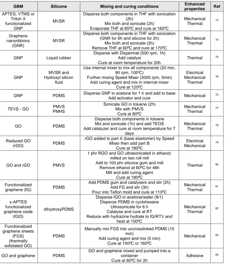
Most research work performed combining silicone elastomers and GBMs focuses on the production of composite materials intending to achieve a reinforcement of mechanical, thermal, electrical, or adhesive properties of the base silicone material, either with graphene nanoplatelets (GNP), graphene oxide (GO) or graphene. As Table 4 shows, GBMs incorporation in silicone elastomer is commonly achieved by dispersing both silicone and the GBMs independently by sonication in a solvent, such as tetrahydrofuran (THF) or toluene, followed by mixing the GBM dispersion to the base elastomer or the recently cured elastomer. The mixture must be heated to evaporate the solvents before curing.
12
Other strategies avoid the use of solvents and consist on adding the GBM in liquid rubber or to the base elastomer using a high-speed mixer before curing. The use of silanes for functionalization of GBMs is generally employed to enhance their dispersion in the polymer matrix by changing the physical and chemical properties of the surface of these materials.69
Although considerable research has been developed in reinforcing the mechanical properties of silicone with GBMs, little work has been done combining both to achieve a suitable material for implants or medical devices. Graphene nanoplatelets have been combined with silicone in heart valve prosthetics to produce a more mechanically fail-resistant silicone material. The incorporation did not significantly change hemocompatibility or induce cytotoxicity.70
Overall, the addition of GBMs to silicone rubber results in the reinforcement of mechanical properties. This may be considered a negative outcome if these materials are employed to develop a novel dialysis catheter, as peritoneal dialysis catheters must be soft and flexible.30 For this reason, a solution based on surface modification of silicone with
GBMs might be an alternative approach to this issue. For instance Lin, et al71, deposited a
single layer of graphene by transfer coating on different substrates, including PDMS with two degrees of increasing stiffness. All substrates coated showed good cytocompatibility, although the stiffer the substrate, the strongest was the adhesion and the proliferation of fibroblasts. Min, et al72 also used rGO in an ethanol dispersion for spray coating in PDMS
electrodes as a substitute for carbon paste, which yielded much more flexible electrodes. This work suggests GBM coatings do not alter the physical properties of the base PDMS material.
13
Table 4 - Silicone elastomer and GBMs composites: preparation and properties.
APTES: (3-Aminopropyl)triethoxysilane; MVSR: Methyl-vinyl silicone rubber; PDMS: Polydimethylsiloxane; PMVS: Polymethylvinylsiloxane; PMHS: Polymethylhydrosiloxane. TEVS: Triethoxyvinylsilane; VTMS: Vinyltrimethoxysilane.
GBM Silicone Mixing and curing conditions Enhanced
properties Ref APTES, VTMS or Triton X functionalized GNP MVSR
Disperse both components in THF with sonication (2h)
Mix both and sonicate (2h) Evaporate THF at 65ºC and cure at 165ºC
Mechanical Thermal 73 Graphene nanoribbons (GNR) MVSR
Disperse both components in THF with sonication (GNR for 6h and silicone for 2h)
Mix both and sonicate (2h) Remove THF at 60ºC and cure at 170ºC
Mechanical Thermal
74
GNP Liquid rubber
Disperse with Dispermat (500 rpm, 1h) Add catalyst
Cure at room temperature for 20h
Thermal 75
GNP
MVSR and Hydroxyl silicon
fluid
Use internal mixer to mix all components (20 min, 90 rpm, 105ºC)
Further mixing Speed Mixer (3000 rpm, 5min) Add curing agent and mix in internal mixer
Cure at 120ºC
Electrical Mechanical
Thermal
76
GNP PDMS Disperse GNP in acetone for 1 h and add to base
Add activator and cure Mechanical
70 TEVS - GO PMVS PMHS Sonicate GO in toluene (2h) Mix with PMVS Cure at 80ºC Mechanical Thermal 77 GO PDMS
Disperse both components in toluene Mix and sonicate (1h) and add TEOS Add catalyzer and cure at room temperature for 7
days Mechanical Thermal 78 Reduced GO (rGO) PDMS
rGO added to part A (base elastomer) by Speed Mixer then add part B
Cure at 180ºC
Electrical Mechanical
79
GO and rGO PMVS
1 phr RGO and GO ultrasonicated in ethanol; milled on two roll mill
Add to 100 phr silicone gum and mill Remove ethanol at 60ºC for 48h
Mill and add curing agent Cure at 180ºC
Thermal 80
Functionalized
graphene (fG) PDMS
Add PDMS gum and catalysers and stir (2h) Add FG and stir (3h)
Pour into Teflon mold and cure at 110ºC
Mechanical Thermal 81 γ-APTES functionalized graphene oxide (fGO) dihydroxyPDMS
Disperse fGO in acetone/water (9/1) Disperse PDMS in cyclohexane
Ultrasonicate for 6 h Catalyse and cure at RT
Reduce with hydrazine hydrate to fG/RTV and heat at 100ºC Mechanical Thermal 82 Functionalized graphene sheets (FGS) (thermally exfoliated GO) PDMS
Manually mix FGS into uncrosslinked PDMS (15 min)
Add curing agent and mix (5 min) Cure at 150ºC or 160ºC
Mechanical 83
GO and graphene PDMS
GO and graphene mixed and pumped into a container
Cure at 80ºC for 2h
14
3. Graphene and its derivatives as antimicrobial
materials
3.1. GBM antimicrobial properties
Intrinsic physicochemical properties of the GBMs such as state of dispersion, size, shape, and layer number are possible factors influencing their antimicrobial activity. Furthermore, the mechanisms by which graphene based materials exert their antibacterial action is also controversial.
It is not surprising that the activity of GBMs highly depends on disaggregation of the materials.85 As they possess high surface energies, their separation is vital to maintain
shape and high surface area. Concerning size, the results are conflicting. Different results are reported especially when comparing GBMs in solution and in coatings. When in solution, larger graphene and GO sheets appear to entrap bacteria, causing the decrease in viability.86 This is the so-called membrane-wrapping mechanism. However, it is likely that
this process is reversible, as live bacteria were found inside aggregated sheets of graphene after sonication.87 But when present in coatings, small-sized sheets are more effective and
pierce the cell membrane on contact86. This blade-like action of sharp edges present on
GBMs is one of the most agreed mechanisms which cause bacterial death. This happens due to the leakage of intracellular materials and cell death.85 However, when present in thin
films, the edges may merge together and this effect is limited.88 Regarding the number of
layers, typically thinner, few-layered GBMs are easier to disperse and readily act as a membrane-piercing structure.85
The production of ROS and death by oxidative stress is also viewed as a favorable mechanism although no consensus has been achieved among the scientific community. The presence of O2 in oxidized GBMs and the introduction of these groups in the bacteria
generate ROS and subsequent lipid peroxidation, mitochondrial dysfunction and protein inactivation.85 Additionally, bacteria are capable of reducing the oxygen species present in
the materials and passively contributing to their own death; this is called the self-killing effect.85 However, other groups support the theory of oxidative stress induced by electron
transfer rather than by ROS production.88
Other conditions unrelated to the materials, such as the presence of analytes, ions and different pH in the solution, and the microbial strain used is also believed to influence the outcome of the activity of GBMs on bacteria.85 The thickness of the bacterial wall
(Gram-positive or Gram-negative) and the cell shape (coccus or bacillus) may affect the sensibility of one strain of bacteria more than another. Typically, Gram-positive bacteria react differently to the same material when compared to Gram-negative bacteria. The use
15
of GBMs in nanocomposites as a way of improving their antibacterial action has been extensively explored using a range of materials, including metals, metal oxides and polymers, as described in various reviews.87,89–91 These nanocomposites have been tested in
solution, films, composite matrices and coatings.
3.3. Antimicrobial activity of GBMs in coatings
Both GBM and GBM nanocomposites have been used in coatings for various materials for electrical, sensing or protective purposes, using diverse coating methods.60
The antimicrobial activity attributed to GBMs has also been explored in coatings.
As reviewed in the previous sections, small, few-layer GBMs tend to work best in coatings. Ideally, their sharp edges should be exposed. However, other factors are important when considering developing antibacterial coatings. One of the factors is hydrophobicity. Bacteria tend to attach to hydrophobic surfaces which implies more adhesion to a surface with graphene and less adhesion to amphiphilic GO-coated surfaces.86 Another factor is the roughness of the coated surface, as an increasing in
roughness attracts more bacteria. This can be positive if the surface is bactericidal but undesirable if the surface is designed to avoid bacterial adhesion.86
Ultimately, with all varying features and mechanisms, it is crucial to pick the best GBM for a specific application. The following section reviews work performed on GBMs used in coatings.
3.3.1. Coatings with GBMs
GBMs can be coated on surfaces by themselves deposited as a thin film. CVD-deposited graphene (G) on conductive Cu and Ge substrates and isolating SiO2 substrates
produced different results on antibacterial properties.88 While G-Cu and G-Ge induced
membrane damage and hindered E. coli and S. aureus proliferation, G-SiO2 did not.
Because the latter is isolating, the theory of charge transfer was proposed as the main disruptor of the bacterial membrane. However, other work performed by Parra, et al92
discredited the theory of charge transfer, as a single-layer graphene conductive layer coated over a Cu substrate suppressed charge interaction with bacteria, contrarily to what was detected on the uncoated Cu substrate and similarly to what was seen on an isolating hexagonal-boron nitride coating. Titanium-niobium (Ti-Nb) alloy coated with graphene oxide (GO) by dip coating also produced antibacterial action against E. coli.93 The authors
discarded the charge transfer theory and proposed bacterial reduction of GO as the main killing mechanism. Still concerning GO, Perreault, et al94 proposed that when present in
16
planes is enhanced. The importance of oxidative stress was also denoted, although the differences in coating and in suspension still remain unclarified.
Various work has been performed using polymers to embed GBMs in membranes and coatings. Generally, as the GBM content increases, the antibacterial activity is enhanced.86
Santos, et al developed a coating for metal surfaces based on GBMs and poly(N-vinylcarbazole) (PVK).95,96 The authors propose a synergistic antibacterial effect of PVK
possibly derived from the better state of dispersion of graphene, and morphological and electronic modifications due to interaction with the polymer. PVK-GO induced 90% more bacterial death and PVK-G inhibited 80% of biofilm formation. GO-sheets embedded in an alkyd resin also inhibited E. coli, P. aeruginosa, and S. aureus viability for at least 69% after 24 h and 85% after 48 h of contact.97
Because of the successful immobilization of GBMs in coatings, nanocomposites with GBMs also began to be tested with the same purpose. GO and gelatin-functionalized GO deposited on nitinol inhibited E. coli growth, with the membrane integrity being successfully documented in SEM images.98 The combination of GBMs with materials which
are already antibacterial is a common way of achieving the enhancement of these properties. Graphene conjugated with TiO2 nanoparticles deposited on cotton fabric
improved the antibacterial activity of the nanoparticles, possibly by increasing contact with the bacteria due to its high surface area.99 GO combined with antibacterial silver
nanoparticles (AgNPs) lead to an inactivation of E.coli and damaging of its cellular membrane after contact for 2 h.100 Silver/hydroxyapatite/graphene composite coatings
electrodeposited on titanium reduced bacterial growth of S. aureus and E. coli after only 3 h of exposure.101
3.3.2. GBMs coatings on silicone
To this date, very few research has been published related to work on the combination of silicone and graphene towards an antimicrobial material. Nonetheless, one article published by Correa, et al102 reports the successful production of an antimicrobial
PDMS containing two fillers: titanium dioxide and/or graphene oxide. GO and TiO2 were
added to the silicone by dripping a solution of absolute ethanol on the top of the material, producing a coating, or by homogenizing both silicone and solution by stirring. Although PDMS/GO showed one of the two best results for antimicrobial and antifungal activity, the authors did not clarify which method of GO exposure produced the best outcome.
17
4. Testing antimicrobial properties of biomaterial
surfaces
4.1. Catheter-related infection
Infection related to medical devices is a relevant issue, accounting for 25,6% of healthcare associated infections in 2011.103 Dialysis catheter-related infection in particular
is related to as much as 26% of these cases.104 The main routes of infection for catheters
are depicted in Figure 2.
Because of its inherent hydrophobicity, PDMS-based devices (including catheters) have the disadvantage of having low wettability and biofouling due to non-specific analyte adsorption and bacterial adhesion.105
Figure 2. Potential routes of infection in catheters.
The formation of a biofilm on a device or an implant is also a relevant event which culminates in the development of infection.106 A biofilm can be defined as an aggregate of
a single or multiple microbial species attached to a surface.107 The formation of a biofilm
in intravenous or urinary catheters is no exception.55,108
Several approaches have been developed to avoid bacterial attachment and proliferation on biomaterial surfaces in order to lower the risk of infection.109 These
strategies are briefly described in the following section.
4.2. Types of antimicrobial materials
Antibacterial surfaces can be divided into categories depending on their operating mechanism: bactericidal surfaces for killing bacteria, bacteria-resistant surfaces for avoiding attachment, and bacteria-release surfaces for reducing adhesion and enabling the
18
release of the bacteria already attached by an external force.110 The different categories
are depicted in Figure 3.
Figure 3. Different operating mechanisms of antibacterial surfaces.
However, depending on the application, the antibacterial surface requirements are different for distinct types of biomaterials.111 A few antimicrobial strategies based on
surface modification of silicone - either by rendering the surface more hydrophilic or by means of a coating - have been described in a previous section.
Nonetheless, it is vital to make use of techniques which evaluate the antimicrobial properties of all developed materials to validate their further use in clinical trials. The following section will review the strategies currently used to test these properties in biomaterial surfaces.
4.3. Antimicrobial surface testing
4.3.1. Testing on bacteria in suspension
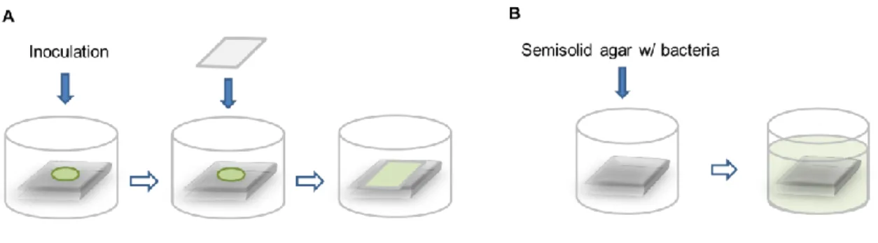
4.3.1.1. Standardized testingISO 22196 is a standard procedure for the measurement of antimicrobial activity on the surface of plastics and non-porous materials.112 The procedure starts with the
preparation of pre-culture using predefined bacterial strains and preparation of the specimens. The surfaces to test, both treated and untreated and with no more than 10 mm of thickness, are cut into 50 mm x 50 mm specimens and subsequently cleaned. Testing should be performed on no less than three specimens for each material tested. The test material surfaces are prepared on Petri dishes and later inoculated with 0,4 mL of a bacterial inoculum with the concentration of 6 x 10⁵ cells/mL. A piece of film that
measures 40 mm x 40 mm is used on top of the specimen to force contact between bacteria and the surface and avoid evaporation (Figure 4A). The surface is subsequently incubated at (35±1) ºC for 24 hours. Bacteria are recovered from the surface immediately after inoculation and after incubation. In the first case, the recovery rate of the bacteria is investigated. The process is equal for both recovery processes. Firstly, the volume of 10 mL of a neutralizer broth is used to wash the surface by collecting and release this volume (pipetting up-and-down) at least four times. The supernatant is then collected and 10-fold
19
dilutions are prepared to inoculate agar plates. The evaluation of bacterial viability is performed as described in 4.3.1.2.
The ASTM E2180 test is used as a standard method for the determination of antimicrobial activity in polymeric or hydrophobic materials.113 The surfaces, both treated
and untreated, are inoculated with 1 mL of the test organism mixed with a semi-solid agar (1 mL of 1-5 x 10⁸ cells/mL suspension in 100 mL of agar) to achieve surface interaction
(Figure 4B). The surfaces must be 30 mm x 30 mm and pre-wetted with a cotton swab dipped in 0,85% saline. The samples are then incubated for 24h at the optimal temperature for the test organism. The procedure for recovery of bacteria from the surface is similar to the ISO 22196 test – one happens immediately and the following at the end of the incubation period. Following the end of incubation, the samples are removed to a container with neutralizing broth and subsequently sonicated and mechanically vortexed to allow complete release of the agar slurry. Subsequent serial dilutions are performed and plated. The evaluation of bacterial viability is performed as described in 3.3.1.2.
Figure 4. Schematics of the contact method used on: (A) ISO 22196 and (B) ASTM E2180 standards.
4.3.1.2. Colony forming units (CFU) counts
Colony Forming Units (CFU) counting on plates is the gold standard used for bacteria quantification.114 The determination of absolute bacterial number is obtained from plating
a bacterial suspension with subsequent incubation. The bacterial colonies resulting from the incubation of the plates are then counted. The results are reported as CFU/mL of suspension.
4.3.1.3. Direct contact test
Correa, et al102 evaluated antimicrobial activity by a direct contact test. Disks of
the material are incubated with bacteria on 96-well plates for 24h at 37ºC. Once this step finishes, culture medium is added to each well and the plate is shaken. The suspension is
20
recovered, serial dilutions are performed and plated. CFU are then counted after 24 h of incubation at 37ºC.
4.3.1.4. Microcalorimetry
Using this method the production of heat by bacteria due to the presence of metabolic activity is being monitored in a continuous manner by a microcalorimeter.115
In the work of Rio et al,116 the correlation between the concentration of MRSA and
the peak heat was obtained by measuring the heat flow curves and finding the time-to-peak heat in suspension for materials incubated with serial dilutions (107 to 102 CFU) by
inserting the samples into an ampoule containing broth. The data acquired is reproducible and consistent with growth rate and lag phase data obtained by usually performed OD readings and CFU counts.117
After incubating MRSA on a concentration of 106 CFU on Cu-unsputtered and
antibacterial Cu-sputtered polyester, the peak heat was lower than expected and the time-to-peak heat was delayed for the antibacterial material, showing a reduction of at least 4 times-log10 reduction from the initial bacterial concentration in one hour.
4.3.1.5. Metabolic activity assays
Some metabolic activity assays used for eukaryotic cells like the MTT, XTT, or the Resazurin assay can be used or adapted to determine bacterial viability.
The MTT assay relies on the reduction of a tetrazolium salt, MTT (3-(4,5-dimethylthiazol-2-yl)-2,5-diphenyltetrazolium bromide) by growing cells to produce a formazan product, which is blue. The absorbance of the solution containing the blue product is then measured at the wavelengths between 500 and 600 nm.118 However, with
bacteria the results may vary even when made routinely. The group of Wang, et al119 found
that the formazan crystals quickly produced by bacterial reduction aggregate on the bottom of the wells and may entrap cells in the process and compromise the reduction of the remaining reagent. Another tetrazolium salt, the 2,3-bis(2-methoxy-4-nitro-5-sulfophenly)-5-[(phenylamino) carbonyl]-2H-tetrazolium hydroxide (XTT) is also used for bacterial viability assays, equally relying on the production of the formazan product and absorbance measuring.120 XTT does not form formazan crystals, as its product of reduction
is soluble.121
Resazurin is a blue dye that becomes pink and fluorescent when reduced to resorufin by viable cells.122 A resazurin test has been used for decades to trace bacterial
and yeast contamination of milk.123 For this reason, resazurin has also been used in
21
4.3.2. Testing on bacteria adhered on surfaces
4.3.2.1. Standardized testingIn ISO 22196, when the recovery of bacteria is not sufficient, bacteria must be detached from the surface.112 Mechanical agitation performed by stomaching, vortexing or
sonicatingshow a recovery rate similar to the one described in 4.3.1.1.
4.3.2.2. Direct transfer to agar plates
With this method, the pre-inoculated surfaces are placed in agar plates with the side with the adhered bacteria facing the agar. Some pressure is applied to the surface for 1 minute. The agar plates are then incubated for 16h at 37 ºC and the colonies transferred are counted.116
4.3.2.3. Fluorescence-based assays
Fluorescent dyes which bind to nucleic acids can be used to count bacteria adhered to the surface of materials. The 4′6-diamidino-2-phenylindole (DAPI) dye has been used for this purpose as a way of screening antifouling agents.125 DAPI produces a bright blue
fluorescence when bound to DNA, having a great affinity for the A-T rich regions of the latter.126 The Hoechst stain has also been used to stain and evaluate the attachment of
planktonic bacteria.127 It also stains the cells blue and possesses high affinity for the A-T
rich regions of DNA, but it is less toxic and more permeable than DAPI.126 Both dyes are
compatible with living cells, although DAPI can also be used with fixed cells.126,128
Fluorescence-based viability assays are based on stains designed for labelling living and dead cells. One example is the BacLight® LIVE/DEAD kit which uses two fluorophores:
SYTO9 which attaches to membranes of living cells and stains the cells with green, and propidium iodide (PI) which has affinity for nucleic acids of dead cells, with compromised membranes, and stains them with red fluorescence.129 This stain can be applied on any
type of sample with no incubation needed. Hoechst can also be used with PI to assess cell viability.126
The use of PNA FISH fluorescent probes directed towards rRNA has also been tested for evaluation and discrimination of the different populations of microorganisms in a biofilm grown on various materials including silicone rubber.130
22 4.3.2.4. Cell morphology evaluation
The effect of antimicrobial agents may produce changes in bacterial morphology indicative of cell death such as bulging or filamentation, which can be observed on surfaces by optical microscopy, scanning electron microscopy (SEM) or transmission electron microscopy (TEM).131
4.3.2.5. Biofilm formation assays
Several methods have been used to assess biofilm formation on the surface of biomaterials. These methods include SEM and viable cell counts,132 and both have also
been described here as methods to evaluate bacterial adhesion.
The crystal violet assay uses a crystal violet solution to stain and detect biofilm formation. This assay assesses the total amount of biofilm formed including cell polymeric substances and dead cells.132
The previously described MTT assay has also been used to assess biofilm formation.133
23
CHAPTER III: Materials and Methods
1. Materials Production
1.1. Graphene Nanoplatelets (GNP)
1.1.1. Graphene Nanoplatelets
Graphene nanoplatelets (xGnP® Graphene Nanoplatelets Grade M) were purchased
from XG Sciences (Lansing, USA). GNP grade M (GNP-M) have a surface area of around 120-150 m2/g and an average thickness of 2 to 10 layers of graphene. GNP-M with 5 μm of
diameter (GNP-M5) was used in this work.
1.1.2. Oxidation of Graphene Nanoplatelets
The widely applied modified Hummer’s method134 (MHM) was used to oxidize
GNP-M5 to GNP-GNP-M5ox. The GNP to KMnO4 ratio used was of 1:6.
4 g of GNP-M5 were added to a mixture of 160 mL of 95-97% H2SO4 (VWR, Germany)
and 40 mL of H2PO4, as proposed by Marcano135 for an improved oxidation, while stirring at
room temperature. The solution was cooled down using an ice bath to the temperature of 0°C before adding 24 g of KMnO4, as this reaction is highly exothermic. The resultant
mixture was kept under stirring for 2 hours at no more than 35°C. 600 mL of distilled water was then slowly added to the cooled mixture under stirring. Temperatures above 35°C should be avoided. This step was followed by the addition of 35% H2O2 until no gas (oxygen)
was released. After overnight resting, the mixture was decanted to separate the acidic solution from the oxidized GNP (GNP-M5ox). Other decantations were performed after the M5ox fully deposited on the bottom of the flask over the next day. The obtained GNP-M5ox was washed by sequentially resuspending in distilled water and centrifuging (4000 rpm, 20 minutes) until the decanted washing water had a pH close to the one of the distilled water (usually around 5). Approximately 6 to 8 washes were required to achieve this pH. The washed GNP-M5ox was then kept in distilled water in a plastic flask.
The concentration in water of the final oxidized GNP was determined by drying a known volume of GNP suspension in a vacuum oven (MMM Group, Germany) at 70 °C overnight. The container of said volume was weighed previous and following the evaporation of the water in the solution to evaluate the mass of GNP-M5ox present.