Porto, 22 de maio de 2017
Mestrado Integrado em Medicina
Dissertação – Artigo de Investigação
ANATOMICAL STUDY OF THE ANTEROLATERAL STABILIZING
LIGAMENTS OF THE HUMAN KNEE
Pedro Cristiano Pinto Ribeiro
Orientador
Adélio Justino Machado Vilaça Co-Orientadores
Filipa Isabel de Almeida Moreno João Cardoso Esteves
2
ANATOMICAL STUDY OF THE ANTEROLATERAL STABILIZING
LIGAMENTS OF THE HUMAN KNEE
Pedro Pinto Ribeiro1, Filipa Moreno2, João Esteves3 and Adélio Vilaça4
1 [email protected], Anatomy Department, Universidade do Porto, Porto, Portugal
2 [email protected], Department of Anatomic Pathology, Centro Hospitalar do Porto, Porto, Portugal 3[email protected], Department of Orthopaedics, Centro Hospitalar do Porto, Porto, Portugal
4[email protected], Department of Orthopaedics, Centro Hospitalar do Porto, Porto, Portugal
Corresponding author: Adélio Vilaça, Department of Orthopaedics, Centro Hospitalar do Porto, Porto,
Portugal.
E-mail: [email protected] Telephone number: +351 912 293 219
Abstract
Purpose: The iliotibial tract in association with the surrounding muscles has the
capacity to stabilize the knee. The purpose of the study is to describe the structure of the iliotibial tract, as well as its insertions and relationship with other structures of the lateral region of the knee showing the anatomic basis for its functional and biomechanical importance.
Methods: Were performed eight anatomic dissections in knees from formalin fixed fetus.
Histological investigation of the iliotibial tract was executed in six other legs and knees from formalin fixed fetus, using paraffin embedded serially cut axial segments. Results: The authors identified, on the anatomic dissections, the different components of the iliotibial tract as well its proximal and distal insertions, being described as: fascia lata, gluteus maximums muscle, linea aspera, Gerdy’s tubercle, patella and femoral condyle. Histologically, the continuity through the band and the intermuscular septum until the linea aspera was demonstrated and a fragility zone was identified when the iliotibial tract dips posteriorly to the vastus intermedius muscle.
Conclusions: The macroscopic and histologic findings corroborated the data published in
scientific literature. The authors were able to demonstrate that the major anatomic components of the iliotibial tract, its continuity with the intermuscular septum and its insertion at the lower segment of the femur are present even in fetus as young as 22 weeks. There is a clear continuity through this band and the intermuscular septum until the linea aspera. According to this, the fragility zone found in all specimens deserve further investigation.
Keywords: Iliotibial tract, knee stabilization, linea aspera, fetus knee, cadaveric study,
3
Introduction
The study of the Iliotibial tract (ITT) has been developed through the years. In 1543, Vesalius described it and called it the sixth muscle of the tibia. Later, in 1958, Kaplan developed a comparative study and showed that there is no other animal among the mammals with a distinct ITT, and that it likely represents an adaptation to the erect posture and bipedal gait [11].
In 2013, Huang et al. [8] described the proximal anatomy of the ITT as a tract of three layers: the superficial layer, the intermediate layer and the deep layer. These layers fuse around the region of the greater trochanter to form the proximal ITT. The superficial layer arises from the ilium, superficial to the tensor fascia lata. The intermediate layer arises from the ilium slightly below the origin of the tensor fascia lata and lies deep to the muscle. These two layers merge at the distal end of the tensor facia lata, serving as the tendon for this structure. The deep layer arises from the supraacetabular fossa between the hip capsule and the tendon of the head of the rectus femoris and merges just distal to where the superficial and intermediate layers fuse. The gluteus maximus and the gluteal aponeurotic fascia contribute with tendinous fibers to the posterior part of the ITT and the deep fibers of the inferior gluteus maximus continue toward the femur to insert onto the gluteal tuberosity of the linea aspera. The gluteal aponeurosis arises from the posterior iliac crest and extends distally, covering the anterior two-thirds of the gluteus medius and inserts into the posterior ITT and onto the gluteal tuberosity. After the fusion of the three layers in the region of the greater trochanter, the ITT attaches through the intermuscular septum (which arises from the ITT) to the linea aspera of the femur.
Studies conducted by Vieira et al. (2017) [19] corroborated the presence of the linea aspera insertions and showed that, as it flows distally, the ITT forms a ribbon-shaped insertion arround the infracondylar tubercle of the tibia (Gerdy’s tubercle), an insertion at the epicondyle, that is done through a strong ligament enclosing at the upper edge of the lateral epicondyle, and, near the lateral collateral ligament, a patellar insertion that fuses itself to the lateral retinaculum, contributing to the lateral patellofemoral ligament complex, and finally a capsular-osseous insertion.
The capsular-osseous insertion was first described by Paul Segond in 1897 [1] as “a pearly, resistance, fibrous band which invariably showed extreme amounts of tension during forced internal rotation” and several terms have been used to describe it without defining the content precisely. In 1982 Muller [5] used the term “lateral femorotibial ligament.” Terry et al. (1986) [18] and Viera et al. (2007) [19] described the existence of capsular-bony fibers of the iliotibial. For Johnson (1979) [10] this was a “lateral capsular ligament,” for Campos et al. (2001) [1] an “anterior oblique band,” and for Hughston et al. (1976) [9], LaPrade and Terry
4 (1997) [12], Haims et al. (2003) [7], and Goldman et al. (1988) [5] the “mid-third lateral capsular ligament”. Recent studies have detailed the descriptive anatomy of this capsular-osseous insertion and in 2012, Vincent et al. [20] nominated it as the anterolateral ligament (ALL). In 2013, Claes et al. [3] conclude that the ALL is a ligamentous structure completely independent from the ITT. Contrary to previous studies, Caterine et al. (2014) [2] proposed a three-stage classification according to the differences in the femoral and tibial insertions: its origin is always in the lateral femoral epicondyle, anterior-distal to fibular collateral ligament (FCL) or posterior-proximal to FCL; its tibial insertion can be posterior to Gerdy’s tubercle or in the medial fibular head.
The ITT serves as a lateral restraint through its bony attachment sites, supporting the knee and preventing subluxation and/or dislocation. The presence of fibrous stands anchoring the ITT to the femur has implications for understanding the functions of tensor fasciae latae. According to Fairclough et al. (2006) [4] their presence divides the ITT into a proximal “tendinous” and a distal “ligamentous” part and they suggest the proximal part as a component of a musculo-tendinous unit and the distal part as a support to limiting internal rotation of the tibia.
In 1987, Lobenhoffer et al. [13] analysed other studies and referred a functional distinction of the ITT in two parts: the ventral tract with radiation in the patella, the patella retinaculum and Gerdy’s tubercle was classified as a dynamic stabilizer of the outside of the knee against varus stress; the dorsal part is fixed to the distal femur and to the Gerdy’s tubercle so it is a static “collateral-band-like” stabilizer of the lateral side of the knee.
It is known that the anterolateral structures of the knee, including the ITT and the ALL, are an important restraint to internal rotation of the knee and anterior translation acting synergistically with the anterior cruciate ligament (ACL) [21]. Accepting the assumption that all the structures described are present in fetus, they can be studied by anatomic macroscopic dissection as well as by histological visualization of axial sections of the leg and knee, giving us a global perspective of the anatomy and origin of this fibrous tendinous tissue. In young fetus, the dense collagenous fibrous tissue is not yet present and the ITT and ALL are represented by continuous cellular bands of immature fibroblasts.
The purpose of the study is to describe the structure of the ITT, as well as its insertions and relationship with other structures of the lateral region of the knee showing the anatomic basis for its functional and biomechanical importance.
5
Methods
After obtaining ethical approval, fourteen formalin-fixed cadaveric human knees were used in the study, eight masculine and six feminine; seven left and seven right; with ages ranging from twenty two to thirty eight weeks, with no muscular-osseous pathology or injury identifiable. The dissections were made at the Anatomic Pathology departments of Centro Hospitalar do Porto and Instituto de Ciências Biomédicas Abel Salazar. Gender, weight and gestation age were recorded.
Anatomic dissection
The anatomic dissection was performed in eight knees, with the cadaver positioned in the lateral decubitus and with extended knee. The dissection started with the separation of the skin and subjacent subcutaneous cellular tissue. Terry et al. (1986) [18] recommended four different incisions to probe the deep structures of the knee and, with that, a better identification and documentation: an incision across the ITT oriented to the coronal section from the lateral intermuscular septum to the anterior portion of Gerdy’s tubercle; an incision between the ITT and the short head of the biceps femoris muscle; a lateral parapatellar incision with Gerdy’s tubercle osteotomy, obtaining a view from inside out of the entire ITT; and an incision of the superficial portion of the ITT together with the thigh fascia and its lateral reflex, showing the ITT’s relationship with the crural fascia and the lateral intermuscular septum. In order to visualize the highest number of structures, occasionally more than one of those incisions were performed. Dissection of the posterolateral aspect of the knee were performed, and their relationship with the ITT were observed.
Histologic analysis
For each case, the entire fetus specimen was fixed in 10% neutral buffed formaldehyde. Axial sections in different levels of interest of the thigh and knee of three fetus were performed and then were dehydrated with graded alcohols, cleared in xylene and embedded in paraffin. Serial sections were cut along the long axis of the ITT and were mounted on glass slides and stained with hematoxylin and eosin. The glass slides were visualized with an optic Olympus®
6
Results
Cadaveric anatomic investigation
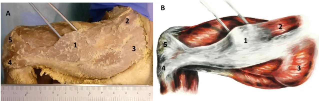
The iliotibial tract was identified in eight cadaveric knees (Figs. 1A and B).
Proximal insertions: after proper dissection, it was possible to perceive the ITT as a structure receiving tendinous fibers of the gluteus maximus and the gluteal aponeurotic fascia, enclosing also the tensor fascia lata. Direct fibers from the iliac crest were observed as well.
Distal insertions: in the region of the greater trochanter, the deep portion of the ITT becomes the intermuscular septum and attaches to the linea aspera of the femur. In the knee, it was possible to identify three insertions. These insertions extend to an extensive periarticular area in the lateral knee, including the distal femur, patella, proximal tibia and join capsule. The stronger and direct one courses over the vastus lateralis, inserting as a ribbon-shaped in the Gerdy’s tubercle and around it. At the upper part of the lateral epicondyle of the femur is another ITT insertion through a strong ligament. The ITT also contributes to the lateral patellofemoral ligament complex by a wide insertion in the patella, which merges itself to the lateral retinaculum.
Histological investigation
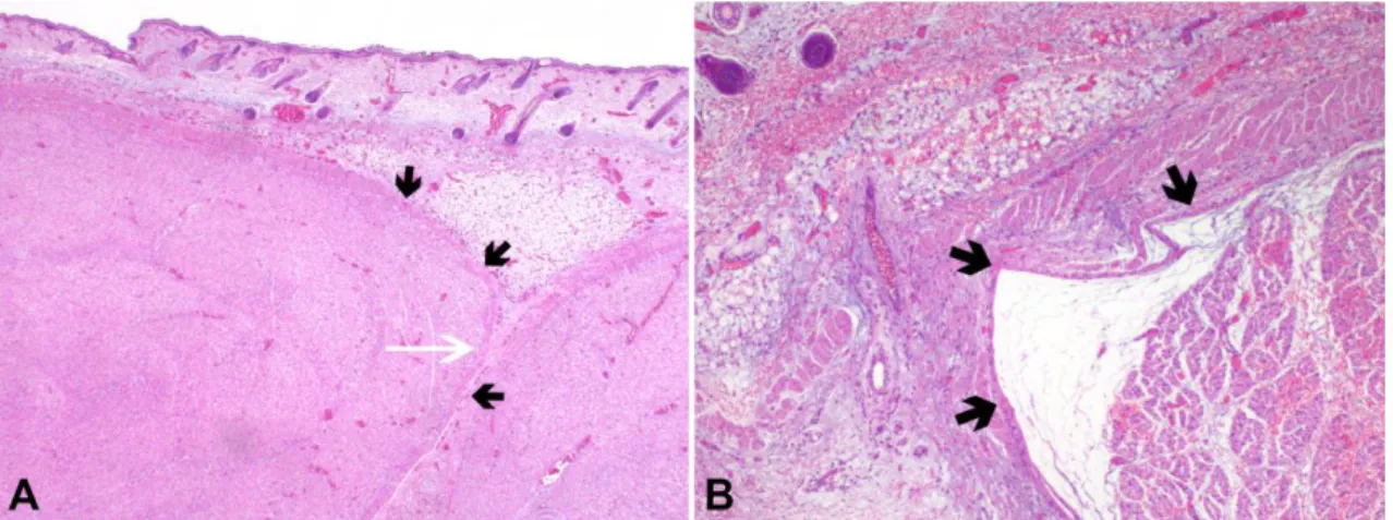
Histologically, the ITT and ALL were constituted by continuous cellular bands of immature fibroblasts (Figs. 2A and B).
The continuity of the ITT with the intermuscular septum and its insertions on the femur and the infracondylar tubercle of the tibia (Gerdy’s tubercle) were demonstrated. Fetal tissue between the ITT and the adjacent muscles and femoral bone was composed of a thin layer of sparsely adipocitic tissue with occasional vessels and myelinated and non-myelinated nerve
Fig. 1 Anatomic dissection of the knee and thigh. Iliotibial band (1) and its insertions at the tensor fascia lata (2),
7 fibers. Occasional oedema and loose thin collagen fibers were present along the fibroblast bands.
It was noted that, in all specimens, the fibroblasts were less numerous where iliotibial tract dipped posteriorly to the vastus intermedius muscle forming the intermuscular septum. This fact was interpreted as an area of fragility of the ITT.
Fig. 2 HE, 100X (A) and 200X (B) – The iliotibial tract is constituted by a continuous cellular band of immature
fibroblasts (black small arrows), extending underneath the fibroadipocitic layer of subcutaneous tissue and continuing with the intermuscular septum. A microscopic area of fragility (white long arrow) was visible where the iliotibial tract dipped posteriorly to the vastus intermedius muscle forming the intermuscular septum.
8
Discussion
Previous embryologic studies demonstrated that the fetal knee is similar to the adult knee in all anatomic structures at nine and a half to ten weeks gestational age. After that stage, there are no major structural changes and only continued growth [6].
According to adult cadaveric studies, the ITT is a lateral thickening that encloses tensor fascia lata and attaches it to the iliac crest. It also receives up to three-quarters of the gluteus maximus. Beyond its proximal insertion on the iliac crest, several bony attachments are being described, which are frequently vague and confusing. Vieira et al. (2007) [19] identified, in anatomic dissections, insertions at the linea aspera, epicondyle, patella, Gerdy’s tubercle and a capsular-osseous insertion, later nominated as anterolateral ligament, which originates on the lateral femoral condyle and inserts midway between Gerdy´s tubercle and the tip of the fibular head. This last ligament is being subject of controversy regarding to its anatomic characteristics. Formerly it was part of the ITT, nevertheless the theory where it is considered an independent structure is nowadays reaching more followers. Even regarding to its origins there are disagreements: some authors describe its origins anterior to the insertion site of the fibular collateral ligament on the lateral femoral epicondyle while others report it to be posterior to the lateral femoral epicondyle.
Our study in fetus partially corroborates this information - we could identify the joining of the ITT with the tensor fascia lata and gluteus maximus. The insertion at the linea aspera was observed as an intimate continuity through the ITT and intermuscular septum until the linea aspera. The insertions at the epicondyle, patella and Gerdy’s tubercle were presents, with the characteristics of former descriptions. However, the anterolateral ligament was not identified, suggesting that it might have developed over time in response to repetitive knee loading [16].
The histologic study was also consistent with previous histological investigations regarding the ITT and was able to demonstrate the continuous nature of the ITT and its continuity with the intermuscular septum.
Differently from what was visualized in adult specimens from anterior published studies [4], fetal tissue between the ITT and the adjacent muscles and femoral bone was not composed of a highly vascularized mass of adipose tissue but of a thin delicate layer of sparsely adipocitic tissue with occasional vessels and myelinated and non-myelinated nerve fibers.
On the other hand, the fragility area identified in all specimens, where iliotibial tract dipped posteriorly to the vastus intermedius muscle forming the intermuscular septum raises some questions. Being a constant finding, it can be argued that it might be a real point of instability of the ITT instead of a histologic artefact and that it can have actual clinical
9 implications. Another hypothesis is that it is present due to the tissue immaturity of the fetal specimens and will eventually develop into a continuous fibrous band in older children, ceasing to exist and leaving behind all the possible consequences of a fragility area.
The rotational stability of the knee is responsibility not of a single stabilizer, but rather, a complex system of anterolateral structures, which include the ITT, the anterolateral capsule and the anterolateral ligament [21]. According to Suero et al. (2011) [17], the magnitude of anterior tibial translation during the pivot shift and the Lachman tests increases when the ITT is removed and suggests that sectioning the ITT results in a significant increase in lateral compartment translation during the pivot shift maneuver. The insertion through the intermuscular septum stabilizes the tract in a defined distance to the femur preventing its ventral dislocation, while the other insertions seem to stabilize the lateral side of the knee. The integration of this with the possible fragility zone we found on the ITT can be important during the clinical evaluation of some traumatic lesions, because lateral instabilities can be complex, including anterolateral, posterolateral, varus, and straight components. Nevertheless, according to Suero et al. (2013) [17] the ITT does not appear to have a greater impact than other lateral structures in the prevention of anterior tibial translation.
For the proper study of the knee instabilities it is important to have a detailed and precise description of the components of the iliotibial tract. The description of its insertions provides a better understanding of the knee instability and supports the development of better and more adequate surgical repairs.
There are several limitations to this research. Our analysis was based only in fourteen corpse’s knees, which is a small number. It is also important to consider possible changes due the post-mortem degeneration of tissue and the formaldehyde preservation, which is associated with extreme rigidity and it severely affects the quality of the cadaveric tissue, particularly soft tissue. During the dissections, it’s difficult to reproduce the anatomy meticulously, because of the high biological variability of the lateral side of the knee. The possibility of injuries during the procedure should be taken in account as well possible artefacts.
10
Conclusions
The ITT has important connections with the femur, the patella and the tibia, which are fundamental to the stabilization of the lateral side of the knee. There is a clear continuity through this band and the intermuscular septum until the linea aspera. The fragility zone found in all specimens may have clinical implications which deserve further investigation.
For some time now, the ITT is considered an anatomic structure, however its anatomy was not always well accepted by all the authors. Nowadays, its descriptions have become more and more detailed, regarding to their proximal and distal insertions as well its contribution to the knee stability. This way it is possible a better correlation with the clinical presentations and under these circumstances proceed to the most correct diagnosis. This way the treatment offered to the patient will be the most appropriate.
Acknowledgment: the authors would like to thank the Department of Anatomy of the
Instituto de Ciências Biomédicas Abel Salazar and the Departments of Anatomic Pathology and Orthopaedics of Centro Hospitalar do Porto the appreciated contributions during the cadaveric dissections and Carlos Costa Reis for the illustrations of the anatomic dissections. The authors have no conflict of interest to declare regarding the material presented in this manuscript.
11
References
1. Campos JC, Chung CB, Lektrakul N, et al. (2001) Pathogenesis of the Segond fracture: anatomic and MR imaging evidence of an iliotibial tract or anterior oblique band avulsion. Radiology, 219(2):381–6. doi: 10.1148/radiology.219.2.r01ma23381
2. Caterin S, Litchfield R, Johnson M, Chronik B, Getgood A (2014) A cadaveric study of the anterolateral ligament: re-introducing the lateral capsular ligament. Kee Surg Sports Traumatol Arthrosc. doi: 10.1007/s00167-014-3117-z
3. Claes S, Vereecke E, Maes M, Victor J, Verdonk P, Bellemans J (2013) Anatomy of the anterolateral ligament of the knee. J. Anat., 223:321-328. doi: 10.1111/joa.12087 4. Fairclough J, Hayashi K, Toumi H, Lyons K, Bydder G, Phillips N, Best TM, Benjamin
M (2006) The functional anatomy of the iliotibial band during flexion and extension of the knee: implications for understanding iliotibial band syndrome. J. Anat. 208:309-316. doi: 10.1111/j.1469-7580.2006.00531.x
5. Goldman AB, Pavlov H, Rubenstein D (1988) The Segond fracture of the proximal tibia: a small avulsion that reflects major ligamentous damage. AJR Am J Roentgenol, 151:1163–7. doi: 10.2214/ajr.151.6.1163
6. Gray DJ, Gardner E (1950) The early development of the knee join in staged human embryos. Am J Anat, 86(2):235-299.
7. Haims AH, Medvecky MJ, Pavlovich Jr R, Katz LD (2003) MR imaging of the anatomy of and injuries to the lateral and posterolateral aspects of the knee. AJR Am JRoentgenol, 180:647–53. doi: 10.2214/ajr.180.3.1800647
8. Huang BK, Campos JC, Peschka PGM, Pretterklieber ML, Skaf AY, Chung CB, Pathria MN (2013) Injury of the Gluteal Aponeurotic Fascia and Proximal Iliotibial Band: Anatomy, Pathologic Conditions, and ME Imaging. RadioGraphics 33:1437-1452. doi: 10.1148/rg.335125171
9. Hughston J, Andrews J, Cross M, Moschi A (1976) Classification of knee ligament instabilities. Part I. Medial compartment and cruciate ligaments. J Bone Joint SurgAm, 58:159–72.
10. Johnson LL (1979) Lateral capsular ligament complex: anatomical and surgical considerations. Am J Sports Med, 7:156–60. doi: 10.1177/036354657900700302 11. Kaplan EB (1958) The Iliotibial Tract. The Journal of Bone and Join Surgery vol40-A,
no4
12. LaPrade RF, Terry GC (1997) Injuries to the posterolateral aspect of the knee. Association of anatomic injury patterns with clinical instability. Am J Sports Med, 25:433–8. doi: 10.1177/036354659702500403
13. Lobenhoffer P, Posel P, Witt S, Piehler J, Wirth CJ (1987) Distal Femoral Fixation of the Iliotibial Tract. Arch Orthop Truma Surg, 106:285-290
14. Lutz C, Sonnery-Cottet B, Niglis L, Freychet B, Clavert P, Imbert P (2015) Behavior of the anterolateral structures of the knee during internal rotation. Orthopaedics & Trumatology: Surgery & Research 101:524-528. doi: 10.1016/j.otsr.2015.04.007 15. Muller W (1983) The Knee. New York: Springer Verlag
16. Shea KG, Polousky JD, Jacobs JC Jr, Yen YM, Ganley TJ (2016) The Anterolateral Ligament of the Knee: An Inconsistent Finding in Pediatric Cadaveric Specimens. J Padiatr Orthop, 36(5):51-4. doi: 10.1097/BPO.0000000000000616
17. Suero EM, Njoku IU, Voigt MR, Lin J, Koenig D, Pearl A (2013) The role of the iliotibial band during the pivot shift test. Kee Surg Sports Traumatol Arthrosc 21:2096-2100. doi: 10.1007/s00167-012-2257-2
18. Terry GC, Hughston JC, Norwood LA (1986) The anatomy of the iliopatellar band and iliotibial tract. Am J Sports Med,14:39–45. doi: 10.1177/036354658601400108
12 19. Vieira ELC, Vieira EA, Silva RTC, Berlfein PAS, Abdalla RJ, Cohen M (2007) An Anatomic Study of the Iliotibial Tract. The Journal of Arthroscopic and Related Surgery, 23:269-274. doi: 10.1016/j.arthro.2006.11.019
20. Vincent JP, Magnussen RA, Gezmez F, Uguen A, Jacobi M, Weppe F, Al-Saati MF, Lustig S, Demey G, Servien E, Neyret P (2011) The anterolateral ligament of the human knee: an anatomic and histologic study. Kee Surg Sports Traumatol Arthrosc. doi: 10.1007/s00167-011-1580-3
21. Zens M, Feucht MJ, Ruhhammer J, Bernstein A, Mayr HO, Südkamp NP, Woias P, Niemeyer P (2015) Mechanical tensile properties of the anterolateral ligament. Journal of Experimental Orthopaedics 2:7. doi: 10.1186/s40634-015-0023-3