Vol.54, n. 5: pp. 901-906, September-October 2011
ISSN 1516-8913 Printed in Brazil BRAZILIAN ARCHIVES OF
BIOLOGY AND TECHNOLOGY
A N I N T E R N A T I O N A L J O U R N A L
Effects of Catuaba Cristal
®on the Testis of Wistar rats
Karine Moura de Freitas
1*, Kyvia Lugate Costa
2, Pamela Kelly Araújo Campos
2, Fabiana
Cristina Silveira Alves de Melo
3, Tarcízio Antônio Rego de Paula
4and Sérgio Luis Pinto da
Matta
21Departamento de Anatomia, Biologia Celular, Fisiologia e Biofísica; Instituto de Biologia; Universidade Estadual
de Campinas; Campinas – SP - Brasil. 2Departamento de Biologia Geral; Universidade Federal de Viçosa;Viçosa - MG - Brasil. 3 Ciências Biologicas; Universidade Federal de Goiás; Jataí – GO - Brasil. 4Departamento de Veterinária;Universidade Federal de Viçosa; Viçosa – MG - Brasil.
ABSTRACT
The aim of this study was to evaluate the possible beneficial effects of Catuaba Cristal® (CC), an alcoholic drink made from wine and Erythroxylum catuaba Ar. Cam on testis. Wistar rats either received CC solution (n=8) or water (n=9). Results showed significant body weight reduction within the CC group, although, no weight changes were observed for liver, kidney, testis, epididymis, seminal vesicle and prostate. The volumetric proportion and volume of interstitial tissue and lymphatic space were reduced in the treated group. In the CC group, although the nuclear volume of Leydig cells (LC) decreased, the number of LC per testis increased. These results suggested that CC had no beneficial effect on spermatogenesis of Wistar rats.
Key words: Aphrodisiac drink, medicinal plants, morphometry, spermatogenesis, testis
*Author for correspondence: [email protected]
INTRODUCTION
Several plants have traditionally been used as medicine, although there are few studies regarding their effects on different organs (Monteiro et al., 2008; Monteiro et al., 2009; Predes et al., 2009) Most of the plants and their derivatives are used in popular medicine without prior investigation. Erythroxylum catuaba Ar. Cam., Erythroxylaceae, is a Brazilian native species that is known as “catuaba” (Fonseca, 1932; Silva, 2004a). E. catuaba is used as a stimulant for the central nervous system and against sexual impotence (Silva, 2004b). The stem cortex of E. catuaba is used as a phytotherapic substance, in the form of
fluid extract, tincture, infusion, syrup and wine (Silva, 2004b). Catuaba Cristal® (CC) is an alcoholic drink (15% alcoholic content), made from E. catuaba, dry red wine and other associations (exact information was not provided by the producer). Despite the fact that this drink is widely consumed as an aphrodisiac and a stimulant drink, no study has ever been performed to confirm these popular beliefs. Therefore, the present study aimed to evaluate whether CC drink affected the testis of Wistar rats.
MATERIAL AND METHODS
Adult male Wistar rats (Rattus norvegicus) (120 days) were obtained from the Central Biotery of Biological and Health Science Center (Federal University of Viçosa, Viçosa, MG, Brazil). The animals were housed in individual cages, under standard conditions with 12 h light: 12 h dark, and an average temperature of 24.7º C. The laboratory chow was available, ad libitum. They received pure drinking water (control group, n=9) or a solution of drinking water and CC (0.1%) ad libitum (CC-treated group, n=8). Each animal in CC-treated group received 100 mL of CC solution daily. The consumption of CC solution was measured daily to verify if the animals ingested the minimum volume (40 mL). The animals were treated according to the Manual on care and use of laboratory animals of the National Research Council and in agreement with the ethics principles for the use of laboratory animals, recommended by Brazilian Society of Science in Laboratory Animals – SBCAL/COBEA.
Euthanasia, Biometry and Tissue Collection After 54 days of treatment, the animals were euthanized by carbon dioxide asphyxia and weighed. Testes, epididymis, seminal vesicle and prostate were collected and weighed. The tunica albuginea was removed from one testis to obtain the weight of the testicular parenchyma and of the albuginea. The relative weight (somatic index) of the organ corresponded to the percentage of body weight represented by that organ.
Preparation of Tissue for Microscopy
One testis was dissected and fixed by immersion in Karnovsky’s fixative (paraformaldehyde 4% and glutaraldehyde 4% in sodium phosphate buffer 0.1M, pH 7.4). Testicular fragments were dehydrated and embedded in hydroxyethyl methacrylate (GMA, Leica). Sections (3 m) were obtained and stained with toluidine blue-sodium borate 1%.
Morphometry and Stereology
Morphometrical and stereological analyzes were performed using the software Image-Pro Plus 4. For this purpose, images were captured with a light microscope (Olympus-AX-70). Ten digital images were randomly taken at 200x magnification and used for estimating the volumetric proportion of the testicular parenchyma
elements (interstitium and seminiferous tubules - tunica propria, epithelium and lumen) using a grid with 494 points per image. In the interstitium, the volumetric proportions of Leydig cells (nucleus and cytoplasm), the connective tissue cells and fibers, macrophages, blood vessels and lymphatic space were obtained. For this measurement, 1000 points were counted in the interstitium, using images (400x magnification).
The total volume of each testicular component, expressed in mL, was estimated by multiplying the volumetric proportion of each component by the testicular volume, divided by 100. Assuming that the testicular density is approximately 1 (França, 1991), thus, testicular volume has the same value as the testicular parenchyma weight. The tubule-somatic index (TSI) was calculated by the ratio of seminiferous tubule volume and body mass, the result being multiplied by 100.The diameter of seminiferous tubules was measured on 10 round seminiferous tubule sections per animal. Total seminiferous tubule length was estimated, in meters, from the volume occupied by seminiferous tubules within the testis and mean tubular diameter for each animal (Attal and Courot, 1963; Dorst and Sajonski 1974).
Leydig cell (LC) nuclear and cytoplasmic volumetric proportions were estimated counting 500 points over the nuclei and cytoplasm of this cell type (400x magnification images). Mean diameter of LC nuclei was estimated by the measurement of nuclear diameter of 30 nuclei per animal, in the same images. The nuclear volume was calculated by the formula 4/3πr3, in which “r” corresponded to the nuclear radius. Nuclear volume and nucleus/cytoplasm proportion were used to obtain the cytoplasm and individual volume of LC. LC number was estimated by the total volume occupied by these cells in the testis divided by the individual volume of LC. The Leydig-somatic index (LSI) was calculated by the ratio of the volume occupied by LC on the testes and the body mass, the result being multiplied by 100.
Statistical Analysis
Effects of Catuaba Cristal on the Testis 903
RESULTS
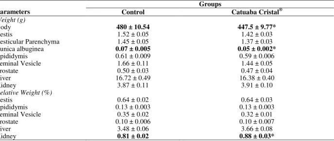
The CC group showed a significant decrease in the body weight relative to control (p=0.04). The weight of testis, epididymis, seminal vesicle, prostate, liver and kidney did not vary in the CC treated group (Table 1). Testicular parenchyma weight was not altered in the treated group. The tunica albuginea weight was significantly reduced (p<0.001), though this reduction did not influence the testicular total weight (Table 1). The organs’
relative weights did not vary (Table 1), except the kidney relative weight that increased in CC-treated group (Table 1). The volumetric proportion of seminiferous tubules in the testis increased in the CC group, whereas interstitium was reduced (p<0.001) (Table 2). The volume of seminiferous tubules did not vary among the experimental groups. However, the volume of interstitial tissue was significantly reduced in the CC group (p=0.005) (Table 2).
Table 1 - Biometrical parameters of control Wistar rats and those treated with Catuaba Cristal®. The values are means ±SEM.
Groups
Parameters Control Catuaba Cristal®
Weight (g)
Body 480 ± 10.54 447.5 ± 9.77*
Testis 1.52 ± 0.05 1.42 ± 0.03
Testicular Parenchyma 1.45 ± 0.05 1.37 ± 0.03
Tunica albuginea 0.07 ± 0.005 0.05 ± 0.002*
Epididymis 0.61 ± 0.009 0.59 ± 0.006
Seminal Vesicle 1.66 ± 0.11 1.44 ± 0.05
Prostate 0.50 ± 0.03 0.47 ± 0.04
Liver 16.72 ± 0.49 16.38 ± 0.40
Kidney 3.87 ± 0.11 3.91 ± 0.10
Relative Weight (%)
Testis 0.64 ± 0.02 0.64 ± 0.03
Epididymis 0.13 ± 0.003 0.13 ± 0.003
Seminal Vesicle 0.35 ± 0.02 0.32 ± 0.01
Prostate 0.10 ± 0.006 0.10 ± 0.007
Liver 3.48 ± 0.06 3.66 ± 0.08
Kidney 0.81 ± 0.02 0.88 ± 0.03*
* Indicates significant difference (p<0.05), according to to t-test.
Table 2 - Morphometrical and Stereological parameters of testis of control adult Wistar rats and those treated with Catuaba Cristal®. The values are means ± SEM.
Groups
Parameters Control Catuaba Cristal®
Volumetric proportion in testis (%)
Seminiferous tubule 84.08 ± 0.35 86.87 ± 0.84*
Interstitium 15.91 ± 0.35 13.13 ± 0.84*
Lymphatic space 9.78 ± 0.43 6.90 ± 0.60*
Blood vessels 2.35 ± 0.41 1.82 ± 0.26
Leydig cells 2.89 ± 0.26 3.76 ± 0.35
Macrophages 0.25 ± 0.06 0.35 ± 0.06
Connective tissue 0.64 ± 0.19 0.30 ± 0.04
Volume(mL)
Seminiferous tubule 1.22 ± 0.04 1.19 ± 0.03
Interstitium 0.23 ± 0.019 0.18 ± 0.01*
Lymphatic space 0.14 ± 0.017 0.09 ± 0.01*
Blood vessels 0.03 ± 0.01 0.02 ± 0.003
Leydig cells 0.04 ± 0.004 0.05 ± 0.005
Macrophages 0.004 ± 0.001 0.005 ± 0.001
Connective tissue 0.009 ± 0.003 0.004 ± 0.001
TSI 0.51 ± 0.02 0.53 ± 0.02
Tubular Diameter(µm) 301.37 ± 6.67 315.72 ± 5.80
Length of seminiferous tubules (m) per testis 17.27 ± 0.96 15.31 ± 0.63
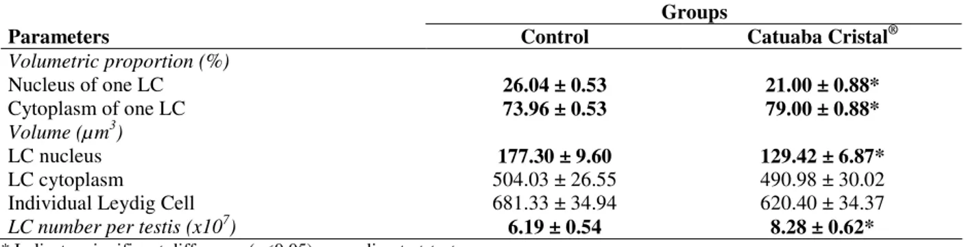
Volumetric proportion and volume of blood vessels, Leydig cells and macrophages in the testis did not vary among the groups, whereas lymphatic space volumetric proportion and volume were significantly reduced (p=0.001, p<0.001 respectively) (Table 2).The tubule-somatic index, diameter and total length of seminiferous tubules did not vary in the treated group (Table 2). Leydig cells (LC) nuclei volumetric proportion diminished
in the treated group, whereas the cytoplasm increased (p<0.001) (Table 3). The volume of LC nuclei was also significantly reduced (p=0.0012). However, the cytoplasm volume and the single LC volumes did not vary significantly among the groups (Table 3). The number of LC per testis was 25.25% higher in the treated group when compared to the control (Table 3).
Table 3 - Stereological parameters of Leydig cells (LC) of control adult Wistar rats and those treated with Catuaba Cristal®. The values are means ± SEM.
Groups
Parameters Control Catuaba Cristal®
Volumetric proportion (%)
Nucleus of one LC 26.04 ± 0.53 21.00 ± 0.88*
Cytoplasm of one LC 73.96 ± 0.53 79.00 ± 0.88*
Volume (µm3)
LC nucleus 177.30 ± 9.60 129.42 ± 6.87*
LC cytoplasm 504.03 ± 26.55 490.98 ± 30.02
Individual Leydig Cell 681.33 ± 34.94 620.40 ± 34.37
LC number per testis(x107)
6.19 ± 0.54 8.28 ± 0.62*
* Indicates significant difference (p<0.05), according to t-test.
DISCUSSION
The dose of Catuaba Cristal® utilized in the present study was calculated according to the popular use.
Furuya et al., (2003) observed that Wistar rats treated with 0.5, 1.5, 3 and 7% alcoholic solution had no changes in the body weight.
In contrast, a significant reduction of body weight in the CC group was observed in the present study. Since the alcoholic concentration of CC solution was lower than 0.5%, alcohol was not likely to be responsible for the weight loss observed in the CC group.The slight reduction in body weight (7% less in the CC-treated group) was not an indicative of health problems, as the animals were within the normal weight range for this species. Moreover, no weight change was observed for both liver and kidney within the CC group. Since liver and kidney were responsible for the detoxification and excretion of toxic substances (Stevens and Lowe, 1999), the absence of significant variation in these weights indicated that CC solution was non toxic for the animals at the utilized dose. Previous study showed that animals treated with Heteropterys aphrodisiaca, a popular aphrodisiac plant, did not show changes in weight of testis, epididymis, seminal vesicle, ventral and dorsolateral prostate
Effects of Catuaba Cristal on the Testis 905
seminiferous tubules (Fawcett et al., 1973). Thus, the reduction of lymphatic space could damage the seminiferous tubules, although no variation of seminiferous tubule morphology and the analyzed parameters were observed.
According to França and Russel (1998), the tubular diameter has usually a positive correlation with spermatogenic activity. Therefore, despite having increased the seminiferous tubule volumetric proportion, CC might not have influenced the spermatogenic activity in the CC group. Gomes (2007) also did not observe any alteration in both tubular diameter and length of seminiferous tubules after treatment with Catuama® or T. catigua. Mori and Christensen (1980) showed that the normal volume of LC nucleus was approximately 150 µm3. In the CC treated group, although the nuclear volume mean was smaller (129.47 µm3) than the observed by the authors mentioned above, the same parameter was larger for the control group (177.30 µm3). Therefore, a significant reduction of 26.98% for the LC nuclear volume was found in the CC-group compared to the control group. These results were similar to the ones reported by Gomes (2007) which showed a significant reduction of single LC nuclear volume without any significant alteration of an individual LC cytoplasm and total volume. The reduction of nuclear volume indicated lower cell activity.
Studies performed by Castro et al., (2002) showed a positive correlation between the testosterone plasma levels and the total number of LC per testis and the percentage of LC nuclei volume. Although there was an increase in the total number of LC per testis, a reduction in single LC nucleus volume was observed in the present study. Stereology of LC along with the absence of alteration of accessory glands weight indicated that there was no change in testosterone levels within the treated groups.
In conclusion, the present study showed that Catuaba Cristal® in the administrated dose was not beneficial to spermatogenesis of Wistar rats, although it did not appear to be toxic to the treated animals. Since alcohol is reportedly harmful to spermatogenesis, Catuaba Cristal® consumption should be carefully moderated.
ACKNOWLEDGEMENTS
We thank Dr. Mary Anne Heidi Dolder, MS Rodrigo Paula Leite and MS Marcos de Lucca Moreira Gomes for reviewing this manuscript. Grant sponsor: Fundação de Amparo à Pesquisa do Estado de Minas Gerais (FAPEMIG).
REFERENCES
Attal, J.; Courot, M. (1963), Developpement testiculaire et etablissement de la spermatogeneses chez le taureau. Ann Biol Anim Biochim Biophys., 3, 219-241 Castro, A. C. S.; Berndton, W. E.; Cardoso, F. M.
(2002), Plasma and testicular testosterone levels, volume density and number of Leydig cells and spermatogenic efficiency of rabbits. Braz J Med Biol Res., 35, 493-498
Chieregatto, L. C. (2005), Efeito do tratamento crônico com extratos de Heteropterys aphrodisiaca O. Mach e Anemopaegma arvense (Vell.) Stellf no testículo de ratos Wistar adultos. MS Dissertation, Federal University of Viçosa, Viçosa, Brazil
Dorst, V. J.; Sajonski, H. (1974), Morphometrische untersuchunhen am tubulussystem des schweinehodens während der postnatalen entwicklug. Monotsh Ver Med., 29, 650-652
Fawcett, D. W.; Neaves, W. B.; Flores, M. N. (1973), Comparative observations on intertubular lymphatics and the organization of the interstitial tissue of the mammalian testis. Biol Reprod., 9, 500-532
Fonseca, E. T. (1932), Indicador de Madeiras e Plantas Úteis do Brasil. Oficinas Graphicas VILLAS-BOAS & C. Rio de Janeiro, Brazil
França, L. R. (1991), Análise morfofuncional da espermatogênese de suínos adultos da raça Piau. PhD Thesis, Federal University of Minas Gerais, Belo Horizonte, Brazil
França, L. R.; Russell, L. D. (1998), The testis of domestic animals. In: Male Reproduction. A Multidisciplinary Overview, eds J. Regadera, R. Martinez-Garcia, Churchill Livingstone, Madrid, pp. 197–219
Furuya, D. T.; Binsack, R.; MachadoLow, U. F. (2003), Low ethanol consumption increases insulin sensitivity in Wistar rats. Braz J Med Biol Res., 36(1), 125-130
Melo, F. C. S. A. (2007), Efeito da infusão do caule de cipó-cravo (Tynnanttus fasciculatus Miers, Bignoniaceae) sobre as características morfométricas de componentes testiculares de ratos Wistar adultos. PhD Thesis, Federal University of Viçosa, Viçosa, Brazil
Monteiro, J. C.; Predes, F. S.; Matta, S. L. P.; Dolder, H. (2008), Heteropterys aphrodisiaca Infusion Reduces the Collateral Effects of Cyclosporine A on the Testis. Anat Rec., 291, 809–817
Monteiro, J. C.; Matta, S. L. P.; Predes, F. S.; Toledo, T. O. (2009), Liver Morphology and Morphometry and Plasma Biochemical Parameters of Wistar Rats that Received Leaf Infusion of Rudgea viburnoides Benth. (Rubiaceae). Braz Arch Biol Technol., 52(2), 407-412
Mori, H.; Christensen, A. K. (1980), Morphometric analysis of leydig cells in the normal rat testis. J Cell Biol., 84, 340-354
Predes, F. S.; Matta, S. L. P.; Monteiro, J. C.; Toledo, T. O. (2009), Investigation of Liver Tissue and Biochemical Parameters of Adult Wistar Rats treated with Arctium lappa L. Braz Arch Biol Technol.,
52(2), 335-340
Silva, A. J. (2004a), Estudo botanico e chimico da catuaba (Erythroxylaceae Catuaba do Norte) – Parte I. Rev Bras Farmacogn., 14(1), 67-77
Silva, A. J. (2004b), Estudo botanico e chimico da catuaba (Erythroxylaceae Catuaba do Norte) – Parte II. Rev Bras Farmacogn., 14(2), 145-151
Stevens, A.; Lowe, J. (1999), Human Histology. Mosby, Barcelona, Spain.