BROILERS INFECTED EXPERIMENTALLY WITH EIMERIA SPP AND FED WITH DIETS CONTAINING DIFFERENT SUPPLEMENTS
A.P.V. Cassenego 1, P.A. d'Azevedo 2, A.M.L. Ribeiro 3, J. Frazzon 4, S.T.Van Der Sand 5, A. P. G. Frazzon 5*
1
Programa de Pós-Graduação em Microbiologia Agrícola e do Ambiente, Universidade Federal do Rio Grande do Sul, Porto
Alegre, RS, Brasil; 2 Departamento de Ciências Básicas da Saúde, Microbiologia, Universidade Federal de Ciências da Saúde de
Porto Alegre, Porto Alegre, RS, Brasil; 3 Faculdade de Veterinária, Departamento de Zootecnia, Universidade Federal do Rio
Grande do Sul, Porto Alegre, RS, Brasil; 4 Instituto de Ciência e Tecnologia de Alimentos, Universidade Federal do Rio Grande do
Sul, Porto Alegre, RS, Brasil; 5 Instituto de Ciências Básicas da Saúde, Departamento de Microbiologia, Universidade Federal do
Rio Grande do Sul, Porto Alegre, RS, Brasil.
Submitted: May 24, 2010; Returned to authors for corrections: August 30, 2010; Approved: January 13, 2011.
ABSTRACT
Resistant bacteria in animal can be spread to environment and to humans. Poultry feed and infections caused
by Eimeria spp. are important factors in determining the intestinal microbial communities. The aim of this
study was to verify the prevalence of species and antimicrobial susceptibility of Enterococcus isolated from
broilers fed with different supplements and infected experimentally with Eimeria spp. Broilers were divided
in eight groups, fed with diets supplemented with a combination of antimicrobial, ionophore-coccidiostatics,
probiotic, essential oil. At 14 days old all birds, except the control, received a solution containing oocysts of
Eimeria spp. Samples of cloacal swabs from broilers were collected. A total of 240 Enterococcus sp. strains
were isolated, confirmed genus by PCR, classified as species, tested for antimicrobial susceptibility and
screened by PCR for the presence of tet(L), tet(M) and erm(B) genes. The overall distribution of species
isolated from fecal samples was E. faecalis (40%), followed by E. casseliflavus/E. gallinarum (10.8%), E.
mundtii (10.8%), E. faecium (10.8%), E. columbae (5.8%) and E. gallinarum (4.2%). Changes in the
composition or frequency of Enterococcus species were observed in all dietary supplementation.
Antimicrobial susceptibility tests showed resistance phenotypes a range of antibiotics, especially used in
humans such as, streptomycin, penicillin, rifampicin and vancomycin. There was no correlation between
different supplementation for broilers and antimicrobial resistance and the presence of tet(M), tet(L) and
erm(B) genes. Dietary supplementation had effect on the Enterococcus sp. colonization, but did not have
significant effect on the phenotype and genotype of antimicrobial resistance in enterococci.
Key words:Enterococcus sp.; broilers feed; antimicrobial resistance; resistance genes.
INTRODUCTION
The poultry industry worldwide has increased over the last
40 years. In 2009, world chicken meat production was 91.9
million tonnes (ton), and Brazil, United States and Europe are
the world’s largest exporters (12). Modern technologies of
poultry production use massive doses of antimicrobial as
therapeutics and for growth promotion, during all stages of
production (23). The use of antimicrobial as growth promoter
in animal feed has caused an increase in resistant bacteria
isolated from animal (36). Almost all poultry dietary contain of
2 to 40 g/ton of any type of antimicrobial agent. In 2006, the
European Union banned several antimicrobial growth
promoters, however in Brazil, some growth promoters are used
to feed commercial poultry and broilers chickens (43). The
demand of products free of antibiotics and chemotherapy has
led to the exploration of news to growth promoters, such as
probiotics, prebiotics, and essential oils. In broiler nutrition,
probiotic species belonging to genera Lactobacillus and
Enterococcus are utilized to modulate intestinal microbial
communities and pathogen inhibition (34). On the other hand,
studies testing the addition of essential oils in the diet of broiler
chickens have demonstrated in vitro antimicrobial,
anti-inflammatory, antioxidant and anticoccidiostatic activity of
many plants (29, 43). Hume et al. (21) observed that essential
oil supplemented in the diets avoided the drastic shifts after a
mixed challenge of Eimeria spp. and the microbial
communities were better modulated than monensin.
The feeding is an important factor in determining the
intestinal microbial communities. Commensal enteric bacteria
from chickens are frequently objects of researches, for
supervision of animal products for human consumption and
marketing. Enterococci are gram-positive bacteria widely
distributed in the environment, inhabiting mainly the
gastrointestinal tract of humans and animals (33). Furthermore,
enterococci are opportunistic pathogens commonly associated
with in immunocompromised patients and nosocomial
infection (15). Enterococcus sp. resistant to several
antimicrobials were isolated from clinical specimens and
environment (5,8,15,18,37). These microorganisms are
intrinsically resistant against many antimicrobial agents
commonly prescribed for Gram-positive cocci and exhibit
resistance to a wide variety of other antimicrobials, by
acquisition of resistance genes via transposons or plasmids,
such as, tetracycline and macrolides (25). Resistance to
tetracycline are mediated by two majors groups, the first
includes the tet(M), tet(O) and tet(S) genes that encoding
ribosomal protection proteins, and the second is represented by
tet (L) and tet (K) genes encodes tetracycline efflux pumps
proteins (3). One of the most widespread mechanism of
resistance to macrolides is mediated by methylation of a
specific adenine residue in 23S rRNA and is associated with
the erm (B) gene (29).
In Brazil, few studies have investigated the presence of
enterococci resistant in poultry (28, 43, 44, 47). The aim of this
study was to verify the effect of diets containing different
supplements and the presence or not of Eimeria spp. on the
specific colonization and antimicrobial susceptibility of
Enterococcus sp. in the gastrointestinal tract of broilers.
MATERIAL AND METHODS
Animals
The study was conducted using a total of 250 day-old
male Cobb 500. The broilers were housed indoors and
distributed in a completely randomized design divided into
eight treatment groups with five replicates each (12 birds/box).
Broilers were fed with a diet based on corn-soybean meal,
vegetable oil, minerals and vitamins and supplemented with a
combination of ionophore-coccidiostatics (monensin) at
90g/ton, probiotic (Enterococus faecium 3,5 x 10¹⁰ UFC/g) at
35g/ton, essential oil (extracted of thyme and clove) at
100g/ton and/or antimicrobial (bacitracin methylene
the control, received a solution containing 5x10⁴ and 1x10⁴
oocysts/chicken of Eimeria maxima and Eimeria acervulina,
respectively. The broilers were maintained until 28 days old at
the Departamento de Zootecnia, Universidade Federal do Rio
Grande do Sul. The nutritional requirements used are as
indicated in Rostagno et al. (40).
Isolation and confirmation of enterococci genus
Five cloacal swabs samples from each group were
collected and it was avoided environmental contamination
(Table 1). Each swab was inserted into Cary-Blair medium and
sent to the laboratory. The samples were inoculated in Broth
Azide (Himedia, Mumbai, India) media for 24 hours at 35º C.
One hundred microliters of broth were inoculated on agar
plates with Brain Heart Infusion agar (BHI) (Himedia,
Mumbai, India) supplemented with 6.5% NaCl and incubated
as described above. Phenotypic criteria such as size/volume,
shape, color, hemolytic profile, Gram staining, catalase
production, and esculin hydrolysis tests were used to separate
the enterococci and the non-enterococcal strains. Enterococci
genus was confirmed by polymerase chain reaction (PCR)
technique. DNA extraction and amplification using tuf gene
followed the protocols described by Ke et al. (27) and Riboldi
et al. (37). Thirty enterococci were randomly select from each
group.
Phenotype characterization of enterococci resistant
The phenotype characterization was determined according
to the protocol determined by Facklam et al. (12).
Enterococcus faecalis ATCC 51299 and Staphylococcus
aureus (ATCC 25923) were used as control strains.
PCR-RFLP of 16S ribosomal DNA to confirm the Enterococcus gallinarum and Enterococcus casseliflavus
Isolates biochemically classified as E. gallinarum or E.
casseliflavus were confirmed by PCR-RFLP using primers
specific region of 16S ribosomal DNA, as described by
Medeiros et al. (32). Extraction of genomic DNA followed the
method of Riboldi et al. (37). The PCR product of 661 bp
amplified was submitted to digestion with the restriction
enzyme HinfI (Jena Bioscience GmbH, Germany) following
the manufacturer instructions.
Antimicrobial susceptible testing
All isolates were subjected to antimicrobial susceptibility
assay by disk diffusion method, as recommended by the
Clinical and Laboratory Standards Institute (6). The following
antimicrobials were tested: ampicillin (AMP 10 g), penicillin
(PEN 10 U), erythromycin (ERI 15 g), streptomycin (STR 10
mg), chloramphenicol (CLO 30 g), ciprofloxacin (CIP 5 g),
nitrofurantoin (NIT 300 g), vancomycin (VAN 30 g),
rifampicin (RIF 5 g) and tetracycline (TET 30 g). As
positive and negative controls strains E. faecalis ATCC 51299
and E. faecium ATCC 53519 were used, respectively.
Detection of tet(M), tet(L) and erm(B) genes:
All strains were tested for the presence of tet(M), tet(L)
and erm(B) genes by PCR. Enterococcus sp. genomic DNA
was extracted by the boiling method as described by Riboldi et
al. (37). The primer of tet(M), tet(L) and erm(B) genes and
PCR amplifications were performed as described by Frazzon et
al. (15).
Statistical analysis
The experimental data was submitted to analysis of
variance using the Program SASM-AGRI for Statistical
Significant Differences at P<0.05 and P<0.01 (2).
RESULTS AND DISCUSSION
Identification of enterococci isolates
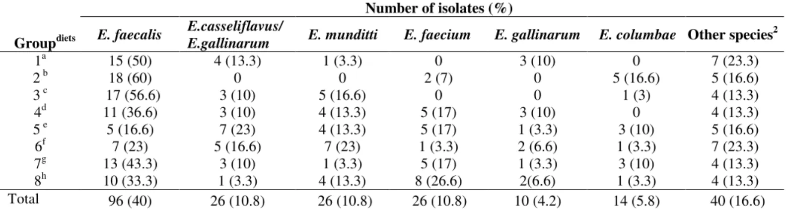
A total of 240 Enterococcus were isolated and confirmed
the genus through PCR. Table 1 shows the overall distribution
different species were identified and E. faecalis was the most
prevalent specie (40%), followed by E. casseliflavus/E.
gallinarum (10.8%), E. mundtii (10.8%), E. faecium (10.8%),
E. columbae (5.8%)and E. gallinarum (4.2%). Other species
identified were E. pseudoavium, E. saccharolyticus, E. avium,
E. hirae, E. durans, E. cecorum, E. sulfureus, E. asini, E.
malodoratus and E. raffinosus characterizing 10% of the
isolates. Sixteen isolates (6.6%) could not be identified to the
species level, and were classified as Enterococcus sp. The
species identified in the current study were also reported by
Hwang et al. (23), Fracalanzza et al. (14), Debnam et al. (9) for
broilers and chicken meat. Enterococci are part of the
gastrointestinal microbiota of chickens (17). In chickens, the
presence of Enterococcus species varies with age of birds. In
young birds the most prevalent species are E. faecalis and E.
faecium, with the maturity of the birds there is a decline in E.
faecium, followed by E. faecalis, allowing the growth of other
species (10). The prevalence of E. casseliflavus/E. gallinarum
was relatively higher in the present study compared to other
countries (9, 23). Meanwhile, some studies conducted in Brazil
have been observed high prevalence of these species from
samples of chicken meat (15, 14).
Table 1. Groups, supplements and distribution of Enterococcus species from broilers feed with different dietary supplemented.
Number of isolates (%)
Groupdiets E. faecalis
E.casseliflavus/
E.gallinarum E. munditti E. faecium E. gallinarum E. columbae Other species 2
1a 15 (50) 4 (13.3) 1 (3.3) 0 3 (10) 0 7 (23.3)
2 b 18 (60) 0 0 2 (7) 0 5 (16.6) 5 (16.6)
3 c 17 (56.6) 3 (10) 5 (16.6) 0 0 1 (3) 4 (13.3)
4d 11 (36.6) 3 (10) 4 (13.3) 5 (17) 3 (10) 0 4 (13.3)
5 e 5 (16.6) 7 (23) 4 (13.3) 5 (17) 1 (3.3) 3 (10) 5 (16.6)
6f 7 (23) 5 (16.6) 7 (23) 1 (3.3) 2 (6.6) 1 (3.3) 7 (23.3)
7g 13 (43.3) 3 (10) 1 (3.3) 5 (17) 1 (3.3) 3 (10) 4 (13.3)
8h 10 (33.3) 1 (3.3) 4 (13.3) 8 (26.6) 2(6.6) 1 (3.3) 4 (13.3)
Total 96 (40) 26 (10.8) 26 (10.8) 26 (10.8) 10 (4.2) 14 (5.8) 40 (16.6)
Diets: a) unmedicated infected control; b) ionophore coccidiostatic infected group; c): probiotic infected group; d) ionophore coccidiostatic and probiotic infected group; e) essential oil infected group; f) ionophore coccidiostatic and essential oil infected group; g) growth promoter infected group; h) unmedicated uninfected control 2:E. pseudoavium, E. saccharolyticus, E. avium, E. hirae, E. durans, E. cecorum, E. sulfureus, E. asini, E. malodoratus and E. raffinosus and Enterococcus
spp.
Of the 39 isolates classified as E. casseliflavus and E.
gallinarum and tested by PCR-RFLP, only two isolates did not
showed DNA fragments pattern expected to E. gallinarum and
E. casseliflavus, and were reclassified as Enterococcus sp. The
correct identification of these species is very important to
distinguish between intrinsic and acquired resistance to
vancomycin.
Species distribution according to dietary supplement employed
Changes in the composition or frequency of Enterococcus
species were noticed (Table 1). The species distribution
between all groups was comparable with the control. The
unmedicated, uninfected control (group 8) the most prevalent
species were E. faecalis (33.3%), E. faecium (26.6%),
E.mundtii (13.3%), E. gallinarum (6.6%), E. casseliflavus/E.
gallinarum (3.3%), E. columbae (3.3%), and Enterococcus sp.
(13.6%). Changes in the composition of Enterococcus species
were detected in groups 1, 2, 3 and 4. In group 1
(unmedicated, infected control) no E. faecium, E. columbae
and E.mundtii were isolated. One reason for this change in the
species frequency should be due the intestinal dysbacteriosis
caused by Eimeria spp. Fukata et al. (16) reported that
composition of the intestinal microbiota, compared to
uninfected chickens. In group 2 (ionophore-coccidiostatic,
infected) E. casseliflavus/E.gallinarum, E. gallinarum, and E.
mundtii were isolated. Monensin is a polyether antibacterial of
the ionophore group, and demonstrate an anticoccidial action
against E. tenella, E. acervulina, E. mivati, E. brunetti, E.
maxima, E. necatrix in chickens. Since monensin is
antibacterial agent, it may have an effect against some
enterococcus species (41). Debnam et al. (10) verified that
antibiotic virginiamycin had a negative effect of some
enterococcus species. In swabs from group 3 (probiotic,
infected) no E. faecium and E. gallinarum were isolated and
from group 4 (ionophore-coccidiostatic and probiotic, infected)
E. columbae was detected, but E. faecium and E. gallinarum
were observed. The diarrhea caused by Eimeria spp. in the
broilers from group 3 may have been impaired the colonization
of the gut by E. faecium (probiotic) and eliminated the
probiotic and, may possibly be the reason why this species was
not detected in group 3. The association
ionophore-coccidiostatic and probiotic appeared to have a positive effect
on this species. The reduction in the prevalence of some
species detected in groups 3 and 4 should be caused by the
presence of probiotics in the diet of broilers, since probiotic
microorganisms has the ability to produce inhibitory
substances (antagonize other microorganism) and also
competitive capacity (hydrophobic cell wall, that facilitate
adhesion to the epithelium intestinal) (44). Changes in the
frequency of Enterococcus species were perceived in groups 5,
6 and 7.In groups 5 (essential oil, infected) and 6
(ionophore-coccidiostatic and essential oil, infected) an increase of 20%
and 13% in the frequency of E. casseliflavus/E.gallinarum,
respectively, was observed. In the group 6, the highest
prevalence of E. pseudoavium (20%) was identified, showing
that the association ionophore-coccidiostatic and essential oil
have a positive effect on this specie. Alterations in the
frequency of some enterococcus species observed in groups 5
and 6 should be caused by essential oil. Antibiotic and
anticoccidial activity of plants oil against Eimeria spp. are well
known (29, 43). In the broilers receiving antibiotic and were
infected with Eimeria spp. (group 7), variations in the
frequency of Enterococcus species were noted. The antibiotic
in the supplement, should selected the most adapted organisms
inside the gut of broilers. Leme et al. (28) have demonstrated
that broilers fed with feed supplemented with avoparcin as
growth promoter showed an increase of E. faecium when
compared with control group.
Antimicrobial susceptibility according to dietary supplement used
Table 2 shows the antibiotic susceptibility of Enterococcus
species isolated from cloacae of poultries. The prevalence of
antimicrobial resistance between all groups was compared, and
an increase or decreases in frequency of resistance as observed.
Absence of susceptibility to at least one of the antimicrobials
tested was detected, except to the antimicrobial ampicillin.
Enterococcus sp. ampicillin-susceptible has been related in
isolated from chickens and food (38, 44).
Resistance to tetracycline was verified in all groups
(56.7%-100%). Although tetracycline is an antimicrobial not
approved by the European Union as a food supplement for
chickens, therapeutic and prophylactic use in veterinary is
common, and Enterococcus resistant to tetracycline are
frequently found in broilers, animal feed, poultry production
environment and chicken carcasses (7,15,20). The higher
percentage of erythromycin resistant enterococcus was noticed
in groups 1, 3, 4, 5 and 7 (53-70%). Hwang et al. (23) observed
same prevalence of erythromycin resistance in enterococcus
isolated from fecal samples of chicken in Korea. The
erythromycin resistant enterococcus is very interesting, since
this drug is the choice for treatment of several infection and
also enterococcal infection in penicillin-allergic patients.
An increase in the percentage of Enterococcus sp.
resistant to penicillin was observed in groups 1 3, 4, and 7. The
here was highest, contrasting to the results reported by
Aarestrup et al. (1) and Hayes et al. (20), where low levels of
penicillin-resistant enterococcus isolated from chickens were
detected, in spite of the fact that San Martín et al. (42) have
found a high percentage of enterococci resistant to penicillin in
broilers samples. Penicillin-resistant enterococci isolate from
poultries is important, for the reason that the animals can serve
as an important reservoir of resistant bacteria that can be spread
to humans through the food chain and also by the reason that
penicillin is the basis of therapy of enterococcal infection (36).
A lower prevalence of erythromycin-resistant enterococci (7%)
was perceived in group 2. However, Jacob et al. (24) had
reported that ionophore-coccidiostatics (monensin) in pigs
increased prevalence of erythromycin-resistant enterococci.
The difference observed between the previous study and results
showed here, may be caused by the lower concentration of
erythromycin (8 mg) used by Jacob et al. The interactions
ionophore-coccidiostatics and probiotic (group 4) and
ionophore-coccidiostatics and essential oil (group 6) increased
the prevalence of Enterococcus sp. resistant to
chloramphenicol. The presence of enterococci resistant to
chloramphenicol is undesirable, because the use of this drug is
prohibited in Brazil (44). However, chloramphenicol-resistant
enterococci have already been observed in isolates from
broilers (20, 42). Among the 240 enterococcus isolated from
broilers, 183 (76.7%) were resistant to 2 or more antimicrobial
tested. Multi drug resistant Enterococcus sp. has been isolated
from broilers and chicken meat (14, 15, 40).
Table 2. Frequency of antimicrobial resistance patterns in Enterococcus sp. isolated from cloacal swabs of broilers feed with different dietary supplemented
Resistance % to a
Group AMP PEN VAN CIP ST RIF CLO NIT TET ERI
1* 0 67.7 0 0 3.3 23.3 0 0 56.7 50
2* 0 40 10 6.6 3.3 33.3 0 0 100 6.6
3* 0 56.7 3.3 16.6 16.6 60 0 0 100 53.3
4* 0 60 6.6 3.3 46.7 26.7 13.3 0 90 70
5* 0 33.3 3.3 0 43.3 20 0 6.6 100 53.3
6* 0 33.3 0 6.6 13.3 20 6.6 3.3 87 26.7
7* 0 50 6.6 0 23.3 63.3 3.3 0 93 53.3
8 0 30 3.3 6.6 6.6 56.7 0 3.3 97 10
aAntibiotics: ampicillin (AMP), penicillin (PEN),vancomycin (VAN), ciprofloxacin (CIP), streptomycin (ST), rifampicin (RIF), chloramphenicol (CLO),
nitrofurantoium (NIT), tetracycline (TET), erythromycin (ERI). * Broilers inoculated with Eimeria spp.
An interesting observation of this study was
vancomycin-resistant enterococcus (VRE). In Brazil, few studies have been
focused on VRE in broilers. Souza et al. (44) detected a high of
the VRE isolated from commercial broilers, however Xavier et
al. (46-47) have not detected VRE in cloacal swabs collected
from poultry. The differences observed in these studies could
be due to several factors, including stage of production, which
samples were taken.
Dietary supplementations did not influence the resistance
profile in enterococcus isolated from broilers. Some
explanations to this should be: i) resistant bacteria may persist
on production equipments or in other parts of the production
environment that are difficult to decontaminates (7, 19); ii)
there is the possibility the resident birds, small rodents or
insects work as carries of resistant bacteria (35) and also iii) the
resistant enterococcus (7).
Frequency of tet(M), tet(L) and erm(B) genes.
All strains were tested for the presence of tet(M), tet(L)
and erm(B) genes by PCR. The tet(M) was present in 62%,
tet(L) in 3.8 % and tet(M) and tet(L) in 23.3% of isolates. The
prevalence of these genes is consistent with other previous
studies in poultry and chicken meat (1, 2, 12). Five strains
susceptible to tetracycline carried, at least one of the tet genes.
This can be attributed to the lack of expression of the resistance
gene, as described by Martineau et al. (31) and Martel et al.
(30), and Frazzon et al. (15). In our study, 94 % and 30% of
tetracycline resistance strains showed the presence of tet(M)
and tet(L) genes, respectively. Huys et al. (22) observed in
Enterococcus sp. tetracycline resistance strains isolated from
food a prevalence of 95% tet(M) and 35% tet(L) genes.
The erm(B) gene was detected in 39.6 % of all isolates
and 97.9% of erythromycin resistant present the erm(B) gene.
This gene is frequently observed in enterococci of animal
origin and is reported to be the most common gene for
resistance to macrolides (1, 24). Evidence of dissemination of
erythromycin resistance has been shown in Enterococcus sp.
isolated from pigs, chickens and humans (11). Furthermore,
eighty four isolates positive for tet(M) also contained erm(B)
gene. In most cases, tet(M) is carried by conjugative
transposons such as Tn916/Tn1545 and in this plasmids tet(M)
is associated with erm(B) (5, 39).
Dietary supplementation did not have significant effect on
the presence of tet(M), tet(L) and erm(B) genes. Jacob et al.
(24), also were reported that steam-flaked corn diet with wet
distillers grains for cattle, did not affect the presence of either
tet(M) or erm(B) genes, in fecal samples.
To our knowledge, this is the first study to report the
distribution and antimicrobial resistance in Enterococcus sp. in
relation to dietary supplementation for broilers in Brazil.
Dietary supplementation had effect on the Enterococcus sp.
colonization, but did not have significant effect on the
phenotype and genotype of antimicrobial resistance in
enterococci. The presence of resistant enterococci in feces of
broilers is very important, for the reason that these bacteria can
spread to environment and to humans, through the food chain
or dropping or aerosol particles, which can be inhaled by
healthy individuals.
ACKOWLEDGEMENTS
Conselho Nacional de Desenvolvimento Científico e
Tecnológico (CNPq), Coordenação de Aperfeiçoamento de
Pessoal de Nivel Superior (CAPES) and Fundação de Amparo
a Pesquisa do Rio Grande do Sul (FAPERGS).
REFERENCES
1. Aarestrup, F.M.; Agerso, Y.; Gerner-Smidt, P.; Madsen, M.; Jensen, L.B. (2000). Comparison of antimicrobial resistance phenotypes and resistance genes in Enterococcus faecalis and Enterococcus faecium
from humans in the community, broilers, and pigs in Denmark. Diagn. Microbiol. Infect. Dis. 37, 127-137.
2. Canteri, M.G.; Althaus, R.A.; Virgens Filho, J.S.; Giglioti, E.A.; Godoy, C.V. (2001). SASM-Agri: Sistema para análise e separação de médias em experimentos agrícolas pelos Métodos Skoft- Knot, Tukey e Duncan.
Revista Brasileira de Agrocomputação. 1(2), 18-24.
3. Cauwerts, K.; Decostere, A.; De Graef, E.M.; Haesebrouck, F.; Pasmans, F. (2007). High prevalence of tetracycline resistance in Enterococcus
isolates from broilers carrying the erm (B) gene. Avian. Pathol. 36, 1-2. 4. Chopra, I., M. Roberts. (2001). Tetracycline antibiotics: mode of action,
applications, molecular biology, and epidemiology of bacterial resistance. Microbiol. Mol. Biol. Rev. 65, 232-260.
5. Clewell, D.B.; Jaworski, D.D.; Flannagan, S.E.; Zitzow, L.A.; Su, Y.A. (1995). The conjugative transposon Tn916 of Enterococcus faecalis: structural analysis and some key factors involved in movement. In J. J. Ferretti, M. S. Gilmore, T. R.Klaenhammer, and F. Brown (Eds.), Genetics of Streptococci, Enterococci, and Lactococci. Developments in Biological Standardization. 85, p.11-15
7. Costa, P.M.; Oliveira, M.; Bica, A.; Vaz-Pires, P.; Bernardo, F. (2007). Antimicrobial resistance in Enterococcus spp. and Escherichia coli
isolated from poultry feed and feed ingredients. Vet. Microbiol. 120, 122-131.
8. d'Azevedo, P.A.; Dias, C.A.G.; Teixeira, L. M. (2006). Genetic diversity and antimicrobial resistance of enterococcal isolates from southern region of Brazil. Rev. Inst. Med. Trop. S. Paulo. 48, 11-16.
9. Debnam, A.L.; Jackson, C.R.; Avellaneda, G.E.; Barrett, J.B.; Hofacre, C.L. (2005). Effect of growth promotant usage on enterococci species on a poultry farm. Avian Dis. 49(3), 361-5.
10. Devriese, L.A.; Baele, M.; Butaye, P. (2006). The Genus Enterococcus: Taxonomy. Prokaryotes. 4, 163-174.
11. De Leener, E.; Martel, A.; Decostere, A.; Haesebrouck, F.(2004) Distribution of the erm (B) gene, tetracycline resistance genes, and Tn1545-like transposons in macrolide- and lincosamide-resistant enterococci from pigs and humans.(2004) Microb. Drug. Resist. 4, 341-345.
12. Food and Agriculture Organization of the United Nations. 2009. Available at:www.fao.org/docrep/012/ak341e/ak341e09.htm. Accessed 05.02.2010.
13. Facklam, R.R.; Carvalho, M.R.S.; Teixeira. L.M. (2002). History, taxonomy, biochemical characteristics, and antibiotic susceptibility
testing of enterococcis. In: Gilmore, M.S. (ed.). The Enterococci:Pathogenesis, Molecular Biology, and Antibiotic
Resistance. ASM Press, Washington, D.C.
14. Fracalanzza, S.A.P.; Scheidegger, E.M.D.; Santos, P.F.; Leite, P.C.; Teixeira, L.M. (2007). Antimicrobial resistance profiles of enterococci isolated from poultry meat and pasteurized milk in Rio de Janeiro, Brazil. Mem. Inst. Oswaldo Cruz. 102 (7), 853-859.
15. Frazzon, A.P.G.; Gama, B.A.; Hermes, V.; Bierhals, C.G.; Pereira, R.I.; Guedes, A.C.; d’Azevedo, P.A.; Frazzon, J. (2009). Prevalence of antimicrobial resistance and molecular characterization of tetracycline resistance mediated by tet(M) and tet(L) genes in Enterococcus spp. isolated from food in Southern Brazil. World J. Microbiol. Biotechnol. 26, 365-370.
16. Fukata, T.; Kageyama, A.; Baba, E.; Arakawa, A. (1987). Effect of infection with Eimeria tenella upon the cecal bacterial population in monoflora chickens. Poult. Sci. 66, 841-844.
17. Giraffa, G. (2002). Enterococci from foods. FEMS Microbiol. Rev. 26, 163-171.
18. Gomes, B.C.; Esteves, C.T.; Palazzo, I.C.V.; Darini, A.L.C.; Felis, G.E.; Sechi, L.A.; Franco, B.D.G.M.; Demartinis, E.C.P. (2008). Prevalence and characterization of Enterococcus spp. isolated from Brazilian foods.
Food Microbiol. 25, 668-675.
19. Graham, J.P.; Price, L.B.; Evans, S.L.; Graczyk, T.K.; Silbergeld, E.K. (2009). Antibiotic resistant enterococci and staphylococci isolated from flies collected near confined poultry feeding operations. Sci. Total
Environ. 407 (8), 2701-2710.
20. Hayes, J.R.; English, L.L.; Carter, P.J.; Proescholdt, T.; Lee, K.Y.; Wagner, D.D.; White, D.G. (2003). Prevalence and antimicrobial resistance of Enterococcus species isolated from retail meats. Appl. Environ. Microbiol. 69, 7153-7160.
21. Hume, M.E.; Clemente-Hernández, S.; Oviedo-Rondón, E.O. (2006). Effects of feed additives and mixed Eimeria species infection on intestinal microbial ecology of broilers. Poult. Sci. 85, 2106-2111. 22. Huys, G.; D’haene, K.; Collard, J. M.; Swings, J. (2004). Prevalence and
molecular characterization of tetracycline resistance in Enterococcus
isolates from food. Appl. Environ. Microbiol. 70 (3), 1555-1562. 23. Hwang, I.Y.; Ku, H.O.; Lim, S.K.; Park, C.K.; Jung, G.; Jung, S.C.; Nam
H.M. (2009). Species distribution and resistance patterns to growth-promoting antimicrobials of enterococci isolated from pigs and chickens in Korea. J. Vet. Diagn. Invest. 21(6):858-62.
24. Jacob, M.E.; Fox, J.T.; Narayanan, S.K.; Drouillard, J.S.; Renter, D.G.; Nagaraja, T.G. (2008). Effects of feeding wet corn distillers grains with solubles with or without monensin and tylosin on the prevalence and antimicrobial susceptibilities of fecal foodborne pathogenic and commensal bacteria in feedlot cattle. J. Anim. Sci. 86, 1182-1190. 25. Jackson, C.R.; Fedorka-Cray, P.J.; Barrett, J.B.; Ladely, S.R. (2004).
Effects of Tylosin use on Erythromycin resistance in enterococci isolated from swine. Appl. Environ. Microbiol. 70(7), 4205-4210.
26. Kak, V.; Chow, J.W. (2002). Acquired antibiotic resistances in enterococci. In: Gilmore, M.S. The enterococci: pathogenesis, molecular biology, and antibiotic resistance. 355-376.
27. Ke, D.; Picard, F.J.; Martineau, F.; Menard, C.; Roy, P.H.; Ouellette, M.; Bergeron, M.G. (1999). Development of a PCR assay for rapid detection of Enterococci. J. Clin. Microbiol. 37, 3497-3503.
28. Leme, I.L.; Ferreira, A.J.P.; Bottino, J.A.; Pignatari, A.C.C. (2000). Glycopeptides susceptibility among enterococci isolated from a poultry farm in São Paulo, Brazil (1996/1997). Braz. J. Microbiol. 31, 53-57. 29. Manzanilla, E.G.; Perez, J.F.; Martin, M.; Kamel, C.; Baucells, F.; Gasa,
J. (2004). Effect of plant extracts and formic acid on the intestinal equilibrium of early-weaned pigs. J. Anim. Sci. 82, 3210-3218.
30. Martel, A.; Meulenaere, V.; Devriese, L.A.; Decostere, A.; Haesebrouck, F. (2003). Macrolide and lincosamide resistance in the gram-positive nasal and tonsilar flora of pigs. Microb. Drug Resist. 9, 293-297. 31. Martineau, F.; Picard, F.J.; Lansac, N.; Menard, C.; Roy, P.H.; Ouellette,
M.; Bergeron, M.G. (2000). Correlation between the resistance genotype determined by multiplex PCR assays and the antibiotic susceptibility patterns of Staphylococcus epidermidis. Antimicrob. Agents Chemother.
44, 231-238.
32. Medeiros, A.W.; d’Azevedo, P.A.; Pereira, R.I.; Cassenego, A.P.V.; Van Der Sand, S.; Frazzon, J.; Frazzon, A.P.G. (2010). PCR-RFLP of 16S ribosomal DNA to confirm the Enterococcus gallinarum and
samples. Rev. Soc. Bras. Med. Trop. 43, 1-2.
33. Moreno, M.R.; Sarantinopoulos, P.; Tsakalidou, E.; Devuyst, L. (2006). The role and application of enterococci in food and health. Int. J. Food Microbiol. 106, 1-24.
34. Mountzouris, K.C.; Tsirtsikos, P.; Kalamara, E.; Nitsch, S.; Schatzmayr, G.; Fegeros, K. (2007). Evaluation of the efficacy of a probiotic containing Lactobacillus, Bifidobacterium, Enterococcus, and
Pediococcus strains in promoting broiler performance and modulating cecal microflora composition and metabolic activities. Poult. Sci. 86, 309-317.
35. Nesse, L.L.; Nordby, K.; Heir, E.; Bergsjoe, B. Vardund, T.; Nygaard, H.; Holstad, G. (2003). Molecular analyses of Salmonella enterica
isolates from fish feed factories and fish feed ingredients. Appl. Environ. Microbiol. 2, 1075-1081.
36. Poeta, P.; Antunes, T.; Rodrigues, J. (2005). Enterococcus spp. resistentes à vancomicina isolados de fezes de frangos, pombos, gamos e ratos. Arq. Bras. Med. Vet. Zootec. 58, 412-414.
37. Riboldi, G.P.; Mattos, E.P.; Frazzon, A.P.G.; d’Azevedo, P.A.; Frazzon, J. (2008). Phenotypic and genotypic heterogeneity of Enterococcus
species isolated from food in Southern. Brazil. J. Basic Microbiol. 48, 31-37.
38. Riboldi, G.P.; Frazzon, J.; d’Azevedo, P.A.; Frazzon, A.P.G. (2009). Antimicrobial resistance profile of Enterococcus spp. isolated from food in Southern Brazil. Braz. J. Microbiol.40, 125-128.
39. Rice, L.B.; Carias, L.L. (1998). Transfer of Tn5385, a composite, multiresistance chromosomal element from Enterococcus faecalis. J. Bacteriol. 180 (3), 714-721.
40. Rostagno, H.S.; Albino, L.F.T.; Donzele, J.L.; Gomes, P.C.; Oliveira, R.F.; Lopes, D.C.; Ferreira, A.F.; Barreto, S.L.T. (2005). Tabelas
brasileiras para aves e suínos: Composição de Alimentos e Exigências
Nutricionais. 2ª ed. UFV-Departamento de Zootecnia, Viçosa.
41. Russell, J.B. (1987). A proposed mechanism of monensin action in inhibiting ruminal bacterial growth: effects on ion flux and proton motive force. Anim. Sci. J. 64, 1519-1525.
42. San Martín, B.; Campos, L.; Bravo, V.; Adasne, M.; Borie, C. (2005). Evaluation of antimicrobial resistance using indicator bacteria isolated from pigs and poultry in Chile. Intern. J. Appl. Res. Vet. Méd. 2, 171-178.
43. Silva, M.A.; Pessotti, B.M.S.; Zanini, S.F.; Colnago, G.L.; Rodrigues, M. R.A.; Nunes, L.C.; Zanini, M.S.; Martins, I.V.F. (2009). Intestinal mucosa structure of broiler chickens infected experimentally with
Eimeria tenella and treated with essential oil of oregano Cienc. Rural . 5, 1471-1477.
44. Souza, M.R.; Moreira, J.L.; Barbosa, F.H.F.; Cerqueira, M.M.O.P.; Nunes, A.C.; Nicoli, J.R. (2007). Influence of intensive and extensive breeding on lactic acid bacteria isolated from Gallus gallus domesticus
ceca. Vet. Microbiol. 120, 142-150.
45. Sutcliffe, J.; Grebe, T.; Tait-Kamradt, A.; Wondrack, L. (1996). Detection of erythromycin-resistant determinants by PCR. Antimicrob. Agents Chemother. 40, 2562-2566.
46. Xavier, D.B.; Bernal, F.E.M.; Almeida, R.T. (2006). Absence of vanA- and vanB-containing enterococci in poultry raised on non-intensive production farms from Brazil. Appl. Environ. Microbiol. 72 (4), 3072-3073.
47. Xavier, D.B.; Bernal, F.E.M.; Almeida, R.T. (2008). Prevalence of
Enterococcus spp. isolated from free-range chickens in different regions of Distrito Federal, Brazil. Arq. Bras. Med. Vet. Zootec. 6, 1550-1553.