www.scielo.br/aabc
Modulation by context of a scene in monkey anterior
inferotemporal cortex during a saccadic eye movement task
BRUSS LIMA, MARIO FIORANI and RICARDO GATTASS
Department of Neurobiology, Institute of Biophysics Carlos Chagas Filho Federal University of Rio de Janeiro, 21941-900 Rio de Janeiro, Brazil
Manuscript received on December 17, 2002; accepted for publication on December 23, 2002; contributed byRicardo Gattass*
ABSTRACT
We investigated the effect of a scene on the activity of cells in the anterior inferotemporal (AIT) cortex while the monkey performed a saccadic eye movement (SEM) task with and without the context of a scene (gray frame). Most neurons did not code for the presence of a scene when it appeared alone (monkey free viewing) or when the monkey was fixating. Nevertheless, when a peripheral target was turned on and the monkey had to make a SEM to it, some cells were capable of differentially coding the presence of the scene before and after the saccade.
Key words:inferotemporal cortex, spatial coding, object coding,Cebusmonkey, perirhinal cortex.
INTRODUCTION
The underling circuitry and mechanisms subserv-ing visual object recognition in the inferotemporal (IT) cortex is still poorly understood. Nevertheless, since the initial works of Gross and collaborators (Gross et al. 1969, 1972, Gross 1973, Schwartz et al. 1983, Desimone et al. 1984), models have been proposed based mainly on work carried out in the anesthetized preparations. The procedure normally adopted to study the response profile of isolated neu-rons in IT cortex is to select stimuli for which the cell responds preferentially. Using an extensive set of stimuli, Tanaka and collaborators (Tanaka et al. 1991, Tanaka 1996) systematically studied the re-sponse profile of IT units by determining the stim-ulus features necessary and sufficient for the cells maximal activation. Based on results that the
se-*Member of Academia Brasileira de Ciências Correspondence to: Ricardo Gattass E-mail: [email protected]
lectivity of IT cells were rather sharp but not abso-lute, these investigators proposed the ‘‘combinatory code’’ model. Optical imaging data posteriorly cor-roborated the evidence obtained by electrophysiol-ogy of a modular organization in IT cortex (Wang et al. 1996, 1998). Modules specific for particular parameters of the visual stimuli would be the neural substrate for ‘‘combinatory code’’ mechanisms, so that activation of a few of these modules would be capable of representing any natural object.
72 BRUSS LIMA, MARIO FIORANI and RICARDO GATTASS
MATERIALS AND METHODS
All experimental procedures were conducted in ac-cordance with the guidelines for care and use of lab-oratory animals (CAUAP) of the Institute of Bio-physics Carlos Chagas Filho, which conform to the National Institutes of Health (Bethesda, MD, USA) guidelines. OneCebus apellamonkey weighing 4 kg was used in this study. The methods of anesthe-sia, single unit recording and histological process-ing have been described in detail elsewhere (Gallyas 1979, Gattass and Gross 1981). In summary, the an-imal was implanted under sterile conditions with a recording chamber and a head bolt. For monitor-ing the eye position, a scleral search coil was also implanted (Judge et al. 1980). The monkey was trained to gaze a FP and to make a SEM to a pe-ripheral target in conditions with and without a con-textual scene. The fixation window was a 3.5oper 3.5osquare centered on the FP or on the target. Fig. 1a illustrates the behavioral tasks used. In the con-ditions with contextual scene, a gray frame would initially appear alone for 1250 ms in the beginning of the trial. The frame had a thickness of 0.25oand inner dimensions of 23o(width) per 17o(height). A FP (0.3o
×0.3o) would appear and the animal would have to hold fixation during 500 ms. Then, the FP would be turned off at the same time that a peripheral target appeared. The stimulus used as target was a colored disc of 1odiameter. The center of the target was 2.5o away from the closest side of the frame. The animal would have to make a SEM to the tar-get and fixate it during 650 ms to receive a reward. For the conditions without the context of a scene, the task would be identical except for the presence of the gray frame. The FP and the scene could ap-pear in one of two positions and the target could appear in one of eight positions (Fig. 1b). This re-sulted in a total of 48 conditions that had to be each performed 10 times correctly (total of 480 correct trials). Because our aim was to analyze the effect of the frame, the conditions with same context of scene were grouped together, independent of the FP and target position, resulting in 3 groups, each with
16 conditions (Fig. 1b): conditions with no contex-tual scene (NoSc), conditions with centered scene (CenSc) and conditions with shifted scene (ShiftSc). At 4 different periods, the average firing rate of the cell was computed and named: FrON (when a gray frame was on and the monkey was free viewing); FixON (same as FrON but the monkey was fixat-ing); Pre-Sac (when the peripheral target was on but the monkey had not yet moved the eyes) and Post-Sac (after the eye was inside the target fixation win-dow). Considering the neurons response latency, the initial 70 ms of activity after the monkey moved the eyes was usually attributed to Pre-Sac and not to Post-Sac. The average firing rate in each of the 4 periods were used for the analyses. Comparisons between the 3 contexts of scene were made using a one-way analysis of variance (ANOVA) with 5% significance level. Comparisons were always made within the corresponding period of activity.
Using 1.2 Mtungsten microelectrodes, we recorded extracellular potentials from neurons in the AIT cortex isolated with the aid of a spike sorter (SPS – Signal Processing System, Australia). No attempt was made to isolate cells that responded to the frame or to the target.
RESULTS
Fig. 1 – (A): Behavioral task adopted, illustrating one of the 48 possible conditions. In this condition, a gray frame (scene) appears on a black screen for 1250 ms while the monkey is free viewing (FrON period). Two periods of fixation then follow: the FixON (gazing on the FP) and the Post-Sac (gazing on the target). Between the FixON and Post-Sac periods, the Pre-Sac period was also analyzed (not shown). This interval consisted mainly of the latency for the onset of the SEM after the target was turned on. (B) illustrates the two possible FP positions (crosses), the eight possible target positions (upper and lower spots) and the three contexts of scene: context with no scene (NoSc), with centered scene (CenSc) and with shifted scene (ShiftSc). The square with rounded corners represents the border of a 21’’ monitor. The bold arrow represents the trial progression in time.
the 3 contexts of scene. It can be observed, however, a subtle differential modulation just before and after the monkey begins gazing the FP. Among other pos-sibilities, the differential activity spanning the event of fixation could be correlated with the process of SEM, as will be discussed below. It may be argued that the general failure of the neurons on coding the presence of the scene during the FrON period was due to a lack of stabilization of the image on the retina because the animal was free viewing. An ar-gument against this possibility is the observation of other cells (2/8) that failed to code the context of scene during any part of the FixON period, when the animal was fixating. Nevertheless, these same units were capable of coding the presence of the scene after the target was turned on. An example of contextual scene modulation exclusively
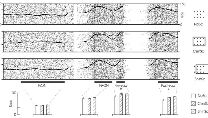
target-dependent is shown in Fig.3. For this neuron there is a general increase in activity after fixation. Nev-ertheless, the modulation by the frame only occurs after the appearance of the target. Thus, it is pos-sible to partially dissociate the phenomena of scene modulation from the engagement of the monkey on the task or from possible attentional processes.
DISCUSSION
fix-74 BRUSS LIMA, MARIO FIORANI and RICARDO GATTASS
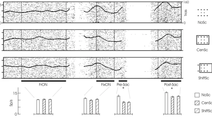
Fig. 2 – Example of a neuron with contextual scene modulation. On the raster plots in the top of the figure, the black dots and the black curve represent the cells firing and the spike density function (gaussian smoothed), respectively. The trial progresses from the left to the right. The FP and the target are turned on after the FrON or FixON periods, respectively. There is a variability in the raster length after these two periods due to the variability of the animal to acquire fixation. The average firing rate and standard error for each of the four periods in the three contexts of scene was computed and plotted in the graph below. One-way ANOVA analyzes showed a statistical significance (asterisks) between the scene contexts only after the target was turned on(P <10−6). The differential modulation by the
scene observed just before and after the monkey begins gazing the PF is discussed in the text. See also legend for Fig. 1.
ation (FixON) periods, we were probably recording from a site with some selectivity to the frame, pos-sibly a module tuned to that object as described by Wang et al. (1996, 1998). For these situations, we would have obtained the neurons response profile to an object as it is customary done for IT cortex. Nevertheless, for the example illustrated in Fig. 3, the differential response only occurred after a be-haviorally relevant target enclosed in the object was turned on. So, even though it has been demonstrated that the modules of IT cortex are tuned in their selec-tivity, there seems to be some interaction between them. It would be interesting to know the intracel-lular membrane potential during the 4 periods an-alyzed and to verify when the modulation actually started; whether before (subthreshold level) or after the behaviorally relevant target was turned on.
We observed the phenomena of modulation by
Fig. 3 – Example of a neuron with contextual scene modulation exclusively target-dependent. Conventions as in Fig. 2. Contrary to the unit illustrated in Fig. 2, this cell does not present differential modulation by the frame prior to the appearance of the target. The neurons firing rate only distinguished the presence of the scene after the target was turned on, as confirmed by one-way ANOVA analyzes between the scene contexts (Pre-Sac:P <0.03 and Post-Sac:P <10−7). See also legends for Figs. 1 and 2.
ACKNOWLEDGMENTS
We wish to thank Edil Saturato da Silva Filho and Theresa Monteiro for skillful technical assistance, and to Paulo Coutinho and Gervasio Coutinho for animal care. This research was supported by grants from CNPq, PRONEX, FUJB and FAPERJ.
RESUMO
Nós investigamos o efeito de uma cena na atividade de
células do córtex inferotemporal anterior enquanto o
ma-caco executava uma tarefa de movimento sacádico dos
olhos, com e sem o contexto de uma cena (moldura
re-tangular cinza). A maioria dos neurônios não codificou a
presença da cena quando ela foi apresentada sozinha no
campo visual e o animal estava livre para mover os olhos
(macaco na condição de visão livre) ou quando o
ani-mal estava fixando um alvo na tela. No entanto, quando
um alvo periférico era apresentado e o animal tinha que
fazer um movimento sacádico para o alvo, algumas células
foram capazes de codificar diferencialmente a presença da
cena antes ou depois de um movimento sacádico.
Palavras-chave: córtex inferotemporal, codificação es-pacial, codificação de forma, macaco cebus, córtex
perirrinal.
REFERENCES
Desimone R, Albright TD, Gross CG and Bruce C.
1984. Stimulus-selective properties of inferior tem-poral neurons in the macaque. J Neurosci 4: 2051-2062.
Gattass R and Gross C.1981. Visual topography of the
striate projection zone in the posterior superior tem-poral sulcus (MT) of the macaque. J Neurophysiol 46: 621-638.
Gattass R, Rosa MGP, Sousa APB, Piñon MCG,
Fio-rani Jr M and Neuenschwander S.1990. Cortical
streams of visual information processing in primates. Brazilian J Med Biol Res 23: 375-393.
Gallyas F.1979. Silver staining of myelin by means of
physical development. Neurol Res 1: 203-209.
Gross CG.1973. Visual functions of the
76 BRUSS LIMA, MARIO FIORANI and RICARDO GATTASS
WR, MacKay DM, Teuber HL(Eds.), Handbook
of sensory physiology: Central processing of visual information, vol. 7. Springer-Verlag, Berlim, pp. 451-482.
Gross CG, Bender DB and Rocha-Miranda CE.1969.
Visual receptive fields of neurons in the inferotempo-ral cortex of the monkey. Science 166: 1303-1306.
Gross CG, Rocha-Miranda CE and Bender DB.1972.
Visual properties of neurons in inferotemporal cortex of the Macaque. J Neurophysiol 35: 96-111.
Judge SJ, Richmond BJ and Chu FC.1980.
Implan-tation of magnetic search coils for measurement of eye position: an improved method. Vision Res 20: 535-538.
Ringo JL, Sobotka S, Diltz MD and Bunce C.1994.
Eye movements modulate activity in hippocampal, parahippocampal, and inferotemporal neurons. J Neurophysiol 71: 1285-1288.
Scheinberg DL and Logothetis NK.2001. Noticing
familiar objects in real world scenes: the role of tem-poral cortical neurons in natural vision. J Neurosci 21: 1340-1350.
Schwartz EL, Desimone R, Albright T and Gross
C. 1983. Shape recognition and inferior temporal
neurons. PNAS 80: 5776-5778.
Sobotka S, Nowicka A and Ringo JL.1997.
Activ-ity linked to externally cued saccades in single units recorded from hippocampal, parahippocampal, and inferotemporal areas of macaques. J Neurophysiol 78: 2156-2163.
Suzuki WA and Amaral DG.1994. Perirhinal and
parahippocampal cortices of the macaque monkey: cortical afferents. J Comp Neurol 350: 497-533.
Tanaka K.1996. Inferotemporal cortex and object
vi-sion. Annu Rev Neurosci 19: 109-139.
Tanaka K, Saito H-A, Fukada Y and Moriya M.1991.
Coding visual images of objects in the inferotemporal cortex of the macaque monkey. J Neurophysiol 66: 170-188.
Ungerleider LG and Mishkin M.1982. Two cortical
visual systems. In: Analysis of visual behavior (Ingle DJ, Goodale MA and Mansfield RJW, eds). The MIT Press, Cambridge, MA pp. 549-586.
Wang G, Tanaka K and Tanifuji M.1996. Optical
imaging of functional organization in the monkey in-ferotemporal cortex. Science 272: 1665-1668.
Wang G, Tanifuji M, Tanaka K.1998. Functional