(Annals of the Brazilian Academy of Sciences) ISSN 0001-3765
www.scielo.br/aabc
The synergetic modulation of the excitability of central gray matter
by a neuropeptide: two protocols using excitation waves in chick retina
VERA M.F. DE LIMA1, JOSÉ R.C. PIQUEIRA2 and WOLFGANG HANKE3 1Departamento de Medicina, Universidade Federal de São João del Rei, Campus de Divinópolis
Rua Sebastião Gonçalves Coelho, 400, 35501-296 Divinópolis, MG, Brasil
2Departamento de Engenharia de Telecomunicações e Controle, Escola Politécnica, Universidade de São Paulo
Avenida Prof. Luciano Gualberto, Trav. 3, 158, 05508-900 São Paulo, SP, Brasil
3Institute of Zoophysiology, Hohenheim University, Paracelsusstr. 91, 70599 Stuttgart, Germany
Manuscript received on February 28, 2008; accepted for publication on June 21, 2008; presented byALEXANDERW.A. KELLNER
ABSTRACT
The isolated chick retina provides anin vitrotissue model, in which two protocols were developed to verify the efficacy of a peptide in the excitability control of the central gray matter. In the first, extra-cellular potassium homeostasis is challenged at long intervals and in the second, a wave is trapped in a ring of tissue causing the system to be under self-sustained challenge. Within the neuropil, the extra-cellular potassium transient observed in the first protocol was affected from the initial rising phase to the final concentration at the end of the five-minute pulse. There was no change in the concomitants of excitation waves elicited by the extra-cellular rise of potassium. However, there was an increase on the elicited waves latency and/or a rise in the threshold potassium concentration for these waves to appear. In the second protocol, the wave concomitants and the propagation velocity were affected by the peptide. The results suggest a synergetic action of the peptide on glial and synaptic membranes: by accelerating the glial Na/KATPase and changing the kinetics of the glial potassium channels, with glia tending to accumulate KCl. At the same time, there is an increase in potassium currents through nerve terminals.
Key words:extra-cellular potassium, migraine, neuronal-glial dynamics, somatostatin, spread depression.
INTRODUCTION
The isolated chicken retina provides an experimental model that have been studied for more than 40 years and basic research on the spreading excitation phenom-enon is well developed (Martins-Ferreira and Oliveira e Castro 1966, Martins-Ferreira and Do Carmo 1987, Fernandes de Lima and Hanke 1997). In this paper, we present two protocols designed to monitor efficacy properties of a peptide against G-protein coupled recep-tors in intact tissue. The neuropeptide used was Somato-statin (SS), a family of homologous integral membrane proteins (receptors SST 1 to 5). The receptors regulate
Correspondence to: José R.C. Piqueira E-mail: [email protected]
protein phosphorylation, control second messenger pro-duction and regulate membrane potential partly through voltage and Ca-dependent potassium channels. Struc-turally, SS receptors belong to the superfamily of G-protein coupled receptors (Moller et al. 2003). All the five receptors have been reported in vertebrate retinas, in neuronal, glial and endothelial cells (Thermos 2003). The SST 2 receptor acts through the NO/c-GMP pathway in neuronal and glial cell lines in the retina (Vasilaki et al. 2002, 2004, Mastrodimou et al. 2006). The protective effects of SS against excitotoxicity appear to be mediated via this receptor (Catalani et al. 2007).
waves have been recorded in this experimental model (Ulmer et al. 1995, Dahlem and Hanke 2005). Thus, one could predict the effects on the electrophysiological potassium wave concomitants (Do Carmo and Martins-Ferreira 1984) recorded in the experiments reported in this paper. The expected results were: lower amplitude of the wave concomitants, acceleration of equilibrium within the neuropil of exogenous potassium pulses, rise in threshold for wave elicitation and decrease in prop-agation velocity of excitation waves (retinal spreading depression waves).
The in vitro chick retina provides an ideal tissue model to observe excitation waves (Fernandes de Lima and Hanke 1997), and the spread of excitation waves are a good model to observe neuronal-glial interactions. Furthermore, the central gray matter of the retina has the widest dynamic range of operation (six log units of input energy from dark adapted to full day light) and thus its network must have very fine control of excitabil-ity, in order to maintain the output under light and in the dark.
Somatostatinergic pharmacology is under investi-gation for a wide range of therapeutic applications rang-ing from anti-proliferatrang-ing agent in cancer and diabetic retinopathy, to analgesic and antiepileptic action (Ther-mos 2003, Binaschi et al. 2003) as well as modulatory action in cognitive syndromes such as Alzeimer’s de-mentia, and as a cognition enhancing agent (Epelbaum 1986, Pittaluga et al. 2001, Kumar 2005).
The two protocols presented here were made such that the central nervous system is observed at very dif-ferent states. In the first, exogenous potassium pulses are applied in a system at rest, or at least with large time intervals between challenges to the ionic homeostasis. On the other hand, in the second protocol, the system is maintained in a state of permanent challenge by trap-ping an excitation wave in a circle of tissue (Martins-Ferreira et al. 1975). The effect of the peptide is ob-served in the extra-cellular potassium and potential drop transients that accompanies the wave propagation, as well as in the propagation velocity within a cycle.
The results of both protocols are interpreted in the context of the conceptual framework derived from the non-linear thermodynamics that describes waves in ex-citable systems (Nicolis and Prigogine 1989).
It must be emphasized that the aim of these ex-periments is to obtain qualitative conclusions about the modulation of oscillations and electrochemical waves and their interactions at the multicelular level of the CNS (Central Nervous System). Consequently, the data are analyzed without statistical techniques, as the sys-tems are considered to respond in a robust way to the stimulus presented (Dahlem and Hanke 2005, Siegel 1996, Epelbaum, 1986).
MATERIALS AND METHODS
The methods used here are described by (Do Carmo and Martins-Ferreira 1984, Martins-Ferreira et al. 1975). The chosen SS concentration was in the micro-molar order of magnitude, three orders of magnitude larger than the used with isolated receptors (Martins-Ferreira and Oliveira e Castro 1966, Martins-Ferreira and Do Carmo 1987, Fernandes de Lima and Hanke 1997).
AGENTS
The Somatostatin 14 and 28 were purchased from Sigma, St. Louis, MO, USA. The ion-exchanger resine used was the Corning 477317 for K.
ANIMALS ANDSLICEPREPARATION
Young chicks with ages from 10 to 15 days were killed by decapitation and the enucleating processes of the eye-cups were performed immediately. The vitreous humors were removed and the eyes were cut in strips of about 4×10 mm, which were immersed in Ringer’s solution
maintained at 30 degrees Celsius.
Only tissue experiments were done, and they are in agreement with all federal regulations of the involved authors’ countries.
RETINARINGER’SSOLUTION
NaCl 100 mM; KCl 6 mM; Na H2PO4 1mM; NaHCO3
30 mM; MgSO4 1 mM; CaCl2 1 mM; Tris 10 mM and
Glucose 30 mM. The pH of the solution was adjusted to 7.4.
ELECTRODES ANDRECORDINGSYSTEM
NaCl. The silanized ion-sensitive tip (filled with Corn-ing 477317) was backfilled with 150 mM KCl. All potentials recorded within the retina were measured against a grounded silver/silver chloride/agar/Ringer’s bridge electrode. The electrodes tips had a diameter be-tween 10 and 20 microns. At the beginning and at the end of the experiments, the electrodes response was cal-ibrated using Ringer’s solutions in which the potassium concentration was raised to 10, 15, 30 and 60 mEq/l substituting for equal amounts of NaCl taken from the Ringer in order to maintain osmolarity. The extra-cellu-lar potential and potassium activity sensing probes were mounted in a micro-drive and the output connected to a two-channel electrometer (WPI-FD-223). The signals were displayed in a Grass paper recording and saved in a magnetic tape for further processing.
PROTOCOLI – EXOGENOUSADMINISTRATION OFHIGH
POTASSIUMRINGER’SSOLUTIONS
According to the description presented in (Do Carmo and Martins-Ferreira 1984), the slices of retina were separated from the sclera preserving the pigment epithe-lium as much as possible and transferred to an exper-imental chamber where they were positioned with the vitreous side up. The chamber had a volume of 0.2 ml and it was perfused with Ringer at a rate of 0.6 ml/min. It was assured a uniform laminar flow with no mechan-ical disturbance. A bypass system coupled to the main superfusing circuit was used to introduce the high potas-sium pulses through the opening of valves, so that no mechanical disturbance was associated with the change in potassium as is the case with pressure injection techniques. The temperature was maintained at 30◦C.
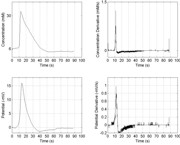
After positioning the electrode at the centre of the slice and within the inner plexiform layer, a control wave was elicited mechanically with a gentle touch with a tungsten needle (tip diameter less than 100 microns). The stimulus was delivered closed to the exit of the perfusion solution such that the wave propagated against the flow. The presence of the wave was verified ob-serving the intrinsic optical signal associated with it. In Figure 1, the extra-cellular potassium concentration and potential associated with the retinal spreading depression wave recorded at the inner plexiform layer is shown.
The sharp profile of these concomitant waves
across the retina makes possible to recognize the elec-trode position (Do Carmo and Martins-Ferreira 1984). After 30 minutes rest, pulses of high potassium solu-tions were introduced in the perfusion system. Concen-trations of 10, 12 and 15 mEq/l were used. Recordings began 10 minutes before the pulse and continued until 5 minutes after the extra-cellular level came back to base-line level. One-hour interval was used between pulses of increasing concentrations. A second series of pulses was applied by adding Somatostatin to the high potas-sium pulses at concentrations of 1 and 4 micro-mol in different slices.
PROTOCOLII – THECIRCLINGWAVEPREPARATION
The method to obtain circling waves has already been published in detail (Martins-Ferreira et al. 1975, Fer-nandes de Lima et al. 1993, FerFer-nandes de Lima and Hanke 1996). In brief, after removing the vitreous, a circular cut was made in the eyecup forming a ring of tissue. The eyecup was positioned in a perfusion cham-ber with 5 ml volume capacity. The rate of perfusion was 1.2 ml/min. In the narrow part of the ring, a wave was elicited by a gentle touch destroying one of the wave fronts by spraying a Ringer solution with a mag-nesium sulfate concentration of 4 mM over it, by using a syringe and flexible needle. The other front was let to propagate and the flow in the system transiently in-creased to 12 ml/min (about 20 seconds). With a wave trapped in the ring of tissue, the electrode was posi-tioned in the inner plexiform layer. When the propaga-tion velocity was stabilized, usually after 10 to 15 cycles, the perfusion solution was changed to a Ringer contain-ing SS at a 4µM concentration for 30 minutes. If the
circling continued, a second exogenous administration of SS was applied.
RESULTS
PROTOCOLI
Fig. 1 – Typical record of electrophysiological wave concomitants at the inner plexiform layer of chick retina. Upper row: Extra-cellular potassium activity and its derivative. Lower row: Extra-cellular potential and its derivative.
similar with either peptide and thus are presented only as SS.
The laminar flow of solution implied a constant rate of rise of potassium in the bulk solution, propor-tional to the added K concentration in the Ringer. The bulk solution reached equilibrium within forty seconds of perfusion change (not shown), but the tissue response was slower and more complex.
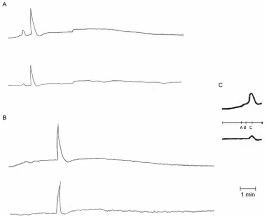
The tissue response can be divided into four phases, the rising phase with two slopes, the non-linear “mass active” phase with or without a full wave, and the falling off phase. In Figure 2C the details of the rising phase of the potassium within the inner plexiform layer are shown. An accelerated one follows a slow linear rise followed by a non-linear increase reaching a peak that usually was above the exogenous applied potassium concentration. This dome shaped “active” response pro-duced potential drops of 200 to 300µV. The potential
drop is shown in the trace bellow the potassium record. In Figure 2A, the two traces show the tissue re-sponse to a pulse of 10 mEq/l of potassium. After the peak of the dome response (11 mEq/l), the extra-cellular
potassium activity falls to 9 mEq/l and slowly rises up to the abrupt explosive growth of a spreading depression wave. In Figure 2B the two traces show the tissue re-sponse in the presence of SS (4µM). There was one
hour interval between the recordings shown in A and B. One can notice that the faster rising phase and the potas-sium decrease in the sequel imply a long latency for the appearing wave.
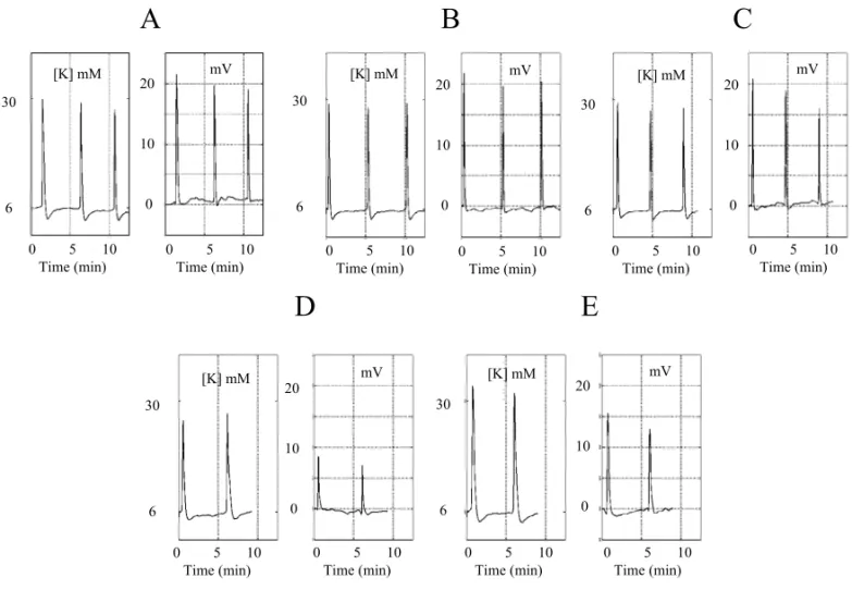
In Figures 3A–E, the sequence of pulses of another experiment is shown. In this experiment, the interval be-tween pulses was also of one hour. The concentration of SS added to the Ringer was of 1µM. The extra-cellular
potassium peaks at the “active” dome were: 10,5 mEq/l for the 10 mEq/l K pulse, 15 mEq/l for the 12 mEq/l and 18 mEq/l for the 15 mEq/l. These values were mea-sured for the potassium challenges in the absence of SS. In the presence of 1µM of SS, the values were 14 mEq/l
Fig. 2 – A – Upper row: extra-cellular potassium concentration versus time, lower row: simultaneous extra-cellular potential (mV) versus time. The pulse of potassium concentration is from 6 to 10 mEq/l. B – Same slice one hour later. C – Detail of the initial response shown in A. The labels A, B and C in C picture show three phases of initial growth, two linear and the last non-linear “active” response.
A B
C
D E
Concentration [mM]
Concentration [mM]
Concentration [mM]
Concentration [mM]
Concentration [mM] Potential
(mV)
Potential (mV)
Potential (mV)
Potential (mV)
Potential (mV) 30
6
30
6
30
6
30
6
30
6 15
10
5
0
15
10
5
0
15
10
5
0
15
10
5
0
15
10
5
0
0 2 4 6 8 10 Time (min)
0 2 4 6 8 10 Time (min) 0 2 4 6 8 10
Time (min)
0 2 4 6 8 10 Time (min)
0 2 4 6 8 10 Time (min)
0 2 4 6 8 10 Time (min)
0 2 4 6 8 10 Time (min) 0 2 4 6 8 10
Time (min)
0 2 4 6 8 10 Time (min)
0 2 4 6 8 10 Time (min)
In the presence of the peptide, the values were 11 mEq/l for the 12 mEq/l pulse and 10.5 mEq/l for the 15 meq/l pulse. The recovery phase was always much slower than the rising phase, and it took between 5 to 6 minutes to return to the baseline values.
Finally, the presence of SS had a marked influ-ence in the exogenous potassium pulses ability of elicit-ing full fledge propagatelicit-ing excitation waves in the reti-nas. As can be seen in Figures 2 and 3, in the pres-ence of SS waves, the propagation of the excitation was elicited with longer latencies. On the other hand, the in-ternal wave dynamics, i.e., the peak extra-cellular potas-sium, the potential drop values and the explosive rate of growth of both variables were not affected by the pep-tide. In Figures 2 and 3 the qualitative changes in the tis-sue response observed in the presence of SS are shown: there was either an increase in the threshold level for the exogenous applied potassium to elicit waves, or the la-tency for the wave was increased. In Figure 3 the thresh-old was increased from 12 to 15 mEq/l. The response to the 15 mEq/l pulse, instead of two, presented only one wave. Also the levels of extra-cellular potassium in the tissue at the end of the pulse (5 minutes) were lower in the presence of SS.
PROTOCOLII
The circling wave experiments were performed in 6 eyecups. The SS concentration used was 4µM in all
experiments. Figures 4 and 5 summarize the results of a typical experiment. In Figure 4 the mean propaga-tion velocity is plotted against the wave cycle number. The recording electrode was positioned very close to the border of the inner circular cut. The length of this circle was 20 mm. Dividing 20 mm by the period in minutes between one wave and next, one obtains the propagating velocity for that period. A total of 45 waves were recorded in this experiment, close to 4 hours re-cording time. Twenty-two waves were recorded before the first exogenous application of SS to the system. This first application happened after a long period of steady state behavior of propagation velocity and extra-cellular potassium and potential drop dynamics. In Figure 5A–E the shape of successive electrochemical wave concomi-tants is displayed.
The propagation velocity before SS administration was (n = 22) 4.22 mm/min±0.04. The minimum value
after 30 minutes of SS was 3.7 mm/min, a reduction of 11%. Washing out the peptide produced partial recov-ery of the propagation velocity (n = 8) 4.02±0.04.
A second application of the peptide (20 minutes) again reduced the velocity to a minimum of 3.58 mm/ min and then the circling stopped at about 30 minutes after the removal of SS application. Under the influence of the peptide the propagation velocity was (n = 14) 3,86 mm/min±0.21.
In Figure 5A–E, we show the temporal evolution of the extra-cellular potassium and extra-cellular potential drop concomitant to the wave passage around the elec-trodes.
The recordings shown in Figure 5 correspond to the waves marked in Figure 4 by dark bars under the plot. The three waves inAandBwere recorded before the application of SS. The waves inCwere recorded at the beginning of the 30 minutes application and inDat the end of it.
The waves inAandBshow the potassium peaks fluctuating near 30 mEq/l and the potential drop around 20 mV. When SS was added to the Ringer (tracesC), the peak potassium at the beginning was not affected; by contrast, the potential drop was decreased by 5 mV. At the end of the pulse (tracesD), the peak potassium was slightly increased compared to the control values as well as the potassium wave duration. The extra-cellular potential drop fell to 12 mV (a reduction of 40% in amplitude). SS was washed off after the second wave inDand there was an immediate recovery of the prop-agation velocity and amplitude of the potential drop. A second application of SS depressed all the wave con-comitants. On SS removal, there was partial recovery of the peak amplitudes whereas the under shoot of the potassium was even more prolonged (traceE).
add Somatostatin
add Somatostatin
remove Somatostatin
remove Somatostatin
Fig. 4 – Wave propagation velocity (mm/min) through a circle of 20 mm length versus the wave number. The intervals A to E show the waves that have the electrophysiological concomitants shown in Figure 5.
A B
C
D E
0 5 10Time (min)
0 5 10 Time (min) 0 5 10
Time (min)
0 5 10 Time (min)
0 5 10 Time (min) 0 5 10
Time (min)
0 5 10 Time (min)
0 5 10 Time (min) 0 5 10
Time (min)
0 5 10 Time (min) 30
6
[K] mM [K] mM [K] mM
[K] mM [K] mM
30
6
30
6
30
6
30
6
mV mV mV
mV mV
20
10
0
20
10
0 20
10
0
20
10
0
20
10
0
DISCUSSION
THESS INFLUENCE IN THEGLOBALRESPONSE OF THE
CENTRALGRAYMATTER
In the presence of SS, the threshold extra-cellular pot-assium concentration needed to elicit excitation waves was increased and the latency of elicited waves was pro-longed. SS also affected the propagation velocity of cir-cling waves slowing down the propagation. A second ob-served change in circling experiments was the decrease in brightness of the intrinsic optical signals that are con-comitant with retinal spreading depression waves – the IOS of RSDs (Fernandes de Lima and Hanke 1997). These qualitative changes suggest synergy of effects in glial and neuronal membranes in the direction of a damp-ening in the tissue excitability. These results were ex-pected and are in agreement with the previous findings with NO donors and the membrane permeable c-GMP derivatives (Ulmer et al. 1995, Dahlem and Hanke 2005). For example, the optical profile recorded in spatial line projection shows acceleration of recovery, a signal of Na/KATPase acceleration (Ulmer et al. 1995) an effect also observed with increasing the temperature (Weimer and Hanke 2005). The overall mean effect on the wave optical signals after 2 hours perfusion with NO donors was depression of amplitude of both peaks of the tem-poral profile of the intrinsic optical signal (Dahlem and Hanke 2005) suggesting both potassium channels and metabolic rate effects. We just measured the close as-sociation of the glial Na/KATPase pumping rate and the temporal profile of retinal waves.
The initial effect on the rising rate in the neuropil case, with exogenous application of potassium, can only be explained by glial membrane effects, because between the neuropil and the vitreous there is only a base mem-brane secreted by glia and the end feet of the Muller cells. The end feet membrane expresses both potas-sium channels and the Na/K ATPase. During long pulses application of the SS in the second protocol, the under shoot of potassium is prolonged from 1.5 to 5 min. The best explanation for this finding is that glia cells are ac-cumulating K by accelerating the pump and changing the kinetics of their K channels.
For this interpretation we use knowledge from previous experiments with patch-clamping glial and neuronal K channels both in situduring the wave pas-sage and with isolated cells (Hanke et al. 1993, 1997),
the effect of Barium on circling waves (Fernandes de Lima et al. 1993), and intracellular recordings on glia upon barium application (Ballany et al. 1987, Chesler and Kraig 1989), i.e., glia hyperpolarizes and the mem-brane resistance rises. Also we use the findings of Rei-chenbach et al. on the Muller cells Na/KATPase behav-ior (Reichenbach et al. 1992), i.e., potential is maxi-mal at extra-cellular potassium concentrations of 10 to 15 mM. Besides, the pump will transport potassium into the cell even with low intracellular sodium. For exam-ple, by elevating potassium in the bath to 15 mEq/l, the optical wave profile will show a depressed second peak. The exogenous potassium accelerates the pump rate to maximum, such that the breakdown of electrochemical gradients at the wavefront is not able to further accelerate it. As a consequence, the glycolysis is not increased in glia, it is at maximum rate before the wave. The second peak of the optical profile follows closely the pH shift in the extra-cellular space and lactate production by glia (Ferreira-Filho and Martins-Ferreira 1992, Hansen and Quistorff 1993).
Throughout the years, it was observed that the power of modulation of the propagation velocity is a good predictor for the efficacy of antimigraine, antiepi-leptic and protection from excitotoxicity (Ulmer et al. 1995, Wiedemann et al. 1996, Wiedemann and Hanke 1997). In dissipative structure context, this effect could be related to the global coupling within the system. The artificial model of bioelectrochemical waves developed by Wussling (Wussling et al. 1999, 2001) in which sarcoplasmic membrane vesicles and mitochondria are embedded in agarose, showed that the addition of mito-condria to the vesicle system promoted the doubling of propagation velocity of calcium waves and the smooth-ing of the wavefront shape. We also observed the wave-front breakdown with agents that slow propagation in the retina, propanolol and Sumitriptan are examples of such compounds. The present results suggest that the peptide SS synergic action uncouples the elements in the tissue, i.e., the glial and synaptic membranes interacting through the extra-cellular matrix.
THESS EFFECTS ON THEEXCITATIONWAVE
CONCOMITANTS
phases: the abrupt rise in potassium and simultaneous drop in potential, followed by a much slower recov-ery that have a potassium and potential under shoot. The time derivatives of both transients have a dominant peak that occurs at the same time for the two variables (Fig. 1). At the concentrations used (1 and 4µM),
Somatostatin did not alter the peak of the time derivative of the extra-cellular potassium and potential transients in both protocols. On the other hand, the peak of the extra-cellular potassium and potential were not affected in the first protocol (Figs. 2 and 3) whereas both peaks were depressed in the second protocol (Fig. 5), with the extra-cellular potential drop more affected than the potassium. The c-GMP build up with the longer pulses can explain the difference of short term and long-term effects of the peptide. In the circling experiments, the potassium transient duration was increased as well as the potassium under shoot (Fig. 5D). This difference is a di-rect verification about the experimental context influence in the whole tissue response to a single pharmacological agent. A change in the dynamics of the neuronal/glial interactions can change the pharmacological result.
This type of verification shows the importance of tissue models in pre-clinical research and their role between the high throughput in vitro receptor affinity measurements and the whole animal response used in toxicology.
CONCLUSIONS
There is enough knowledge about excitation waves in in vitrochicken retina that some results of pharmacolog-ical manipulations can be predicted. In the experiments shown in this paper, the predicted results could be seen by using two different protocols concerning to the same model. We assumed that since the protective effects of SS against ischemia were related to SST2 receptor and c-GMP, then the measurable effects of SS on excitation waves had to be related to c-GMP effects. Potassium ion regulation is one key feature of neuronal-glial in-teraction, and the mechanisms subjacent to this control are present in physiological and pathological situations. The spreading depression wave in the retina is a sensi-tive, highly reproducible and multiparameter event that can be very useful in the pre-clinical drug development process.
RESUMO
Retinas de pinto isoladas proporcionam um modelo de tecidos in vitro, para o qual dois protocolos foram desenvolvidos para verificar a eficácia de um peptídeo no controle da excitabili-dade da matéria cinzenta central. No primeiro, a homeostase do potássio extra-celular é desafiada por intervalos longos (1 hora) e no segundo, uma onda é capturada em um anel de tecido, de tal maneira que o sistema permaneça em estado de desafio auto-sustentado. Dentro daneuropil, o transiente de potás-sio extra-celular observado no primeiro protocolo foi afetado da fase de início de aumento à concentração final, ao final do pulso de cinco minutos. Não há mudanças nos parâmetros concomitantes das ondas de excitação geradas pelo aumento do potássio extra-celular. Entretanto, houve um aumento da latência das ondas geradas e/ou um aumento no nível de con-centração de potássio necessário para gerar a onda. No segundo protocolo, os parâmetros concomitantes da onda e sua veloci-dade de propagação foram afetados pelo peptídeo. Os resulta-dos sugerem uma ação sinergética do peptídeo nas membranas gliais e sinápticas: acelerando o Na/KATPase glial e mudando a cinética dos canais de potássio gliais, com a glia tendendo a acumular KCl. Nesse período, não há aumento nas correntes de potássio nas terminações nervosas.
Palavras-chave: potássio extra-celular, enxaqueca, dinâmica neuronal, dinâmica glial, somatostatina, depressão alastrante.
REFERENCES
BALLANYKP, GRAFEPANDBRUGGENCATEG. 1987. Ion activities and potassium uptake mechanisms of glial cells in guinea pig olphactory cortex slices. J Physiol (London) 382: 159–174.
BINASCHI A, BREGOLAGANDSIMONATOM. 2003. On
the role of somatostatin in seizure control: clues from the hippocampus. Rev Neurosci 14: 285–301.
CATALANI E, CERVIA D, MARTINI D, BAGNOLI P, SIMONATTI E, TIMPERIO AM AND CASINI G. 2007. Changes in neuronal response to ischemia in retinas with genetic alteration of somatostatin receptor expres-sion. Eur J Neurosci 25: 1447–1459.
CHESLERM ANDKRAIGRP. 1989. Intracellular pH tran-sients of mammalian astrocytes. J Neurosci 9: 2011– 2019.
DOCARMORANDMARTINS-FERREIRAH. 1984. Spread-ing depression of Leão probed with ion-selective micro-electrodes. An Acad Bras Cienc 56: 401–421.
EPELBAUMJ. 1986. Somatostatin in central nervous system:
physiology and pathological modifications. Prog Neuro-biol 27: 63–100.
FERNANDES DELIMAVMANDHANKEW. 1996. Observa-tions of non-stationarities in extra-cellular potassium dy-namics within the gray matter neuropil during self-sus-tained spreading depression. J Brain. Res 37: 505–518. FERNANDES DELIMAVMANDHANKEW. 1997.
Excita-tion waves in central gray matter: the retinal spreading depression. Prog in Retin Eye Res 16: 657–690. FERNANDES DE LIMA VM, SCHELLER D, TEGTMEYER
F, HANKEWANDSCHLUEWR. 1993. Self-sustained spreading depression in the chicken retina and short term neuronal glial interactions within the grey matter neuropil. Brain Res 614: 45–51.
FERREIRAFILHOCRANDMARTINS-FERREIRAH. 1992.
Interstitial fluid pH and its changes during spreading de-pression in isolated chicken retina. in Spreading Depres-sion. DoCarmo R (Ed.), Springer-Verlag Berlin, Heidel-berg, NY, p. 75–88.
HANKEW, FERNANDES DELIMAVMANDSCHLUEWR.
1993. Patch-clamp experiments in the intact retina during spreading depression. In: LEHMENKUELERA, GROTE
-MEYER KHANDTEGTMEYER F (Eds.), Migraine:
ba-sic mechanisms and treatment. Urban & Schwarzenberg, Munich, p. 573–582.
HANKEW, MEZLERM, BRANDS, LADEWIGTANDFER -NANDES DELIMAVM. 1997. Patch-clamp experiments at isolated cells and in intact tissue of the chicken retina correlated to potassium channels and the retinal spreading depression. Electrochim Acta 42: 3231–3240.
HANSENAJANDQUISTORFFB. 1993. Glucose metabolism in experimental spreading depression. In: MIGRAINE: BASIC MECHANISMS AND TREATMENT. LEHMEN
-KUELLER A, GROTEMEYER KH ANDTEGTMEYER F (Eds.), Urban-Swarzenberg, Munich, p. 367–378. KUMARU. 2005. Expression of somatostatin receptors
sub-types (STR1-5) in Alzheimers disease brain: an immuno-histochemical analysis. Neurosci 134: 525–538. MARTINS-FERREIRAHANDDOCARMORJ. 1987. Retinal
spreading depression and the extra-cellular millieu. Can J Physiol Pharmachol 65: 1092–1110.
MARTINS-FERREIRAHANDOLIVEIRA ECASTROG. 1966. Light scattering changes accompanying spreading depres-sion in isolated chicken retina. J Neurophysiol 29: 715– 726.
MARTINS-FERREIRA H, OLIVEIRA E CASTRO G AND
STRUCHINER CJ. 1975. Circling spreading depression
in isolated chicken retina. J Neurophysiol 37: 773–784. MASTRODIMOU N, KIAGIADAKI F, HODJAROVAM, EF
-THIMIAKANDTHERMOSK. 2006. Somatostatin recep-tors (SST2) regulate cGMP production in rat retina. Regul peptides 133: 41–46.
MOLLERLN, STIDSENCE, HARTMANNBANDHOLSTJJ. 2003. Somatostatin receptors. Biochem Biophys Acta 1616: 1–84.
NICOLISGANDPRIGOGINEY. 1989. Exploring complexity: An introduction. W.H. Freeman Company, NY, 313 p. PITTALUGAA, FELIGIONIM, GHERSIC, GEMIGNANIA
ANDRAITERIM. 2001. Potentiation of NMDA receptor function through somatostatin release: a possible mecha-nism for the cognition-enhancing activity of GABAb receptor antagonists. Neuropharmachology 41: 301–310. REICHENBACH A, HENKEA, EBERHARDTW, REICHELT
WANDDETTMERD. 1992. K ion regulation in retina. Can J Physiol Pharmachol, Supp 70: S239–S247. SIEGELG. 1996. Connective tissue: more than just a matrix
for cells. In: COMPREHENSIVEHUMANPHYSIOLOGY. GREGERRANDWINDHORSTU (Eds.), Springer 1: 173–
228.
THERMOS K. 2003. Functional mapping of somatostatin receptors in the retina: a review. Vis Res 43: 1805–1815. ULMER HJ, FERNANDES DE LIMA VM AND HANKE W. 1995. Effects of nitric oxide on retinal spreading depres-sion. Brain Res 691: 239–242.
VASILAKIA, MOURATIDOUM, SHCHULZS ANDTHER
-MOS K. 2002. Somatostatin mediates nitric oxide
pro-duction by activating SST2 receptors in rat retina. Neu-ropharmachology 43: 899–909.
VASILAKI A, PAPADAKI T, NOTAS G, KOLIOS G, MAS -TRODIMOU N, HOYERD, TSILIMBARISM, KOUROU
-MALISE, PALLIKARISIANDTHERMOSK. 2004. Effect of Somatostatin on nitric oxide production in human reti-nal pigment epithelium cell cultures. Invest Ophth Vis Sci 45: 1499–1506.
WEIMER MSANDHANKE W. 2005. Correlation between the durations of refractory period and intrinsic optical sig-nal of spreading depression during temperature variations. Exp Brain Res 161: 201–208.
WIEDEMANNMANDHANKEW. 1997. The chicken retina as a model for investigation of central nervous system lesions. Neurosci Lett 232: 99–102.
WIEDEMANNM, FERNANDES DELIMAVMANDHANKE
WUSSLING MHP, KRANICH K, LANDGRAPH G, HERRMANN-FRANK A, WIEDEMANN D, GELLERICH
FN ANDPODHAISK H. 1999. Sarcoplasmic reticulum vesicles embedded in agarose gel exhibit propagating calcium waves. FEBS Lett 463: 103–109.
WUSSLING MHP, KRANICH K, DRYGALLA V AND
PODHASKIH. 2001. Calcium waves in agarose with cell