online | memorias.ioc.fiocruz.br Like several other multidrug-resistant (MDR)
non-diphtherial Corynebacterium species, Corynebacterium striatum has been cited with increased frequency as a pathogen of nosocomial infections (Lee et al. 2005), cluding septicaemias (Martín et al. 2003), pulmonary in-fection (Renom et al. 2007, Wong et al. 2010), meningitis (Weiss et al. 1996), endocarditis (Oliva et al. 2010), os-teomyelitis (Fernández-Ayala et al. 2001), septic arthritis (Scholle 2007), keratitis (Heidemann et al. 1991), skin wounds (Moore et al. 2010) and intrauterine infections (Boltin et al. 2009). The first case of C. striatum in an uncomplicated urinary tract infection in an ambulatory patient without any other predisposing factors was re-cently reported (López et al. 2009).
Outbreaks caused by C. striatum have been reported in long-term hospitalised patients with prolonged expo-sure to broad-spectrum antibiotics and in intensive care units (ICU) (Leonard et al. 1994, Otsuka et al. 2006). The presence of multiple medical devices may facilitate
colonisation by C. striatum of the upper respiratory tract with subsequent invasive infection. Therefore, C. stria-tum should not be simply disregarded as a contaminant, especially when isolated as a pure growth in chronically debilitated patients with multiple medical devices in situ (Brandenburg et al. 1996, Renom et al. 2007).
An increase in the number of cases in which C. stri-atum has been isolated from clinical specimens from patients with varying degrees of immunocompromisa-tion and severe infecimmunocompromisa-tions has been observed in indus-trialised countries that can afford these types of stud-ies. With the aid of molecular-biology-based techniques, the clonal nature of the isolates in nosocomial outbreaks has been unequivocally established (Brandenburg et al. 1996, Martín et al. 2003, Otsuka et al. 2006, Renom et al. 2007, Adderson et al. 2008, Campanile et al. 2009, Martins et al. 2009, Wong et al. 2010).
In South America, only reports from Brazil indicate the sporadic isolation of C. striatum from representa-tive clinical sites of hospitalised patients with signs and symptoms of infection (Camello et al. 2003, Martins et al. 2009, Superti et al. 2009). In contrast to previ-ous data observed by our research group (Camello et al. 2003), an unusual clustering of 14 patients in our teach-ing hospital within a period of nine months produced a sentinel signal that justified the study of a possible outbreak. Therefore, the microbiologic characteristics, resistance profiles and similarities among the genomes of 15 C. striatum strains isolated from these patients were investigated.
Financial support: CAPES, FAPERJ, CNPq, PAPES V-FIOCRUZ/ CNPq, SR-2/UERJ, PNPD-CAPES/MEC, PAPD-FAPERJ/CAPES PVPB and HFM contributed equally to this work.
+ Corresponding author: [email protected] Received 13 February 2012
Accepted 2 October 2012
Clonal multidrug-resistant Corynebacterium striatum
within a nosocomial environment, Rio de Janeiro, Brazil
Paulo Victor Pereira Baio1,4,5,Higor Franceschi Mota1, Andréa D’avila Freitas1,2,
Débora Leandro Rama Gomes1,6,Juliana Nunes Ramos1,4,Lincoln Oliveira Sant’Anna1,
Mônica Cristina Souza1,Thereza Cristina Ferreira Camello1,3, Raphael Hirata Junior1,
Verônica Viana Vieira4, Ana Luíza Mattos-Guaraldi1/+
1Laboratório de Difteria e Corinebactérias de Importância Clínica, Departamento de Microbiologia,
Imunologia e Patologia, Faculdade de Ciências Médicas 2Unidade Docente Assistencial de Doenças Infecciosas e Parasitárias 3Laboratório de Bacteriologia, Hospital Universitário Pedro Ernesto, Universidade do Estado do Rio de Janeiro, Rio de Janeiro, RJ, Brasil
4Departamento de Microbiologia, Instituto Nacional de Controle de Qualidade em Saúde-Fiocruz, Rio de Janeiro, RJ, Brasil 5Laboratório Químico Farmacêutico do Exército, Ministério da Defesa, Rio de Janeiro, RJ, Brasil
6Faculdade de Farmácia, Instituto Federal de Educação, Ciência e Tecnologia do Rio de Janeiro, Rio de Janeiro, RJ, Brasil
Corynebacterium striatum is a potentially pathogenic microorganism with the ability to produce outbreaks of nosocomial infections. Here, we document a nosocomial outbreak caused by multidrug-resistant (MDR) C. striatum in Rio de Janeiro, Brazil. C. striatum identification was confirmed by 16S rRNA and rpoB gene sequencing. Fifteen C. striatum strains were isolated from adults (half of whom were 50 years of age and older). C. striatum was mostly isolated in pure culture from tracheal aspirates of patients undergoing endotracheal intubation procedures. The analysis by pulsed-field gel electrophoresis (PFGE) indicated the presence of four PFGE profiles, including two related clones of MDR strains (PFGE I and II). The data demonstrated the predominance of PFGE type I, compris-ing 11 MDR isolates that were mostly isolated from intensive care units and surgical wards. A potential causal link between death and MDR C. striatum (PFGE types I and II) infection was observed in five cases.
C. striatum in Brazil • Paulo Victor Pereira Baio et al. 24
SUBJECTS, MATERIALS AND METHODS
Bacterial strains - We reviewed the microbiological features of 15 C. striatum strains recovered from repre-sentative clinical sites of 14 hospitalised patients (50% male; 50% with fatal outcomes) with signs and symp-toms of bacterial infection. The patients were hospital-ised between August 2009-April 2010 in seven different wards of a 600-bed teaching hospital in Rio de Janeiro (RJ), Brazil (Table). All C. striatum isolates from the pa-tients included in the study were detected using routine diagnostic cultures.
Bacterial phenotypic characterisation - Coryne-bacterium-like colonies were selected for further iden-tification when they were grown in any quantity from normally sterile body fluid or when they were isolated in significant numbers or in pure culture from other specimens obtained at clinical sites where infection was suspected (Funke & Bernard 2007). All clinical samples yielding more than three organisms were regarded as contaminated and discarded (Thomson & Miller 2007). For quantitative bronchoalveolar lavage (BAL) fluid cul-bronchoalveolar lavage (BAL) fluid cul- fluid cul-tures, a colony count > 103 colony-forming units (CFU) mL-1 of potential pathogens was considered positive. Isolation of three species of microorganisms was classi-fied as a polymicrobial infection of the lower respiratory tract. Microorganisms were identified from the urine cultures in cystine lactose electrolyte-deficient agar (Merck, Darmstadt, Germany) and were considered po-tential pathogens when the growth exceeded 104 CFU mL-1 as the only isolate or > 105 CFU mL-1 as the pre-dominant isolate; in cases of nephropathies, > 103 CFU mL-1 was also considered a potential pathogenic level. Blood cultures were always obtained in pairs, wherein at least one of the samples was collected through the central venous catheter, if present. Blood specimens
were inoculated in Bactec Plus anaerobic⁄aerobic vials
and processed in a Bactec 9240 continuous-monitoring system (Becton-Dickinson Microbiology System, Cock-eysville, MD, USA). Other clinical specimens were in-oculated onto a Columbia agar base with the addition of 5% sheep’s blood and incubated at 37ºC in a 3-5% CO2atmosphere and monitored for 72 h (Camello et al. 2003, Martins et al. 2009). Positive bacterial cultures for irregular Gram-positive rods were preliminarily characterised by colonial morphology, pigmentation, haemolysis, DNase activity and CAMP reaction with Staphylococcus aureus (Camello et al. 2003, Funke & Bernard 2007, Pimenta et al. 2008). Phenotypic charac-terisation was also performed using the semi-automated API Coryne System 3.0 (bioMérieux) with the API web decoding system (apiweb.biomerieux.com) (Almuzara et al. 2006, Funke & Bernard 2007, Campanile et al. 2009, Martins et al. 2009).
Susceptibility testing - Antimicrobial susceptibility profiles were determined by the disk diffusion method in cation-adjusted Mueller-Hinton agar supplemented with 5% sheep blood. Breakpoints for the susceptible strains were used as suggested by the Clinical Laborato-ry Standards Institute (CLSI) for bacteria excluded from
tables 2A-K. As there is not yet a defined standard for interpreting these results, the standard proposed in CLSI document M45-A (ISBN 1-56238-607-7) was used (CLSI 2007). The breakpoints for S. aureus were considered in the cases of penicillin, oxacillin and ampicillin. For the other antimicrobial agents, we used the breakpoints for other microorganisms, but not Haemophilus spp or Neisseria gonorrhoeae, which had been validated by previous studies. Intermediate results were considered resistant (Camello et al. 2003, Martins et al. 2009). The antibiotics (Oxoid SA, Spain) tested included penicillin (10 U), ampicillin (30 µg), methicillin (5 µg), cefotaxime (30 µg), cefepime (30 µg), ceftriaxone (30 µg), imipenem (10 µg), erythromycin (15 µg), clindamycin (2 µg), lin-ezolid (30 µg), ciprofloxacin (5 µg), moxifloxocin (5 µg), tetracycline (30 µg), gentamicin (10 µg), rifampin (5 µg), fosfomycin (200 µg), vancomycin (30 µg), mupirocin (200 µg), tobramycin (10 µg), nitrofurantoin (300 µg) and ticarcillin/clavulanate (75 µg/10 µg).
Gene amplification and sequencing - C. striatum identification was confirmed by 16S rRNA and rpoB gene sequencing. Each strain was grown in Brain Heart Infusion broth by incubation for 24/48 h at 30ºC and centrifuged for 5 min at 3,000 rpm. The pelleted bac-teria were suspended in 500 µL of sterile water and subsequently boiled for 15 min for DNA extraction. Cell extracts were then immediately stored at -20ºC for use in polymerase chain reaction (PCR) reactions. 16S rRNA gene was amplified using universal primers pA (5’-AGAGTTTGATCCTGGCTCAG) and pH (5’-AAG-GAGGTGATCCAGCCGCA), as described by Watts et al. (2000). The purified PCR product was sequenced by primer walking with the oligonucleotides using the fol-lowing primers for sequencing: 1831 (5’-GAGGAAC- ACCGATGGCGAAGGC), 1832 (5’-GCCCCCGTCA- ATTCCTTTGAGTT) (Watts et al. 2000) and 519r G(AT)ATTACCGCGGC(GT)GCTG) and 1242f (5’-CACACGTGCTACAATGG) (Johnson 1994). The rpoB gene was amplified and sequenced using primers fol-lowing procedures described previously (Khamis et al. 2004). The sequencing reactions were performed using a BigDye Terminator v 3.1 cycle sequencing kit (Applied Biosystems) on an ABI-3730 Automated DNA Sequenc-er (Applied Biosystems) following standard protocols. The 16S rRNA gene sequences were compared to those available in the National Center for Biotechnology Infor-mation (ncbi.nlm.nih.gov) using the BLAST algorithm and the Ribosomal Database Project II (rdp.cme.msu. edu/html). The rpoB gene sequences were only com-pared in the GenBank database.
2
5
Mem Inst Oswaldo Cruz
, Rio de Janeiro, V
ol.
108
(1), F
ebruary 2013
Characteristics of 15 Corynebacterium striatum strains isolated from infected patients within nosocomial environment
Number of patient/strain
Gender/age (years-old)
Date of bacteriological exams
(month/year)
Hospital wards
Outcome (month/year)
Clinical
specimens Culture
API Coryne code
Phenotypic profiles
PFGE type
1/2023BR-RJa M/66 Aug/09 General ICU Death
Oct/09
Blood Pure (NI)
0100105 Nit-/Pyz-/Suc+; MDR
I
2/2032BR-RJ F/23 Aug/09 General ICU Cure Sep/09
Urine Pure (1.0 x 10⁶ CFU)
2100105 Nit-/Pyz+/Suc+; MDR
I
3/2038/39BR-RJ F/54 Sep/09 Infectious diseases Nov Bloodb Pure
(1.0 x 10⁶ CFU)
0100104 Nit-/Pyz-/Suc-; MDR
II
4/1954BR-RJa M/NI Oct/09 Thoracic surgery NI Surgical wound
secretion
Pure (NI)
3100105 Nit+/Pyz+/Suc+; MDS
IV
5/1958BR-RJa M/51 Nov/09 Thoracic surgery NI Tracheal aspirateb (1.0 x 10⁶ CFU)
+ Klebsiella pneumoniae
(1.0 x 10⁵ CFU)
0100105 Nit-/Pyz-/Suc+; MDR
I
6/1959BR-RJa M/85 Dec/09 Cardiac ICU Cure
Mar/10
Tracheal aspirateb Pure
(1.0 x 10⁶ CFU)
0100105 Nit-/Pyz-/Suc+; MDR
I
7/1961BR-RJa F/37 Dec/09 Infectious diseases NI Urine Pure
(2.5 x 10⁵ CFU)
3100105 Nit+/Pyz+/Suc+; MDS
III
8/1974BR-RJa M/59 Dec/09 Urology Death
Mar/10
Tracheal aspirateb Pure
(4.0 x 10⁴ CFU)
0100105 Nit-/Pyz-/Suc+; MDR
I
9/1987BR-RJa F/50 Dec/09 Nursery 18 Death
Dec/09
BAL Pure
(2.0 x 10⁵ CFU)
2100105 Nit-/Pyz+/Suc+; MDR
I
10/2369BR-RJa M/NI Dec/09 General ICU Cure
Apr/10
Tracheal aspirateb Pure
(1.0 x 10⁶ CFU)
2100104 Nit-/Pyz+/Suc-; MDR
II
11/2539BR-RJ F/NI Dec/09 Nursery 18 Death Dec/09
Tracheal aspirateb Pure
(1.0 x 10⁶ CFU)
0100105 Nit-/Pyz-/Suc+; MDR
I
12/2063BR-RJa F/NI Jan/10 General ICU Death
Jan/10
Tracheal aspirateb Pure
(1.0 x 10⁶ CFU)
2100105 Nit-/Pyz+/Suc+; MDR
I
13/2061BR-RJ M/57 Feb/10 Thoracic surgery Cure Feb/10
Tracheal aspirateb Pure
(1.0 x 10⁶ CFU)
2100105 Nit-/Pyz+/Suc+; MDR
I
14/2083BR-RJ; 2084BR-RJ
F/38 Apr/10 ICU II Death Apr/10
Tracheal aspirateb;
CSF
Pseudomonas + Staphylo-coccus sp.;
Pure (NI)
2100105 Nit-/Pyz+/Suc+; MDR
I
C. striatum in Brazil • Paulo Victor Pereira Baio et al. 26
Pulsed-field gel electrophoresis (PFGE) - Genomic DNA was prepared following a method described previ-ously (García-Crespo et al. 2005). The DNA was cleaved with SwaI (New England BioLabs) according to the man-ufacturer’s instructions. PFGE was carried out in 0.5X TRIS-borate-EDTA-1.2% agarose gels at 13ºC with a CHEF DRII system (Bio-Rad). The pulse times were 1-30 s over 22 h. A lambda DNA concatemer (New England Bi-oLabs) was used as a molecular size marker. Similarities among macrorestriction patterns were identified accord-ing to the criteria established by Tenover et al. (1995).
Ethics - This study was developed in compliance with the Brazilian Government’s Ethical Guidelines for research involving human beings (resolution of the Na-tional Health Council/Ministry of Health) and approved by the ethical research committee (INCA-CEP 008/06).
RESULTS
Epidemiological aspects - All 15 C. striatum clinical samples and the patients’ ages, genders, hospital wards, outcomes, sites of isolation and associated microorganisms are displayed in chronological order in the Table. With the exception of four patients for whom age was not avail-able, C. striatum strains were isolated only from adults (7 males and 7 females; half of whom were 50 years of age or older). Most of the strains were grown in pure cul-ture (n = 13) and from tracheal aspirates (n = 9). Numeri-cally predominant colonies of C. striatum were observed in two polymicrobial cultures from tracheal samples: one co-infected with Klebsiella pneumoniae and another with Pseudomonas sp. and Staphylococcus sp. C. striatum strains were also isolated from blood (n = 1), cerebrospi-nal fluid (CSF) (n = 1), BAL (n = 1), surgical wounds (n = 2) and urine (n = 2) samples. From the deceased patient, number 14, C. striatum strains were isolated from both the tracheal aspirate and the CSF (Table).
When considering bacterial growth in pure culture and the proximity between microbiological diagnosis and death of the patients, the possibility of a causal link between death and C. striatum infection was observed in five cases (3, 9, 11, 12 and 14) (Table). At least half of the patients previously infected with C. striatum died, indicating a high severity of illness and/or immune de-ficiency. All seven deaths during the period of study oc-curred in patients from whom C. striatum was isolated in a pure culture (3 in December 2009).
Fig. 1 presents the epidemic curve of the infections caused by C. striatum, suggestive of a nosocomial out-break. Most of the C. striatum strains (n = 6) were iso-lated in December 2009 from patients from various hos-pital wards. The pathogen was found circulating mostly among inpatients admitted to the ICU and surgical wards (n = 10) from August 2009-April 2010 (Table). C. stria-tum infections were observed in the general ICU (n = 4), cardiac ICU (n = 1), ICU II (n = 2), thoracic surgery (n = 3), nursery 18 (n = 2), infectious diseases (n = 2) and urology (n = 1) wards.
Antimicrobial susceptibility, phenotypic and geno-typic properties of C. striatum clinical isolates - MDR profiles for 21 of the antimicrobial agents tested were
observed in 87% of the C. striatum strains that were sus-ceptible only to vancomycin, linezolid and tetracycline. Two strains (1954BR-RJ surgical wound isolate and 1961BR-RJ urine isolate) were susceptible to most of the tested drugs (MDS) except mupirocin, fosfomycin and ticarcillin/clavulanate. Deaths occurred only in patients from whom MDR C. striatum strains were isolated in the blood, CFS, BAL and/or tracheal aspirate.
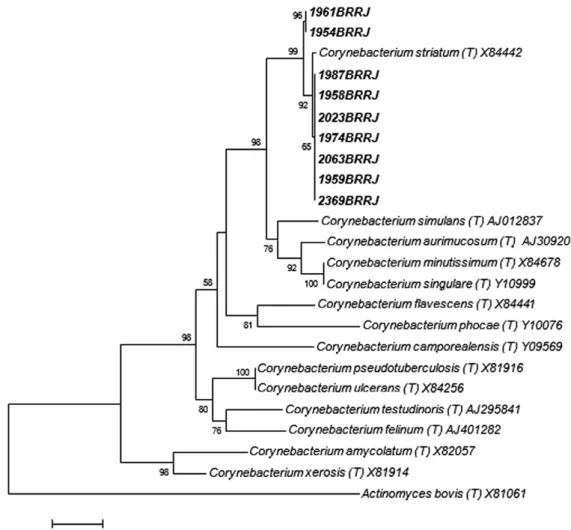
The phenotypic analysis of C. striatum strains revealed variability in the results of nitrate reductase and pyrazin- pyrazin-amide (pyz) activities and/or sucrose fermentation (Table). To genotypically confirm the suspected C. striatum iso-lates, we performed 16S rRNA and rpoB gene sequenc-ing. GenBank accessions for the 16S rRNA and rpoB genes sequences were deposited: JF342692-JF342700 and JF342701-JF342709, respectively. The 16S rRNA se-quences from the clinical isolates exhibited the highest similarity values to the type strain of C. striatum (ATCC 6940), ranging from 99.32-99.84%. The phylogenetic ana- lysis, based on the 16S rRNA sequences, unambiguously demonstrated that the clinical isolates belonged to the C. striatum species, as illustrated in Fig. 2. A comparative analysis of the partial rpoB sequences for the clinical iso-lates revealed sequence similarities in the ranges of 97.08-97.15% with the type strain C. striatum (ATCC 6940).
PFGE analysis - The restriction endonuclease SwaI revealed four distinct PFGE profiles among the C. stria-tum isolates, which were designated I, II, III and IV. The PFGE profile I was the most frequently observed among the 15 strains (78.57%) isolated from the 14 patients (71.42%) included in this study (Fig. 3). PFGE analysis confirmed the isolation of a single clone of C. striatum (PFGE I) from the respiratory tract and CSF of the pa-tient of case 14. Profiles II, III and IV differed by more than three bands from PFGE profile I by visual inspec-tion. According to the interpretation criteria by Tenover et al. (1995), MDR clinical isolates belonging to profile II exhibited six bands of difference from the outbreak MDR strain and were considered potentially related to the out-break. The MDS isolates belonging to profiles III and IV exhibited more than six bands of difference, indicating that they were unrelated to the outbreak strain (Fig. 3).
DISCUSSION
In Brazil, previous investigations revealed that 1.9% of samples isolated from varied clinical sources of can-cer patients treated in a reference centre in RJ during a one-year period were positive for C. striatum, corre-sponding to one case of upper and three of lower respira-tory tract infections and two cases of surgical wounds (Martins et al. 2009).
In the present study, we documented a nosocomial outbreak that includes a fatal case of systemic infection caused by C. striatum in a 600-bed teaching hospital in RJ. PFGE analyses of 15 C. striatum strains indicated the presence of four PFGE profiles, including two re-lated clones of MDR strains (PFGE I and II). Our data demonstrated the predominance of PFGE type I com-prising 11 MDR isolates that were mostly isolated from the ICU and surgical wards. The tracheal aspirate speci-mens obtained from patients with ventilator-associated respiratory tract colonisation or pneumonia were mostly (6 out of 7) PFGE type I. The possibility of a causal link between death and C. striatum (PFGE types I and II)
Fig. 2: phylogenetic tree based on neighbour-joining method using 16S rRNA gene sequences. Distance estimations were calculated by using the Kimura two-parameter model. Bootstrap percentages after 1,000 simulations are shown. The Actinomyces bovis (T) X81061sequence was used as outgroup.
Fig. 3: pulsed-field gel electrophoresis (PFGE) profiles of Brazilian
Corynebacterium striatum isolates. Lane 1: λ DNA ladder PFGE
C. striatum in Brazil • Paulo Victor Pereira Baio et al. 28
infection was observed on five occasions. Similar to our findings, previous studies also revealed ventilator sup-port as a relevant risk factor for acquiring C. striatum in-fection (Brandenburg et al. 1996, Campanile et al. 2009, Wong et al. 2010).
Investigations dealing with infections caused by coryneform bacteria in paediatric oncology patients of St. Jude Children’s Research Hospital (Tennessee, USA) indicated that most bacteraemia cases were due to C. stri-atum. These patients had complications related to their infections, including infection relapse and septic arthritis (Adderson et al. 2008). In the present study, C. striatum strains were not isolated from infants and new-borns and/or cancer patients. The pathogen was isolated from adults, with half of the patients (n = 7) 50 years of age or older. The isolation of pure, heavy-growth C. striatum in the absence of other pathogens together with clinical deterioration provided strong evidence for its pathogenic role in our patients. However, we could not ascertain how many of the fatalities could be attributed to C. striatum infection rather than to underlying conditions.
Except for the unvarying activity of vancomycin against corynebacteria, the variability in resistance to other classes of antimicrobial agents emphasises the need for the continuous surveillance of their resistance patterns. Although most reported C. striatum isolates have been susceptible to a wide range of antibiotics (Martínez-Martínez et al. 1995, 1996, Weiss et al. 1996), it has been suggested that the selective pressure exerted by prior antimicrobial treatment favours the overgrowth of C. striatum as a secondary coloniser in immunocom-promised hosts. In this context, the emergence of MDR strains is of particular concern (Leonard et al. 1994, Campanile et al. 2009). Herein, the MDR phenotype of C. striatum strains was immediately observed and was responsible for the alarm that led to the subsequent laboratory surveillance of these strains. Most (87%) C. striatum strains were only susceptible to vancomycin, linezolid and tetracycline.
In Japan, Otsuka et al. (2006) reported variable rates of the susceptibility of C. striatum to β-lactams and aminoglycosides, with high levels of resistance to erythromycin, tetracycline, rifampin and ciprofloxa-cin, although all strains were sensitive to vancomycin. PFGE procedures identified 14 patterns of C. striatum, with types A, D and E associated with nosocomial out-breaks of respiratory origin and with subtypes A1, A2, D2 and E associated with resistance to a broad range of antibiotics. Moreover, Renom et al. (2007) observed in their samples that the criterion of multidrug resistance (resistance to 3 or more antibiotics of different families) applied to 100% of the strains isolated in nosocomial outbreaks, of which 65% were resistant to four or five different antibiotic groups, 6.9% were sensitive only to imipenem and vancomycin and 11% were sensitive only to vancomycin. According to the sensitivity patterns ob-tained by Otsuka et al. (2006), we observed in our sam-ples that the criterion of multidrug resistance (resistance to 3 or more antibiotics of different families) applies to 87% of C. striatum (PFGE types I and II) strains isolated in the present nosocomial outbreak.
For C. striatum, no publically available database ex-ists, such as PulseNet (cdc.gov/pulsenet), to enable the comparison of PFGE patterns observed in the different nosocomial outbreaks. Campanile et al. (2009) observed that the SwaI-PFGE profiles of C. striatum exhibit bands ranging in size from 48.5-533.5 kb. In our study, we ob-tained SwaI-PFGE profiles with bands ranging in size from over 97.0-533.5 kb. The absence of bands ranging in size from 48.5-97.0 kb suggested that the MDR C. striatum strains isolated in this nosocomial outbreak in Brazil were different from those isolated in Italy by Campanile et al. (2009). Moreover, the analysis of the phenotypic profiles of C. striatum indicated that the Bra-zilian strains were different from those isolated in the Netherlands (biotype: nitrate/pyz-positive and sucrose-negative; API code 3100104) (Brandenburg et al. 1996).
To our knowledge, this is the first Brazilian nosoco-mial outbreak caused by MDR C. striatum described in the literature. With the support of PFGE techniques, the clonal nature of the outbreak isolates was established, although a common source and the mode of transmis-sion could not be determined. The present findings also highlight the importance of C. striatum as an emerging MDR nosocomial pathogen worldwide and the fact that different clones may be responsible for these nosoco-mial outbreaks.
REFERENCES
Adderson EE, Boudreaux JW, Hayden RT 2008. Infections caused by coryneform bacteria in pediatric oncology patients. Pediatr Infect Dis J27: 136-141.
Almuzara MN, De Mier C, Rodríguez CR, Famiglietti AM, Vay CA 2006. Evaluation of API Coryne System version 2.0 for diphthe-roid gram-positive rods identification with clinical relevance.
Rev Argent Microbiol38: 197-201.
Boltin D, Katzir M, Bugoslavsky V, Yalashvili I, Brosh-Nissimov T, Fried M, Elkayam O 2009. Corynebacterium striatum - a classic pathogen eluding diagnosis. Eur J Intern Med20: 49-52. Brandenburg AH, van Belkum A, van Pelt C, Bruining HA,
Mou-ton JW, Verbrugh HA 1996. Patient-to-patient spread of a single strain of Corynebacterium striatum causing infections in a surgi-cal intensive care unit. J Clin Microbiol34: 2089-2094. Camello TCF, Mattos-Guaraldi AL, Formiga LCD, Marques EA
2003. Nondiphtherial Corynebacterium species isolated from clinical specimens of patients in a University hospital, Rio de Ja-neiro, Brazil. Braz J Microbiol34: 39-44.
Campanile F, Carretto E, Barbarini D, Grigis A, Falcone M, Goglio A, Venditti M, Stefani S 2009. Clonal multidrug-resistant Coryne-bacterium striatum strains, Italy. Emerg Infect Dis15: 75-78. CLSI - Clinical Laboratory Standards Institute 2007. Methods for
an-timicrobial dilution and disk susceptibility testing of infrequently isolated or fastidious bacteria. M45-A, CLSI, Wayne, 15 pp. Fernández-Ayala M, Nan DN, Fariñas MC 2001. Vertebral
osteomy-elitis due to Corynebacterium striatum. Am J Med111: 167. Funke G, Bernard KA 2007. Coryneform gram-positive rods. In PR
Murray, EJ Baron, JH Jorgensen, ML Landry, MA Pfaller, Manu-al of clinicManu-al microbiology, 9th ed., ASM Press, Washington DC, p. 485-514.
Heidemann DG, Dunn SP, Diskin JA, Aiken TB 1991. Corynebacte-rium striatum keratitis. Cornea10: 81-82.
Johnson JL 1994. Similarity analysis of rRNAs. In P Gerhardt, RGE Murray, WA Wood, NR Krieg, Methods for general and molecu-lar bacteriology, American Society for Microbiology, Washing-ton DC, p. 691.
Khamis A, Raoult D, La Scola B 2004. rpoB gene sequencing for identification of Corynebacterium species. J Clin Microbiol42: 3925-3931.
Lee PP, Ferguson Jr DA, Sarubbi FA 2005. Corynebacterium stria-tum: an underappreciated community and nosocomial pathogen.
J Infect50: 338-343.
Leonard RB, Nowowiejski DJ, Warren JJ, Finn DJ, Coyle MB 1994. Molecular evidence of person-to-person transmission of a pig-mented strain of Corynebacterium striatum in intensive care units. J Clin Microbiol32: 164-169.
López AB, Gil Ruiz MT, Vega PL, Fajardo OM 2009. Cystitis and haematuria due to Corynebacterium striatum. A case report and review. Actas Urol Esp33: 909-912.
Martín MC, Melon O, Celada MM, Alvarez J, Mendez FJ, Vazquez F 2003. Septicaemia due to Corynebacterium striatum: molecular confirmation of entry via the skin. J Med Microbiol52: 599-602. Martínez-Martínez L, Pascual A, Bernard K, Suárez AL 1996. An-timicrobial susceptibility pattern of Corynebacterium striatum. Antimicrob Agents Chemother 40: 2671-2672.
Martínez-Martínez L, Suárez AI, Winstanley J, Ortega MC, Bernard K 1995. Phenotypic characteristics of 31 strains of Corynebac-terium striatum isolated from clinical samples. J Clin Microbiol 33: 2458-2461.
Martins CAS, Faria LMD, Souza MC, Camello TCF, Velasco E, Hirata Jr R, Thuler LCS, Mattos-Guaraldi AL 2009. Microbio-logical and host features associated with corynebacteriosis in cancer patients: a five-year study. Mem Inst Oswaldo Cruz 104: 905-913.
Moore K, Hall V, Paul A, Morris T, Brown S, McCulloch D, Richard-son MC, Harding KG 2010. Surface bacteriology of venous leg ulcers and healing outcome. J Clin Pathol63: 830-834.
Oliva A, Belvisi V, Iannetta M, Andreoni C, Mascellino MT, Lichtner M, Vullo V, Mastroianni CM 2010. Pacemaker lead endocarditis due to multidrug-resistant Corynebacterium striatum detected with sonication of the device. J Clin Microbiol48: 4669-4671.
Otsuka Y, Ohkusu K, Kawamura Y, Baba S, Ezaki T, Kimura S 2006. Emergence of multidrug-resistant Corynebacterium striatum as a nosocomial pathogen in long-term hospitalized patients with underlying diseases. Diagn Microbiol Infect Dis54: 109-114. Pimenta FP, Souza MC, Pereira GA, Hirata Jr R, Camello TC,
Mattos-Guaraldi AL 2008. DNase test as a novel approach for the routine screening of Corynebacterium diphtheriae. Lett Appl Microbiol 46: 307-311.
Renom F, Garau M, Rubi M, Ramis F, Galmes A, Soriano JB 2007. Nosocomial outbreak of Corynebacterium striatum infection in patients with chronic obstructive pulmonary disease. J Clin Mi-crobiol45: 2064-2067.
Scholle D 2007. A spontaneous joint infection with Corynebacterium striatum. J Clin Microbiol45: 656-658.
Superti SV, Martins DS, Caierao J, Soares F, Prochnow T, Cantarelli VV, Zavascki AP 2009. Corynebacterium striatum infecting a malignant cutaneous lesion: the emergence of an opportunistic pathogen. Rev Inst Med Trop Sao Paulo51: 115-116.
Tamura K, Dudley J, Nei M, Kumar S 2007. MEGA4: Molecular Evo-lutionary Genetics Analysis (MEGA) software version 4.0. Mol Biol Evol24: 1596-1599.
Tenover FC, Arbeit RD, Goering RV, Mickelsen PA, Murray BE, Pers-ing DH, Swaminathan B 1995. InterpretPers-ing chromosomal DNA restriction patterns produced by pulsed-field gel electrophoresis: criteria for bacterial strain typing. J Clin Microbiol33: 2233-2239. Thompson JD, Gibson TJ, Plewniak F, Jeanmougin F, Higgins DG
1997. The CLUSTALX windows interface: flexible strategies for multiple sequence alignment aided by quality analysis tools.
Nucleic Acids Res25: 4876-4882.
Thomson Jr RB, Miller M 2007. Specimen collection, transport and processing: bacteriology. In PR Murray, EJ Baron, JH Jorgensen, ML Landry, MA Pfaller, Manual of clinical microbiology, ASM Press, Washington DC, p. 286-330.
Watts JL, Lowery DE, Teel JF, Rossbach S 2000. Identification of
Corynebacterium bovis and other coryneforms isolated from bo-vine mammary glands. J Dairy Sci83: 2373-2379.
Weiss K, Labbé AC, Laverdière M 1996. Corynebacterium striatum
meningitis: case report and review of an increasingly important
Corynebacterium species. Clin Infect Dis23: 1246-1248. Wong KY, Chan YC, Wong CY 2010. Corynebacterium striatum as