Familial Dysautonomia: Mechanisms and Models
Paula Dietrich
1and Ioannis Dragatsis
11
Department of Physiology, The University of Tennessee, Memphis, TN, USA
Abstract
Hereditary Sensory and Autonomic Neuropathies (HSANs) compose a heterogeneous group of genetic disorders characterized by sensory and autonomic dysfunctions. Familial Dysautonomia (FD), also known as HSAN III, is an autosomal recessive disorder that affects 1/3,600 live births in the Ashkenazi Jewish population. The major features of the disease are already present at birth and are attributed to abnormal development and progressive degeneration of the sensory and autonomic nervous systems. Despite clinical interventions, the disease is inevitably fatal. FD is caused by a point mutation in intron 20 of theIKBKAP gene that results in severe reduction in expression of IKAP, its encoded protein.In vitro and in vivo studies have shown that IKAP is involved in multiple intracellular processes, and suggest that failed target innervation and/or impaired neurotrophic retrograde transport are the primary causes of neuronal cell death in FD. However, FD is far more complex, and appears to affect several other organs and systems in addition to the peripheral nervous system. With the recent generation of mouse models that recapitulate the mo-lecular and pathological features of the disease, it is now possible to further investigate the mechanisms underlying different aspects of the disorder, and to test novel therapeutic strategies.
Keywords: Familial Dysautonomia, HSAN, IKAP, Ikbkap, ELP1.
Received: December 30, 2015; Accepted: March 16, 2016.
Introduction
Hereditary Sensory and Autonomic Neuropathies (HSANs) compose a heterogeneous group of rare periph-eral neuropathies characterized by loss of temperature and pain perception, in combination with other sensory and au-tonomic abnormalities. HSANs are classified clinically into five major types based on age of onset, mode of inheri-tance, and major clinical features (Dyck, 1993). Up to now, HSAN disease-causing mutations have been identified in over 10 genes. For instance, HSAN type IV, also known as Congenital Insensitivity to Pain with Anhidrosis (CIPA), is caused by mutations in the nerve growth factor (NGF) re-ceptor (TrkA/NTRK1), and HSAN type V is caused by mu-tations in NGF that either prevent its processing, secretion, or downstream signaling (Rotthieret al., 2012; Capsoni, 2014; Indo, 2014).
Familial Dysautonomia (FD, MIM 223900), also known as “Riley-Day syndrome”, or HSAN type III, is the most prevalent HSAN. FD is an autosomal recessive con-genital sensory and autonomic neuropathy that affects al-most exclusively individuals of Ashkenazi, or Eastern Eu-ropean, Jewish extraction, although non-Jewish cases have also been reported (Guzzetta et al., 1986; Leyne et al.,
2003; Silveiraet al., 2012; MIM 223900). FD was first de-scribed in 1949 (Rileyet al., 1949), based on reports of five children, all Jewish, who presented with diminished pro-duction of tears, excessive sweating and salivation, red blotching of the skin, reduced deep tendon reflexes, and marked arterial hypertension. Furthermore, two of the chil-dren did not complain when their feet were immersed in ice-cold water, suggesting impaired temperature percep-tion. Over the following decade, additional cases with marked similarities were described, all of them of children with Jewish parents, suggesting that the disease was geneti-cally inherited. The findings in the first few FD patients ini-tially pointed to a central disturbance of autonomic func-tion. Further investigations led to the recognition that several of the FD features, such as reduced pain and tem-perature perception, and cardiovascular abnormalities were caused by peripheral autonomic and sensory deficits. De-spite marked similarities with other HSANs, FD has unique features that distinguish it from the other hereditary neuro-pathies. The clinical diagnosis of FD is based on the pres-ence of the following cardinal features: abspres-ence of fungi-form papillae on the tongue, absence of axon flare after intradermal histamine injection, decreased or absent deep tendon reflexes, absence of overflow emotional tears, and Ashkenazi Jewish descent (Axelrod and Pearson, 1984). Several symptoms of the disease are already present at birth, and worsen over time, suggesting that FD is a con-genital progressive disorder. Initially, FD patients did not
Send correspondence to Paula Dietrich. Department of Physiology, The University of Tennessee, Health Science Center, 71 South Manassas Street, Translational Research Building, rm 325, Mem-phis, TN 38163, USA. Email: [email protected]
survive past childhood, but today with early intervention and supportive treatment the mean age of the FD popula-tion is approximately 15 years of age, with a 50% chance of surviving up to 40 years of age (Axelrod et al., 2002; Gold-von Simson and Axelrod, 2006). Still, current treat-ments for FD are highly invasive, far from optimal, and there is no cure for this devastating disease. The most com-mon causes of death are acute aspiration, chronic pneumo-nia and sudden death during sleep (Axelrodet al., 2002). With the identification of the genetic mutation that causes FD, the last decade has been one of great advances in terms of understanding the mechanisms underlying FD, and also for unraveling the essential roles of its mutated gene, IKBKAP, and its encoded protein, IKAP, in multiple bio-logical processes.
Genetics and epidemiology
FD affects 1:3,600 Ashkenazi Jewish live births, with United States and Israel each having about 33% of the ex-isting total population (Gold-von Simson and Axelrod, 2006). Carrier frequency ranges from 1/27 to 1/32 in the general Ashkenazi Jewish population, with individuals from Polish descent having a higher carrier frequency of 1/18 (Blumenfeldet al., 1999; Donget al., 2002; Lehaviet al., 2003). While it became clear early on that FD was a he-reditary recessive disorder, the genetic basis of FD was only identified in 2001. By studying 26 families with multi-ple affected members, the FD gene (originally called “DYS”), was mapped to chromosome 9q31. Haplotype analyses of 441 FD chromosomes revealed the presence of a major haplotype observed in 435 (98.6%) of the cases (Blumenfeldet al., 1999), indicating that almost all FD car-riers share a common ancestor. The remaining six chromo-somes revealed the existence of three other haplotypes: minor haplotype 1, detected in two unrelated families, mi-nor haplotype 2 present in three families, and mimi-nor haplo-type 3 in only one family. Importantly, in all cases these haplotypes were observed in individuals that were com-pound heterozygotes for the major haplotype.
FD major haplotype
With the candidate gene mapped to a 471 kb interval (Blumenfeldet al., 1999), two research groups independ-ently screened for mutations by performing overlapping RT-PCR on mRNAs encoded by this region, using control and FD patient-derived lymphoblast and fibroblast cell lines. Primers to the transcript that encodesIKBKAP (inhib-itor of kappa light polypeptide enhancer in B cells, kinase complex-associated protein, NCBI Reference Sequence: NM_003640.3, with mRNA length of 6129 bp) generated the predicted 218 bp RT-PCR product using mRNAs from control cells, and a 144 bp product when FD mRNA was used. Sequence comparison between the FD and control RT-PCR products revealed that theIKBKAP mRNA de-rived from FD cells does not contain exon 20. Sequence
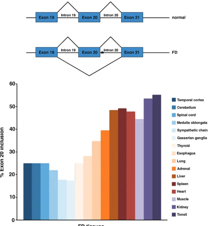
analysis ofIKBKAPgene from FD chromosomes, revealed a T to C transition in position 6 of the donor splice-site of intron 20. While normalIKBKAPencodes a full-length pro-tein (IKAP) of about 150 kDa, the FD mutation, (IVS20+6T > C), results in the generation of an mRNA in which exon 20 (74 bp) is spliced out, causing a frameshift that results in a premature stop codon (Andersonet al., 2001; Slaugenhauptet al., 2001; IKAP, NCBI Reference Sequence: NP_003631.2, protein length 1332 aa). Signifi-cantly though, in FD patients there is variable tissue-specific skipping of exon 20, leading to reducedIKBKAP full-length mRNA levels in all tissues, with nervous system tissues displaying the most severe reduction in exon 20 in-clusion (Figure 1; Cuajungco et al., 2003; Hims et al., 2007). Although the predicted truncated IKAP protein of 79 kDa has been detected in FD-derived lymphoblasts (An-dersonet al., 2001), other groups failed to detect such prod-uct in other FD cell lines or tissues. It is currently believed that at least part of the abnormally splicedIKBKAP tran-scripts is degraded by nonsense-mediated mRNA decay (NMD), as demonstrated in FD-derived olfactory stem cells (Booneet al., 2010) and that the truncated protein might be unstable due to its inability to dimerize (Xuet al., 2015).
To determine the underlying mechanism leading to alternative splicing and preferential skipping of exon 20 in neuronal tissues, an in silico analysis was performed to identify potential sequences that might also be involved in the process. Computational analyses showed that se-quences 5’ and 3’ of exon 20, and the sequence of exon 20 itself provided an environment where the definition of exon 20 is weakened (Ibrahimet al., 2007). In addition, differen-tiation of FD-derived induced pluripotent stem cells (iPSCs) into neuronal cells resulted in further reduction of exon 20 inclusion compared to non-differentiated cells, suggesting that the splicing machinery changes as the cells differentiate into neurons (Leeet al., 2009).
FD minor haplotypes
mother (Leyneet al., 2003). The phenotype caused by these mutations is currently unknown. The mutation responsible for minor haplotype 3 has not yet been characterized.
Genetic testing and current trend of FD population
With the identification of the mutation responsible for most FD cases, carrier testing began. Carrier testing, based on DNA analyses, is available for the two most common mutations (major haplotype and minor haplotype 2), with
an accuracy of 99%. In 2009, only five new FD cases were diagnosed in the world, representing a major reduction in new cases, considering that 15 or 20 were diagnosed per year in the 1990s (Couzin-Frankel, 2010).
Although the FD population at this moment is rela-tively small, there is still no cure for the existing patients, and the mechanisms underlying the disease are still poorly understood, precluding the development of more efficient therapies. In addition, since FD shares many similarities
with other neuropathies, understanding the mechanisms of the disease may shed light into other related disorders and increase our understanding of normal peripheral and central nervous system development and maintenance.
Clinical aspects and pathological findings
Although FD is genetically homogeneous and fully penetrant, clinical and pathological aspects of the disease are variable among individuals, and largely dependent on the age of the patient.
Clinical Presentation
FD babies are born significantly smaller than their unaffected siblings with births weights ranging from 70 to 90% of the normal range, indicative of intrauterine growth retardation (Axelrod and Dancis, 1973). Poor suck and un-coordinated swallow is observed in 60% of FD infants, and, together with a high metabolism, can lead to failure to thrive and malnutrition. In fact, FD children and adoles-cents display very low fat content, even with high caloric intake. Difficulty swallowing may persist in older children, and appears to be the underlying cause of excessive drool-ing, a prominent feature of FD patients (Margulieset al., 1968, Wolffet al., 2002). Along with dysphagia, FD pa-tients also display dysarthria due to oral incoordination (Gold-von-Simson and Axelrod, 2006). FD infants are typ-ically hypotonic, and the average age for independent walk-ing is about 26 months of age. Other gross milestones, including sitting unsupported, rolling over, and jumping are also significantly delayed. Decreased deep tendon re-flex is also a typical characteristic in FD patients. Several of these features, including poor suck and uncoordinated swallow, and dysphagia have been attributed to either brainstem dysfunction or reduced muscle innervation and control. Hypothalamic neuroendocrine dysfunction has been hypothesized as a possible explanation for poor weight gain.
Although most FD patients appear to have normal in-telligence, in general they tend to be literal and have diffi-culty extrapolating, and visual intellect often exceeds verbal abilities (Weltonet al., 1979). About 30% of FD children have signs of attention deficit disorder (ADD) characterized by short attention span and easy distrac-tibility. Emotional lability is also among the prominent fea-tures of FD, and intensifies during episodes of crisis (see below). All these characteristics suggest central nervous system impairment.
FD patients share an unusual facial appearance, de-scribed as “trigonal face” with facial asymmetry (Rileyet al., 1949; Axelrod and Pearson, 1984). Cephalometric measurements showed that in FD patients the maxilla and mandible are retrognathe to the cranial base, a feature that is significantly more pronounced in the mandible. In addition, horizontal mandibular growth is also distinctive in FD. To-gether, these alterations give the impression of small jaws,
and may contribute to difficulties in oral coordination and speech (Mass et al., 1998). It has been suggested that chronic progressive denervation leading to differences in coordinated muscle function might be the primary cause of abnormal facial expression.
The most common orthopedic manifestation in FD is spinal deformity, with a prevalence of 48% at the age of 10, and 86% at the age of 15. About 50% of the spinal deformi-ties are scoliosis only, 44% are kyphoscoliosis, and the re-maining 4% kyphosis only (Kaplanet al., 1997; Bar-onet al., 2000; Laplazaet al., 2001). Charcot joints and foot de-formities (including equinovarus and cavovarus) develop during childhood in about 10-15% of FD patients (Bar-on et al., 2000). In addition, FD patients sustain a much higher incidence of fractures compared to the normal age-matched population (63% versus 34%, respectively), most of them occurring before the time of skeletal maturation. Intrigu-ingly, the number of fractures per individual is also higher than in the general population, despite the fact that FD chil-dren and adolescents are significantly less active than nor-mal age-matched individuals. Progressive denervation is believed to be the cause of spinal deformity, while reduced pain perception is generally thought to be the underlying reason for increased fractures.
Ataxic gait is also one of the hallmarks of FD. FD pa-tients adopt a wide stance, and exhibit unsteady walking, often requiring assistance to prevent falling when turning. The ataxia progressively worsens over time: by the age of 20, about 5% of the patients require walking aids, and the need progresses linearly so that by the age of 40, about 30% of the patients require assistance for walking (Bar-Onet al., 2000; Macefieldet al., 2011). Cerebellar dysfunction or loss of muscle spindle sensory afferents leading to reduced proprioceptive acuity have been proposed as possible un-derlying causes of ataxic gait (Anderson et al., 2001; Macefieldet al., 2013).
A smooth tongue is observed in virtually all FD pa-tients and represents one of the cardinal features of FD. In infants, the number and size of fungiform papillae on the tongue are already reduced (Pearson et al., 1970), and fungiform papillae cannot be observed by the naked eye in older patients (Riley and Moore, 1966). The absence of filiform papillae, the reduced number and rudimentary de-velopment of fungiform papillae, together with the absence of taste buds, give the tongue a smooth appearance (Gold-berget al., 1968; Pearsonet al., 1970).
young and older individuals and temperature perception did not worsen in a five-year follow-up. In the same group of patients, pain perception was normal in 20% of the young patients, and severely impaired in about 12% of the remaining patients of this age-group. Significantly, all older patients exhibited impaired pain perception, with se-vere impairment observed in more than 30% of the cases of this age group; in addition, in a five-year follow-up there was also a tendency for worsening (Axelrodet al., 1981). Together, these observations indicate that the increased threshold for temperature perception does not change over time in FD patients, while there is a marked progression in impairment of pain perception. The decrease in pain and temperature perception are indicative of sensory deficits.
Ophthalmologic problems are also observed in FD and become worse as the patient population age. One of the main characteristics of FD is the absence of overflow emo-tional tears, and alacrima that may lead to corneal abra-sions. Absent corneal reflexes, and abnormal pupillary response to metacholine are also typical findings (Axelrod, 2006). In addition, progressive optic nerve atrophy and vi-sual decline are observed in a large fraction of FD patients, starting at the end of the first decade of life. In some pa-tients, visual acuity and color vision deteriorate over time, likely due to progression of optic nerve damage (Rizzo 3rd et al., 1986; Groomet al., 1997; Mendoza-Santiestebanet al., 2012; Mendoza-Santiestebanet al., 2014).
Cardiovascular abnormalities are also prominent in FD patients. Orthostatic hypotension often starts in school-age children and become more pronounced with in-creasing age, in part due to the increase in height. In FD pa-tients, postural hypotension, characterized by a severe decrease in blood pressure without compensatory increase in heart rate, results in episodes of lightheadedness, dizzi-ness and weakdizzi-ness of the legs (Axelrod, 2004). Supine hy-pertension is also common in adolescents and becomes more frequent in older patients. FD patients are hyperten-sive (SBP > 140mmHg) about 20-50% of the time (Norcliffe-Kaufmannet al., 2010; Carrollet al., 2012) and older FD patients have a high incidence of left ventricular atrophy, a sign of end-organ target damage due to chronic hypertension. Renal function also deteriorates as the pa-tients get older, with 20% of adult papa-tients having reduced renal function. The severity of renal disease parallels the extent of blood pressure variability, and most likely hap-pens as a secondary consequence of chronic hypertension (Norcliffe-Kaufmannet al., 2013). The mechanisms lead-ing to blood pressure variability and hypertension in FD have been sequentially attributed to central autonomic dys-function (Solitare, 1991; Gold-von Simson and Axelrod, 2006), to sympathetic and parasympathetic efferent baro-reflex dysfunction (Hilzet al., 1999; Stemperet al., 2004; Goldsteinet al., 2008), or, more recently, to afferent baro-reflex failure due to vagal withdrawal (Norcliffe-Kaufmannet al., 2010; Carrollet al., 2012).
Vomiting attacks (gastroesophageal reflux), often as-sociated with hypertension, tachycardia, diffuse sweating, and red blotching of the skin consist the most prominent manifestation of FD, occurring in about 65-80% of FD pa-tients, and can occur either intermittently as a response to physical or emotional stress, or daily in response to the stress of arousal. This “constellation of signs” is commonly referred to as “dysautonomic crisis” and is the leading cause of frequent hospitalizations of FD children (Axelrod, 2004; Gold-von-Simson and Axelrod, 2006). Although the pathophysiology of FD crisis is still not fully understood, it appears to involve central autonomic dysfunction (Ander-son and Rubin, 2005; Cheishviliet al., 2014b)
Pulmonary problems are also common in FD patients. Aspiration due to misdirected swallow is the major cause of pneumonia in FD. If gastroesophageal reflux is also pres-ent, the risk for aspiration increases significantly and re-peated pulmonary aspiration eventually leads to pulmonary disease (Gold-von-Simson and Axelrod, 2006). Breath-holding episodes, usually triggered by excitement, are also frequent, occurring in about 63% of FD patients, and can happen from 18 months of age up to 6 years of age. Pro-longed breath-holding can be severe, due to insensitivity to hypoxia, and can lead to secondary complications. These episodes are thought to represent a type of seizure activity (Axelrod, 2004; Gold-von-Simson and Axelrod, 2006).
Pathological, physiological and molecular findings
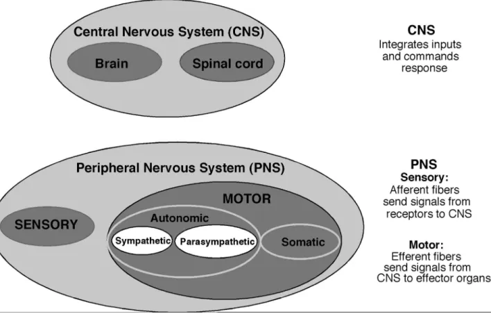
As mentioned above, several of FD major features could potentially be explained by central nervous system and/or peripheral nervous system abnormalities. Most of the pathological and physiological studies on FD have therefore focused on these two systems (Figure 2).
Central nervous system
In initial reports dating from 1945 to 1964, central nervous system (CNS) pathology was described in four FD patients ranging from 2 to 18 years of age. In all four cases, demyelination of the reticular formation of the pons and medulla were consistent findings. In addition, demyeli-nation of spinothalamic tracts, and loss of cranial nerve nu-clei of the brain stem were also reported in two cases (Aring and Engel, 1945; Cohen and Solomon, 1955; Brownet al., 1964). Other abnormalities, including thalamic degenera-tion and brain abscesses were observed in only two cases, but appeared to be secondary to episodes of hypoxia or in-flammation (Solitare, 1991). However, subsequent investi-gations in an additional four FD patients (ranging from 10 months of age to 31 years of age) failed to detect any signs of demyelination or other signs of neuronal loss or atrophy in the CNS (Pearson et al., 1970; Yatsu and Zussman, 1964; Solitare and Cohen, 1965; Fogelsonet al., 1967).
sensory and autonomic disturbances, but also that periph-eral nervous system appeared more consistently altered in FD (see below), subsequent studies focused almost exclu-sively on the evaluation of peripheral nervous system anomalies in FD patients, with a nearly complete disregard to CNS evaluation for almost 40 years.
More recently however, additional studies have pro-vided a strong indication that the CNS is also compromised in FD. For instance, visual impairment in FD patients be-come noticeable after the first decade of life, and are associ-ated with progressive optic atrophy and with predominant loss of papillomacular nerve fibers (Rizzo 3rdet al., 1986; Groom et al., 1997; Mendoza-Santiesteban et al., 2012, 2014). Analyses of brainstem auditory evoked potentials, as well as blink and jaw jerk reflex in FD patients also strongly suggest brainstem dysfunction (Lahatet al., 1992; Gutierrezet al., 2015; Tellez, 2015). MRI studies showed abnormalities that suggest compromised myelination as well as grey matter and white matter micro-structural dam-age in FD brains. Abnormal findings are more evident in optic radiation, middle cerebellar pedunculum, and frontal lobe (Axelrodet al., 2010). Significantly, analyses of FD brains showed that the levels of several transcripts and their respective proteins that are involved in myelination are se-verely reduced in FD brains compared to age-matched healthy individuals (Cheishviliet al., 2007). These
obser-vations indicate that central nervous system abnormalities indeed contribute to FD clinical findings.
Peripheral nervous system
Autonomic nervous system
In one of the first reports, degenerative pigmentary changes and vacuolization in the cytoplasm were observed in thoracic and pelvic sympathetic ganglia of two young FD siblings, ages 2 and 8 (Brown et al., 1964). Subsequent work in three additional young FD infants (ranging from 10 months to 2 years of age) confirmed these initial findings (Solitare and Cohen, 1965; Freytag and Lindenberg, 1967; Pearsonet al., 1970). Post-mortem analyses performed in two of these infants revealed that in the cervical and tho-racic sympathetic ganglia several neurons were small, and overall neuronal numbers were reduced, estimated to be around 30% of normal (Freytag and Lindenberg, 1967; Pearsonet al., 1970; Solitare, 1991). Degenerative changes with signs of inflammation were also observed (Pearsonet al., 1970). Quantitative analyses performed in adult FD pa-tients revealed that the mean volume of superior cervical ganglia (SCG) is reduced to 34% of the normal range, neuronal density to 37% of normal, with neuronal numbers as low as 10% of controls, indicating progressive loss of neuronal cells with age (Pearson and Pytel, 1978). Im-munohistochemistry for tyrosine hydroxylase (a
limiting enzyme of catecholamine biosynthesis) demon-strated that although there was a severe depletion in SCG neurons, the remaining neurons expressed higher levels of tyrosine hydroxylase compared to controls, which has been interpreted as being part of a compensatory mechanism (Pearsonet al., 1979). These analyses clearly indicate that sympathetic neurons are reduced in numbers and are mor-phologically abnormal in FD already at early age, and it is currently assumed that intrauterine development of sympa-thetic ganglia is severely compromised in FD.
Consistent with the reduction in neuronal numbers in sympathetic ganglia, ultrastructural studies of peripheral blood vessels also demonstrated the absence of autonomic nerve terminals. In addition, catecholamine metabolism is altered in FD patients (Smithet al., 1963), and other physi-ological studies indicate that sympathetic deficits also lead to cardiac hypo-innervation (Goldsteinet al., 2008).
In the parasympathetic system, the sphenopalatine ganglion is majorly compromised, with a significant reduc-tion of neuronal numbers, averaging 20% of controls. The ciliary ganglion on the other hand displays only a mild re-duction in total neuronal numbers (Pearson and Pytel, 1978).
Sympathetic and parasympathetic denervation and dysfunction are consistent with several of FD clinical fea-tures, including defective lacrimation, impaired pupillary reflex, impaired body temperature control, skin blotching, and excessive sweating. Hypertension and postural hypo-tension have also been attributed to sympathetic deficits, al-though this is currently under discussion.
Sensory nervous system
Neuronal numbers in dorsal root ganglia (DRG) are already diminished in young FD patients, and can be as low as 10-20% the numbers of normal age-matched individuals (Pearson et al., 1978), although extensive variability has been observed between individuals (Fogelsonet al., 1967; Solitare, 1991). More consistently, demyelination of the posterior columns (dorsal columns) of the spinal cord has been demonstrated in most post-mortem analyses (Fo-gelsonet al., 1967; Pearsonet al., 1978; Solitare, 1991). The finding of severe depletion of DRG neurons early postnatally is suggestive of impaired development during embryogenesis. However, degenerative changes in sensory ganglia (nodules of Nageotte) have been demonstrated in at least some of the cases, suggesting that progressive degen-eration also occurs postnatally (Pearsonet al., 1970, 1978).
Biopsies from the back and calf of FD patients re-vealed significant loss of nerve fibers in the epidermis and subepidermal neural plexus, with epidermal nerve fibers (ENF) densities averaging about 12-15% that of controls. In particular, sensory (substance P (SP) and calcitonin gene-related peptide (CGRP) positive) nerve fibers were virtually absent in FD biopsies, indicating significant loss of sensory skin innervation. In contrast, vasoactive intesti-nal peptide (VIP) staining, usually absent in normal
subepi-dermal plexus, was significantly increased. In addition, a significant number of empty Schwann cell sheaths were also observed, indicating recent denervation (Hilz et al., 2004). The presence of VIP staining and empty Schwann cell sheaths is indicative of inflammation, ongoing dener-vation, with some capability for regeneration, consistent with the clinical observation that FD is a progressive neuro-degenerative disorder. The decreased unmyelinated nerve content with virtual absence of CGRP and SP immunos-taining is compatible with the decrease in pain and temper-ature perception.
Sural nerve biopsies from FD patients exhibit a very consistent pattern that also distinguishes FD from all the other HSANs. Compared to normal individuals, the sural nerve of FD patients has diminished fascicular area (50% the area of controls), displays severe depletion of unmyeli-nated axons, and of small myeliunmyeli-nated fibers. As observed in skin biopsies, there is also a significant number of empty Schwann cell sheaths and occasional presence of macro-phages, indicative of ongoing axonal loss (Aguayoet al., 1971; Pearsonet al., 1975; Guzzettaet al., 1986).
Histopathological analyses from eight FD patients and age-matched controls demonstrated that in FD there is an extreme paucity in the geniculate ganglion neuronal numbers, as well as a significant reduction in neurons in the vestibular ganglion (cranial ganglion VIII), albeit to a les-ser extent (Tokitaet al., 1978). Since the geniculate gan-glion innervates the tongue fungiform papillae taste buds, this reduction in neuronal numbers is consistent with both the reduced sensory innervation and the absence of taste buds demonstrated in FD tongue (Pearsonet al., 1970), which require sensory innervation for their proper develop-ment and survival. Histopathological changes in the vestib-ular ganglion may in turn explain the poor balance and coordination of FD patients (Tokitaet al., 1978). So far, no histopathological analyses have been performed in other cranial sensory ganglia in FD.
growth factor (NGF) as a target field-derived survival fac-tor for sympathetic and sensory nociceptive neurons is well established (Indo, 2014). In the developing rat, NGF ex-pression levels are highest in tissues that are highly inner-vated by sympathetic fibers (Shelton and Reichardt, 1984), while sensory innervation correlates with NGF expression in the developing skin and spinal cord (Davieset al., 1987; Elkabeset al., 1994). Similarly, other types of neurons re-quire appropriate supply and signaling by other neuro-trophins. Neurotrophic support derived from the target field is also required throughout postnatal life for maintenance of innervation and neuronal survival. Hence, disturbance of any of these steps (or a combination of them) could result in depletion of sensory and/or sympathetic neurons and loss of innervation in FD.
IKAP Cellular functions: implications for PNS
deficits
Historically, IKAP was identified as a novel 150 kDa protein that interacts with cytokine-activated IkB kinase (IKK) complexes, which participate in the activation of the transcription factor NF-kB, hence the name IKAP, for “IKK complex associated protein”. IKAP was initially shown to bind directly to IKK-alpha and IKK-beta and, act-ing as a scaffold protein, to assemble IKKs into an active kinase complex (Cohen et al., 1998). Investigations by other research groups confirmed that IKAP is mostly local-ized in the cytoplasm, but failed to identify IKAP as a regu-lar member of IKK complexes and further demonstrated that IKAP is not required for NF-kB signaling (Krappmann et al., 2000). Since then, several other cellular functions of IKAP have been unraveled. IKAP, either as part of the Elongator complex (see below) or possibly acting inde-pendently, has been shown to interact with a variety of nu-clear and cytoplasmic proteins and to play a role in tRNA wobble uridine modification (Linet al., 2013; Karlsbornet al., 2014; Laguesseet al., 2015), cytosolic stress signaling (Holmberget al., 2002), DNA repair (Liet al., 2009), and zygotic paternal DNA demethylation (Okadaet al., 2010), among others. However, so far the link between any of these above-mentioned functions with FD pathology and in particular with PNS deficits still remain to be elucidated.
This section will therefore focus only on the cellular functions of IKAP that have been more thoroughly investi-gated and have led to insights into the mechanisms underly-ing abnormal PNS development and function in FD.
IKAP is a member of the Elongator Complex
Sequence comparison between species showed that IKAP is the human homologue of the yeast elongator pro-tein 1 (ELP1), one of the six subunits of the Elongator com-plex, which was initially identified as a complex essential for RNA polymerase II (RNA pol II) transcription elonga-tion (Oteroet al., 1999). Purification and characterization
of the human Elongator complex revealed that, as in yeast, the human holo-Elongator complex is also composed of six subunits, and confirmed that IKAP is an integral compo-nent of the core of the Elongator complex, which is com-posed of three subunits hELP1/IKAP, hELP2 and hELP3 (Hawkes et al., 2002). Within the Elongator complex, ELP1/IKAP appears to act as a scaffold protein required for Elongator assembly and also serves to dock other proteins that regulate the Elongator function, whereas ELP3 is the catalytic subunit and acetylates histones H3 and H4 (Glatt and Muller, 2013). Consistent with these observations, IKAP protein is also observed in the nucleus, and decreased IKAP expression in FD patient-derived fibroblasts leads to reduced ELP3 levels, reduced Elongator binding, and de-creased histone H3 acetylation in the coding region of genes that are downregulated in FD cells. Moreover, RNA pol II density is significantly decreased at the 3’ end of these genes, but not at the promoter region, implying that the Elongator complex is not required for recruitment of RNA pol II to the promoter region, but affects primarily transcript elongation (Closeet al., 2006). These findings suggested that transcriptional dysregulation might be the underlying cause of the deficits observed in FD.
Decreased IKAP expression impairs cell migration
With the finding that IKAP is part of the RNA pol II transcriptional Elongator complex, much of the subsequent work focused on comparative transcriptome or microarray analyses, aiming at identifying pathways that are affected by IKAP depletion. Using this approach, it was found that gene transcripts involved in cell migration are significantly down-regulated in a variety of FD patient-derived cells, in-cluding fibroblasts (Closeet al., 2006), induced pluripotent stem cells (iPSCs, Lee et al., 2009), and olfactory ecto-mesenchymal stem cells (hOE-MSCs, Booneet al., 2010), as well as inIKBKAPsiRNA- transfected cell lines includ-ing HeLa (Closeet al., 2006), and SHSY5Y neuroblastoma cells (Cohen-Kupiecet al., 2011).
sympathetic ganglia (Georgeet al., 2013; Jackson et al., 2014).
Together, these findings indicate that although reduc-tion or loss of IKAP expression results in impaired cell mi-gration, this impairment is either compensated by external signalsin vivo, or are not severe enough to have a biological significance. In any case, it is clear that peripheral nervous system abnormalities in FD are not caused by impaired cell migration.
IKAP and cytoskeleton organization: impact on neurite outgrowth and intracellular transport.
A much more consistent and biologically relevant cellular function of IKAP appears to be its requirement for cytoskeleton actin filament and microtubule organization and neurite outgrowth. Cytosolic IKAP was shown to co-purify with filamin A in rat cerebellar granule neurons, and immunohistochemistry for IKAP and filamin A showed that they co-localize with membrane ruffles. Depletion of IKAP in these cells did not decrease the expression of genes involved in cell migration, but resulted in actin cytoskeletal disorganization and reduced cell migrationin vitro (Johan-senet al., 2008). These observations suggest that cell mi-gration defects in cells depleted of IKAP may be linked to cytoskeleton disturbances instead of transcriptional dysre-gulation.
Immunostaining of FD-derived fibroblasts for the cytoskeleton component a-tubulin revealed disorganiza-tion of the microtubules (MTs) that resulted in aberrant cell shape. Similar results were obtained with neuroblastoma cells after down-regulation of IKAP. Moreover, in neuro-blastoma cells,a-tubulin was shown to be concentrated in one pole of the cells resulting in abnormal process forma-tion, with concomitant up-regulation of the MT-desta-bilizing protein SCG10 (STMN2). Significantly, RNAi-mediated SCG10 downregulation in FD-derived fibroblasts rescued cytoskeleton organization (Cheishviliet al., 2011).
Microtubules (composed of alpha and beta-tubulin dimers) are particularly abundant in neurons, and are in-volved in multiple intracellular processes, including devel-oping and maintaining cell shape, intracellular transport, cell signaling, and neurite extension (Kapitein and Hoogen-raad, 2015).In vivostudies confirmed the need of IKAP for all these processes.
In the developing mouse brain, Ikbkapsilencing in migrating projection neurons results in altered cell mor-phology and absence of growing apical dendritic tree and processes. In culture,Ikbkapnull cortical neurons also ex-hibited reduced dendrite length and branch numbers (Crep-peet al., 2009). Interaction of IKAP with ELP3 appears to be necessary to regulate alpha-tubulin acetylation, a pro-cess that appears to be required for branching of post-migratory projection neurons (Creppeet al., 2009; Nguyen et al., 2010). Importantly, purified ELP3-enriched fraction promotesa-tubulin acetylationinvitrosuggesting a direct
role of IKAP and ELP3 in this process (Creppe et al., 2009).
Down-regulation ofIkbkapin chick neural crest cell precursors lead to marked disruptions in axonal projections of DRGs (Hunnicuttet al., 2012; Abashidzeet al., 2014). In particular, the major disturbances are observed in the bran-ching and positioning of the distal processes, while the po-sitioning of the main nerves do not appear to be affected. In growth cones of control DRG cultures, IKAP was shown to co-localize with stable tubulin fibers, and to a lesser extent along dynamic tubulin fibers. Significantly, depletion of IKAP in cultured DRGs results in disturbance of tubulin structures (Abashidzeet al., 2014). Similarly, in mice, inac-tivation ofIkbkapgene in neural crest cells leads to 35 to 40% reduction in total neuronal numbers in DRGs (George et al., 2013; Jacksonet al., 2014), and reduction in sympa-thetic neuronal numbers between 42% loss (Jacksonet al., 2014) to 70% loss (Georgeet al., 2013). Similar to chicks, loss of IKAP in neural crest cells in mice leads to a signifi-cant reduction in sympathetic and sensory target tissue innervation, likely due to abnormal axonal branching (Jacksonet al., 2014).
Together these results indicate that IKAP is required for axonal branching and fine-tuning of the innervation process, and suggest that loss of DRG and sympathetic neuronal cells in FD is likely a consequence of failed innervation of target tissues.
Recently, it has also been shown that downregulation of IKAP in cultured chick DRG neurons results in de-creased NGF intracellular signaling. Moreover, IKAP was also shown to interact directly with the motor protein dynein, suggesting that IKAP may also be involved in intracellular transport (Abashidzeet al., 2014). These ini-tial findings were further corroborated by transcriptome analyses in neuronal differentiated FD-derived embryonic stem cells as well as in FD embryonic brains (Lefleret al., 2015). In particular, these analyses revealed that in FD-derived neurons and embryonic brains synaptic vesicular and neuronal transport genes are directly or indirectly af-fected by IKAP depletion (Lefler et al., 2015). Signifi-cantly, among the five types of HSANs, FD (HSAN III), HSAN IV (caused by mutations in NGF receptor), and HSAN V (caused by mutations in NGF) share several simi-larities. All these disorders are congenital and are charac-terized by decreased pain and temperature perception, Charcot joints, decreased skin innervation, and severe de-pletion of unmyelinated fibers and small myelinated fibers in the sural nerve (Capsoni, 2014). Null mutations for NGF or NGF receptor (TrkA/NTRK1) in mice result in severe loss of sympathetic neurons and nociceptive sensory neu-rons (Davies, 2000).
im-paired target innervation, imim-paired neurotrophic signaling might also contribute to PNS neuronal cell death in FD.
What does expression of IKAP in the
developing embryo tell about FD?
Up to now, there is limited information regarding the pattern of expression of IKAP during embryogenesis, and although FD appears to be a developmental disorder, it is currently not known what is the tissue-specific pattern of exon-20 skipping during development, since all the infor-mation regarding alternative splicing in FD has been per-formed in adult-derived tissues and cell lines (see above).
IkbkapmRNA is already detected in early mouse em-bryos (embryonic day 8.5; E8.5), prior to the formation of NCCs, indicating that IKAP may be involved in other de-velopmental processes as well (Chenet al., 2009; Dietrich et al., 2011). High levels of IKAP expression are later on observed in sensory and sympathetic neuroblasts and in the developing brain, as shown by immunohistochemistry (Creppeet al., 2009; Jacksonet al., 2014). RT-PCR analy-ses also detected low-to-moderate levels ofIkbkap expres-sion in peripheral organs, including heart, kidney, and lungs in developing mouse embryos at mid-to-late gesta-tion (Dietrich and Dragatsis, unpublished results).
In the chick, analysis of the pattern of expression of IKAP was focused exclusively in the developing peripheral nervous system (Hunnicuttet al., 2012; Abashidzeet al., 2014). In situ hybridization and immunohistochemistry failed to detect IKAP expression during the period of neural crest migration and DRG formation. Instead, IKAP expres-sion is first observed in nascent and postmitotic neurons, and increases as the neurons mature and differentiate (Hun-nicuttet al., 2012; Abashidzeet al., 2014). In particular, the increase in IKAP expression in DRGs coincides with the onset of peripheral outgrowth and target innervation (Abashidze et al., 2014). In addition, significant immu-nolabeling for IKAP was also observed in the developing spinal cord, in particular in the ventral horn motor neurons and the ventricular zone (Hunnicuttet al., 2012), and in pri-mary sympathetic ganglia (Abashidzeet al., 2014).
A far more detailed analysis has been performed in the developing rat.In situhybridization in mid-to-late ges-tation rat embryos (E15.0 to E21.0) revealed thatIkbkap mRNA is expressed in a wide variety of tissues. At E15.0, IkbkapmRNA is found in the brain, spinal cord, sensory ganglia, eye retina, liver, and intestinal tract. By E17.0, IkbkapmRNA begins to be expressed in additional tissues, including the adrenal gland, Merkel cells, salivary glands, lungs, renal cortex, and cartilage. At E21, in addition to the tissues listed above, a high level ofIkbkapexpression is also detected in the heart, skin, stomach, and carotid body. Notably, at all stages examined, the highest levels of ex-pression were seen in the dorsal root ganglia, trigeminal
ganglia, retina, central nervous system, and kidney cortex (Mezeyet al., 2003).
The wide distribution of IkbkapmRNA expression observed in the developing rat implies that IKAP may play essential roles not only in nervous system tissues but also in non-neuronal cells. In this aspect, it is important to note that expression of IKAP in the kidney correlates well with the relatively high frequency of kidney malformations ob-served among FD patients, and suggests that a need of IKAP in the kidney may also contribute to kidney disease (Norcliffe-Kaufmannet al., 2013). Expression of IKAP in the retina could explain the selective loss of retina ganglion cells (Mendoza-Santiestebanet al., 2014). Likewise, ex-pression of IKAP in cartilage is consistent with the de-crease in bone mineral density observed in FD patients, a possible underlying explanation for the increased fre-quency of fractures (Maayanet al., 2002), and suggests that IKAP may also be required for bone growth. Finally, and most intriguingly, the presence ofIkbkapmRNA in periph-eral target tissues (salivary glands, skin, heart, lungs, stom-ach) suggests that IKAP may also play an essential role in these tissues. An attractive possibility is that IKAP might be required for adequate production of neurotrophins that are retrogradely transported to neuronal cells at the time of innervation as well as postnatally in order to maintain innervation. This possibility is substantiated by the histo-pathological changes in the skin of FD patients where there are signs of denervation and re-innervation, and also by the early biochemical findings that suggested that NGF pro-duced by FD-derived fibroblasts cells demonstrates de-creased neurotrophic activity (Siggers et al., 1976; Schwartz and Breakefield, 1980; Wrathall, 1986).
Animal models for FD
The pattern of IKAP expression and the phenotypic features of FD not related to the peripheral nervous system suggest that FD is a complex disease and could actually be viewed as a multiple system disorder. In order to fully un-derstand the pathogenesis of FD and to develop more com-prehensive and efficient therapies, animal models that faithfully recapitulate the disease would be greatly advanta-geous.
Mouse Ikbkap
mou-se suggest that the function of the gene is also conmou-served be-tween species.
Inactivation of Ikbkap gene in the mouse
In an initial attempt to understand the roles of IKAP in embryogenesis and possibly generate a mouse model for the disease, two research groups generated null mutations in the mouseIkbkapgene. Unexpectedly, it was found that inactivation of theIkbkapgene in mice results in embryonic lethality, between E10.5 and E11.5 (Chen et al., 2009; Dietrichet al., 2011).Ikbkapnull embryos appear grossly normal at E6.5 (Chenet al., 2009), but by E9.5 they display already a significant developmental delay (about 24 hr de-lay) compared to controls. By E10.5, although mutant em-bryos reach several milestones of a typical E9.5 embryo (including neural fold closing, turning of the embryo, and development of the first branchial arch) they display poor vascularization of the extraembryonic tissues, anterior ce-phalic developmental defects, and cardiovascular abnor-malities. In particular, development of the forebrain is com-promised, and the heart does not develop past an E8.5-like primitive heart. Abnormal heart looping and dilated peri-cardial sac in null embryos indicate that heart failure is likely the cause of death ofIkbkapnull embryos (Dietrichet al., 2011). Consistent with a need of IKAP for heart devel-opment, RT-PCR analyses showed that expression of the transcription factorsGata5andSmad4, which are essential for heart morphogenesis, are significantly down-regulated in Ikbkap null embryos (Dietrich et al., 2011). Homo-zygosity for anIkbkapallele lacking exon 20 (IkbkapD20; Figure 3) also resulted in a similar phenotype, indicating that the truncated IKAP protein does not retain significant biological function (Dietrichet al., 2011).
These analyses indicate that inactivation ofIkbkap re-sults in several severe developmental abnormalities that are not seen in FD. Also, inactivation ofIkbkapin neural crest cell precursors results in cleft secondary palate (Jacksonet al., 2014; Dietrich and Dragatsis, unpublished results), a severe developmental malformation that is never observed in FD patients. Keeping in mind that FD has pleiotropic manifestations, and that the IVS20+T > C mutation in FD patients results in reduced levels ofIKBKAPexpression in all tissues, it becomes obvious thatglobal reductionand not tissue-specific elimination ofIkbkapexpression is required to generate a model for FD.
Mouse models of FD
So far, all attempts to generate a mouse model for FD by introducing the FD major haplotype mutation in the mouse genome have been unsuccessful.
Slaugenhaupt and collaborators generated a series of transgenic mice carrying different copy numbers of a hu-manIKBKAPbacterial artificial chromosome (BAC) carry-ing the FD IVS20+6T > C mutation (FD BAC). Characterization of the splicing pattern of humanIKBKAP
mRNA in several transgenic lines by semi-quantitative RT-PCR revealed that they recapitulated faithfully the same tissue-specific splicing pattern seen in FD patients (Hims et al., 2007). As expected, none of the transgenic lines had FD phenotypic features when normal levels of en-dogenous mouse IKAP protein was expressed. In an effort to elicit an FD phenotype, FD BAC mice were crossed into an Ikbkap null background. Unfortunately, none of the compound FD BAC transgenic/Ikbkapnull mice recapitu-lated the FD phenotype. Although the FD BAC mice do not model FD, they can be used to testin vivothe efficacy of compounds that have the potential to correct the FD splic-ing defect (Shettyet al., 2011).
Using another approach, Ast and collaborators gener-ated a mouseIkbkapallele in which exon 20 and its two flanking introns were replaced by the corresponding human FD sequence that contains the IVS20+6T > C mutation (Bochner et al., 2013). Contrary to what was expected, mice homozygous for this mutation were viable and did not exhibit any of the human FD phenotypes. Quantitative RT-PCR analyses revealed that insertion of the FD human mutation resulted in a different tissue-specific splicing pat-tern in the mouse compared to what is observed in FD pa-tients. In the mouse, significant skipping of exon 20 occurred in the liver, but not in the nervous system. This differential pattern of exon 20 skipping most likely results from differences in the mechanisms of splicing sequence recognition between the two species.
Other research groups also attempted to generate an FD mouse model by introducing the FD point mutation into the mouseIkbkapallele and were equally unsuccessful(D. Brenner personal communication; Dietrich and Dragatsis, unpublished results). Although the reasons for this failure are still unclear, it is possible that the sequence and/or size of the introns surrounding the FD point mutation might be the main reason for the technical difficulties, or that differ-ences between the mouse and human splicing machinery might be the underlying impediment for the success of this approach. Since complete inactivation ofIkbkapin mice is embryonic lethal, and FD carriers (heterozygous for the point mutation) expressing 50% the levels of normal full length IKAP protein are asymptomatic, the generation of a successful FD model likely requires that the level of normal IKAP protein be reduced between 0 and 50% the normal levels.
Since FD is caused by severe reduction of full-length IKAP protein, it was reasonable to assume that not only the compoundIkbkapD20/floxmice but also theIkbkapflox/floxmice would represent a model for FD. As it turns out, both Ikbkapflox/floxandIkbkapD20/floxmice recapitulate several of FD major phenotypic features, including reduced IKAP ex-pression, intrauterine growth retardation, reduced birth weight, failure to thrive, reduced number of fungiform papillae on the tongue, progressive ataxia, kyphosis, stress-induced seizures, decreased temperature perception and impaired development and maintenance of sensory and sympathetic neurons (Figure 4 and Table1; Dietrichet al.,
2012). Similar to FD patients, these FD mouse models also display reduction of expression of genes and proteins in-volved in myelination in the CNS (Cheishviliet al., 2014a). Intriguingly, although both Ikbkapflox/flox and IkbkapD20/floxmouse models recapitulate FD, the severity is strikingly higher in the compoundIkbkapD20/floxmodel (Ta-ble 1). In addition to a milder disease phenotype, Ikbkapflox/flox mice also have a significant increase in lon-gevity. This suggests that a slight increase in IKAP expres-sion is sufficient to greatly ameliorate the symptoms of the disease. Slight differences in IKAP expression (possibly due to differences in splicing efficiency) might therefore
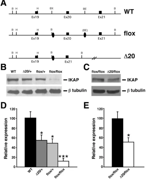
Figure 3- Generation and molecular characterization of FD mouse models. (A) Schematic representation of wild-type allele (WT), Ikbkap flox allele (flox), and Ikbkap allele lacking exon 20 (D20). Exons are represented by black rectangles, and black ovals represent the loxP sites. Restriction sites shown on the schematic are: Bam HI (B), HindIII (H), BstXI (BX) and BstEII (BE). (B) Western blot analyses of total protein lysates from forebrain of 11-month-old WT,IkbkapD20/+,Ikbkapflox/+, andIkbkapflox/floxmice. Upper panel shows detection of IKAP with the polyclonal anti-IKAP antibody (AnaSpec), and lower panel shows anti-b-tubulin staining for loading control. Note that IKAP protein level is severely reduced inIkbkapflox/floxbrain. (C) Western blot analyses of total protein lysates from whole brain of E16.5Ikbkapflox/floxandIkbkapD20/floxembryos. Upper panel shows detection of IKAP
with the polyclonal anti-IKAP antibody (AnaSpec), and lower panel shows anti-b-tubulin staining for loading control. Note that inIkbkapD20/floxbrain IKAP protein expression is reduced compared toIkbkapflox/floxbrain. (D and E) Quantitative analyses of IKAP expression levels in mouse brains of
differ-ent genotypes. (D) IKAP expression levels were normalized over tubulin levels and are expressed as percdiffer-entage of WT. (E) IKAP expression levels were normalized over tubulin levels and are expressed as percentage ofIkbkapflox/flox. Data are represented as Mean±SD; n = 3 experiments. *P < 0.05, ***P <
also explain the variability in phenotypic expression ob-served among FD patients. Another important finding through analyses of these models is that intrauterine devel-opment of sympathetic ganglia is not extensively compro-mised even when IKAP levels are as low as 5% of normal levels, and that degeneration and neuronal cell loss in the
sympathetic ganglia occurs mostly postnatally. Significantly, as seen in young FD patients, a portion of the remaining neurons in the sympathetic ganglia of postnatal FD mouse models appear small and vacuolated, an indica-tion of dysfuncindica-tion and ongoing degeneraindica-tion (Dietrichet al., 2012).
Figure 4- Postnatal characteristics ofIkbkapD20/floxandIkbkapflox/floxmice. (A) Appearance ofIkbkapD20/floxmice at P18. WT (top) andIkbkapD20/flox
(bot-tom) littermates were photographed side by side. Note that the mutantIkbkapD20/floxmouse is significantly smaller than its WT littermate, exhibits abnor-mal posture, and puffy feet. (B and C) Representative histological examinations of tongue fungiform papillae. Tongues of P18 WT (B) andIkbkapD20/flox
(C) littermates were processed for paraffin embedding, and coronal sections of the anterior part of the tongue were stained with H&E. Note that the three fugiform papillae of the WT littermate appear normal (B), while in the mutant the fungiform papilla shown is degenerating (C). (D) Postnatal growth curves of control (blue, n=37) andIkbkapflox/flox(red, n=14) male mice. Similar results were found for female mice. Data are represented as Mean±SEM. (E and F) MicroCT scans of 11-month-old WT (E) and IkbkapD20/flox(F) littermate male mice. Note that the mutant is significantly smaller than control
Taken together, these two findings have major impli-cations for therapeutics, since they suggest that even a slight increase in IKAP expression postnatally might be sufficient to halt the progression of sympathetic neuronal loss, and possibly revert other features as well.
Recently, a phenotypic model of FD in which IKBKAPmRNA splicing and expression can be modulated was generated by introducing the complete human IKBKAPgene (BAC transgene) with the major FD splice mutation (TgFD9; Himset al., 2007) into the IkbkapD20/flox mouse model. The introduction of the humanIKBKAPFD transgene attenuated the severe FD phenotype observed in theIkbkapD20/floxmouse, while still recapitulating FD major features and recreating the same tissue-specific mis-splicing defect seen in FD patients (Moriniet al., 2016). With the availability of mouse models that faithfully reca-pitulate major features of FD, several questions regarding FD disease process as well as testing of novel therapeutic strategies can now be addressedin vivo.
Conclusions and perspectives
With the identification of the genetic cause of FD,in vitroandin vivostudies have provided important informa-tion related to the normal funcinforma-tion of IKAP. In particular, the finding that IKAP plays an essential role in cytoske-leton remodeling and target field innervation provides a plausible explanation for the PNS deficits of the disease. An exciting challenge for the near future is to further eluci-date the essential pathways that are disrupted in FD in neuronal and non-neuronal tissues, so that more compre-hensive and efficient therapies can be developed and applied to the existing patients. In addition, further under-standing of the mechanisms underlying the developmental defects of FD may shed light into other related disorders. For instance, the emerging possible role of IKAP in neuro-trophin retrograde transport and signaling - if proven cor-rect - would link the molecular pathways of FD to at least two other HSANs (HSAN IV and HSAN V). With the availability ofin vitroandin vivomodels for FD, the an-swers to these questions are now close at hand.
Table 1- IkbkapD20/floxand Ikbkapflox/floxmouse models reproduce features of FD.
FD patients IkbkapD20/flox Ikbkapflox/flox
Reduced expression of full-length IKAP protein 5-20% of controls in CNS 5% of controls in CNS tissues 10% of controls in CNS tissues
Intrauterine growth retardation + + +
Low birth weight 80% of controls 70% of controls 85% of controls
Poor suck, uncoordinated swallow at birth + + +
Poor weight gain + + +
Short stature + + +
Reduced life-span + + +
Dysautonomic crisis + N/A N/A
Seizure susceptibility + + +
Gastrointestinal dysfunction + + +
Absence of overflow emotional tears + N/A N/A
Optic neuropathy + + +
Tongue: Reduced numbers of fungiform papillae smooth tongue 45% reduction 30% reduction
Decreased deep tendon reflexes + N/D N/D
Muscle spindle abnormalities Impaired function N/D Reduced sensory innervation Absent axon flare following intradermal histamine
injection
+ N/D N/D
Poor coordination/balance + + +
Spinal abnormalities + + +
Hydronephrosis + + +
Decreased temperature perception + + +
Decreased volume of DRGs + 45% of controls at birth 70% of controls at birth Decreased neuronal numbers in DRGs in adults 10-20% of controls 75% reduction in nociceptive
neurons
20% reduction in nociceptive neurons
Decreased volume of sympathetic ganglia at birth N/D 45% of controls at birth 70% of controls at birth Decreased volume of SCGs in adults 30% of controls N/D 30% of controls
Decreased neuronal numbers in SCGs in adults 10% of controls N/D 20% of controls
Acknowledgments
The authors would like to apologize to all those who-se contributions to the field have been inadvertently ne-glected. The authors were supported by the National Institute of Health [R01 NS061842] and by the Dysau-tonomia Foundation, Inc.
References
Abashidze A, Gold V, Anavi Y, Greenspan H and Weil M (2014) Involvment of IKAP in peripheral target innervation and in specific JNK and NGF signaling in developing PNS neu-rons. PLoS One 9:e113428.
Aguayo AJ, Nair CP and Bray GM (1971) Peripheral nerve abnor-malities in the Riley-Day syndrome. Findings in a sural nerve biopsy. Arch Neurol 24:106-116.
Anderson SL, Coli R, Daly IW, Kichula EA, Rork MJ, Volpi SA, Ekstein J and Rubin BY (2001) Familial dysautonomia is caused by mutations of the IKAP gene. Am J Hum Genet 68:753-758.
Anderson SL and Rubin BY (2005) Tocotrienols reverse IKAP and monoamine oxidase deficiencies in familial dysauto-nomia. Biochem Biophys Res Commun 336:150-156. Aring CD and Engel GD (1945) Hypothalamic attacks with
thala-mic lesions: II anatothala-mic considerations. Arch Neurol Psychiat 54:44.
Axelrod FB (2004) Familial dysautonomia. Muscle Nerve 29:352-363.
Axelrod FB (2006) A world without pain or tears. Clin Auton Res 16:90-97.
Axelrod FB and Dancis J (1973) Intrauterine growth retardation in familial dysautonomia. Am J Dis Child 125:379-380. Axelrod FB and Pearson J (1984) Congenital sensory
neuro-pathies. Diagnostic distinction from familial dysautonomia. Am J Dis Child 138:947-954.
Axelrod FB, Iyer K, Fish I, Pearson J, Sein ME and Spielholz N (1981) Progressive sensory loss in familial dysautonomia. Pediatrics 67:517-522.
Axelrod FB, Goldberg JD, Ye XY and Maayan C (2002) Survival in familial dysautonomia: Impact of early intervention. J Pediatr 141:518-523.
Axelrod FB, Hilz MJ, Berlin D, Yau PL, Javier D, Sweat V, Bruehl H and Convit A (2010) Neuroimaging supports cen-tral pathology in familial dysautonomia. J Neurol 257:198-206.
Bar-On E, Floman Y, Sagiv S, Katz K, Pollak RD and Maayan C (2000) Orthopaedic manifestations of familial dysautono-mia. A review of one hundred and thirty six patients. J Bone Joint Surg Am 82-A:1563-1570.
Blumenfeld A, Slaugenhaupt SA, Liebert CB, Temper V, Maayan C, Gill S, Lucente DE, Idelson M, MacCormack K, Monahan MA,et al.(1999) Precise mapping and haplotype analysis of the familial dysautonomia gene on human chro-moseome 9q31. Am J Hum Genet 64:1110-1118.
Bochner R, Ziv Y, Zeevi D, Donyo M, Abraham L, Ashery-Padan R and Ast G (2013) Phosphatidylserine increases IKBKAP levels in a humanized knock-in IKBKAP mouse model. Hum Mol Genet 22:2785-2794.
Boone N, Loriod B, Bergon A, Sbai O, Formisano-Tréziny C, Gabert J, Khrestchatisky M, Nguyen C, Féron F, Axelrod
FB,et al.(2010) Olfactory stem cells, a new cellular model for studying molecular mechanisms underlyng familial dy-sautonomia. PLoS One 5:e15590.
Brown WJ, Beauchemin JA and Linde LM (1964) A neuro-pathological study of familial dysautonomia (Riley-Day syndrome) in siblings. J Neurol Neurosurg Psychiatry 27:131-139.
Capsoni S (2014) From genes to pain: Nerve growth factor and he-reditary sensory and autonomic neuropathy type V. Eur J Neurosci 39:392-400.
Carroll MS, Kenny AS, Patwari PP, Ramirez JM and Weese-Mayer DE (2012) Respiratory and cardiovascular indicators of autonomic nervous system dysregulation in familial dysautonomia. Pediatr Pulmonol 47:682-691.
Cheishvili D, Maayan C, Smith Y, Ast G and Razin A (2007) IKAP/hELP1 deficiency in the cerebrum of familial dysau-tonomia patiemnts results in down regulation of genes in-volved in oligodendrocyte differentiation and in myelina-tion. Hum Mol Genet 16:2097-2104.
Cheishvili D, Maayan C, Cohen-Kupiec R, Lefler S, Weil M, Ast G and Razin A (2011) IKAP/Elp1 involvment in cytoske-leton regulation and implication for familial dysautonomia. Hum Mol Genet 20:1585-1594.
Cheishvili D, Dietrich P, Maayan C, Even A, Weil M, Dragatsis I and Razin A (2014a) IKAP deficiency in an FD mouse model and in oligodendrocyte precursor cells results in downregulation of genes involved in oligodendrocyte differ-entiation and myelin formation. PLoS One 9:e94612. Cheishvili D, Laiba E, Rekhtman D, Claman A, Razin A and
Maayan C (2014b) Dynamic changes in IKBKAP mRNA levels during crisis of familial dysautonomia. Auton Neu-rosci 180:50-65.
Chen YT, Hims MM, Shetty RS, Mull J, Liu L, Leyne M and Slaugenhaupt SA (2009) Loss of mouse Ikbkap, a subunit of elongator, leads to transcriptional deficits and embryonic lethality that can be rescued by human IKBKAP. Mol Cell Biol 29:736-744.
Close P, Hawkes N, Cornez I, Creppe C, Lambert CA, Rogister B, Siebenlist U, Merville MP, Slaugenhaupt SA, Bours V,et al. (2006) Transcription impairment and cell migration defects in elongator-depleted cells: Implication for familial dysauto-nomia. Mol Cell 22:521-531.
Cohen P and Solomon NH (1955) Familial dysautonomia: Case report with autopsy. J Pediatr 46:663-670.
Cohen L, Henzel WJ and Baeuerle PA (1998) IKAP is a scaffold protein of the IkappaB complex. Nature 395:292-296. Cohen-Kupiec R, Pasmanik-Chor M, Oron-Karni V and Weil M
(2011) Effects of IKAP/hELP1 deficiency on gene expres-sion in differentiating neuroblastoma cells: Implications for familial dysautonomia. PLoS One 6:e19147.
Coli R, Anderson SL, Volpi SA and Rubin BY (2001) Genomic organization and chromosomal localization of the mouse IKBKAp gene. Gene 279:81-89.
Couzin-Frankel J (2010) Chasing a disease to the vanishing point. Science 328:298-300.
Cuajungco MP, Leyne M, Mull J, Gill SP, Gusella JF and Slau-genhaupt SA (2001) Cloning, characterization and genomic structure of the mouse Ikbkap gene. DNA Cell Biol 20:579-586.
Cuajungco MP, Leyne M, Mull J, Gill SP, Lu W, Zagzag D, Axelrod FB, Maayan C, Gusella JF and Slaugenhaupt SA (2003) Tissue-specific reduction in splicing efficiency of IKBKAP due to the major mutation associated with familial dysautonomia. Am J Hum Genet 72:749-758.
Davies AM (2000) Neurotrophins: More to NGF than survival. Curr Biol 10:R374-R376.
Davies AM, Bandtlow C, Heumann R, Korsching S, Rohrer H and Thoenen H (1987) Timing and site of nerve growth factor synthesis in the developing skin in relation to innervation and expression of the receptor. Nature 326:353-358. Dietrich P, Alli S, Shanmugasundaram R and Dragatsis I (2012)
IKAP expression levels modulate disease severity in a mou-se model of familial dysautonomia. Hum Mol Genet 21:5078-5090.
Dietrich P, Yue J, Shuyu E and Dragatsis I (2011) Deletion of exon 20 of the Familial Dysautonomia gene Ikbkap in mice causes developmental delay, cardiovascular defects, and early embryonic lethality. PLoS One 6:e27015.
Dong J, Edelmann L, Bajwa AM, Kornreich R and Desnick RJ (2002) Familial dysautonomia: Detection of the IKBKAP IVS20(+6T > C) and R696P mutations and frequencies among Ashkenazi Jews. Am J Med Genet 110:253-257. Dyck PJ (1993) Neuronal atrophy and degeneration
predomi-nantly affecting peripheral sensory and autonomic neurons. In: Dyck PJ, Thomas PK, Griffin JW, Low PA and Poduslo JF (eds) Peripheral Neuropathy. 3rd edition. WB Saunders Co, Philadelphia, pp 1065-1093.
Elkabes S, Dreyfus CF, Schaar DG and Black IB (1994) Embry-onic sensory development: Local expression of neyrotro-phin-3 and target expression of nerve growth factor. J Comp Neurol 341:204-213.
Fogelson MH, Rorke LB and Kaye R (1967) Spinal cord changes in familial dysautonomia. Arch Neurol 17:103-108. Freytag E and Lindenberg R (1967) Neuropathological findings in
patients of a hospital for the mentally deficient. A survey of 359 cases. Johns Hopkins Med J 121:379-392.
George L, Chaverra M, Wolfe L, Thorne J, Close-Davis M, Eibs A, Riojas V, Grindeland A, Orr M, Carlson GA,et al.(2013) Familial dysautonomia model reveals Ikbkap deletion cau-ses apoptosis of Pax3+ progenitors and peripheral neurons. Proc Natl Acad Sci U S A 110:18698-18703.
Glatt S and Müller CW (2013) Structural insights into Elongator function. Curr Opin Struct Biol 23:235-242.
Gold-von Simson G and Axelrod FB (2006) Familial dysauto-nomia: Update and recent advances. Curr Probl Pediatr Adolesc Health Care 36:218-237.
Goldberg MF, Payne JW and Brunt PW (1968) Ophthalmologic studies of familial dysautonomia. The Riley-Day syndrome. Arch Ophthalmol 80:732-743.
Goldstein DS, Eldadah B, Sharabi Y and Axelrod FB (2008) Car-diac sympathetic hypo-innervation in familial dysautono-mia. Clin Auton Res18:115-119.
Groom M, Kay MD and Corrent GF (1997) Optic neuropathy in familial dysautonomia. J Neuroophthalmol 17:101-102.
Gutiérrez JV, Norcliffe-Kaufmann L and Kaufmann H (2015) Brainstem reflexes in patients with familial dysautonomia. Clin Neurophysiol 126:626-633.
Guzzetta F, Tortorella G, Cardia E and Ferrière G (1986) Familial dysautonomia in a non-Jewish girl, with histological evi-dence of progression in the sural nerve. Dev Med Child Neurol 28:62-68.
Hawkes NA, Otero G, Winkler GS, Marshall N, Dahmus ME, Krappmann D, Scheidereit C, Thomas CL, Schiavo G, Erdjument-Bromage H,et al.(2002) Purification and char-acterization of the human elongator complex. J Biol Chem 277:3047-3052.
Hilz MJ, Axelrod FB, Bickel A, Stemper B, Brys M, Wendels-chafer-Crabb G and Kennedy WR (2004) Assessing func-tion and pathology in familial dysautonomia: Assessment of temperature perception, sweating and cutaneous inner-vation. Brain 127:2090-2098.
Hilz MJ, Stemper B, Sauer P, Haertl U, Singer W and Axelrod FB (1999) Cold face test demonstrates parasympathetic cardiac dysfunction in familial dysautonomia. Am J Physiol 276:R1833-R1839.
Hims MM, Shetty RS, Pickel J, Mull J, Leyne M, Liu L, Gusella JF and Slaugenhaupt SA (2007) A humanized IKBKAP transgenic mouse models a tissue-specific human splicing defect. Genomics 90:389-396.
Holmberg C, Katz S, Lerdrup M, Herdegen T, Jäättelä M, Aronheim A and Kallunki T (2002) A novel specific role for I kappa B kinase complex-associated protein in cytosolic stress signaling. J Biol Chem 277:31918-31928.
Hunnicutt BJ, Chaverra M, George L and Lefcort F (2012) IKAP/ELP1 is required in vivo for neurogenesis and neuro-nal survival, but not for neural crest migration. PLoS One 7:e32050.
Ibrahim EC, Hims MM, Shomron N, Burge CB, Slaugenhaupt SA and Reed R (2007) Weak definition of IKPKAP exon 20 leads to aberrant splicing in familial dysautonomia. Hum Mutat 28:41-53.
Indo Y (2014) Neurobiology of pain, interoception and emotional response: Lessons from nerve growth factor-dependent neu-rons. Eur J Neurosci 39:375-391.
Jackson MZ, Gruner KA, Qin C and Tourtellotte WG (2014) A neuron autonomous role for the familial dysautonomia gene ELP1 in sympathetic and sensory target tissue innervation. Development 141:2452-2461.
Johansen LD, Naumanen T, Knudsen A, Westerlund N, Gromova I, Junttila M, Nielsen C, Bøttzauw T, Tolkovsky A, Westermarck J,et al.(2008) IKAP localizes to membrane ruffles with filamin A and regulates actin cytoskeleton orga-nization and cell migration. J Cell Sci 121:854-864. Kapitein LC and Hoogenraad CC (2015) Building the neuronal
microtubule cytoskeleton. Neuron 87:492-506.
Kaplan L, Margulies JY, Kadari A, Floman Y and Robin GC (1997) Aspects of spinal deformity in familial dysautonomia (Riley-Day syndrome). Eur Spine J 6:33-38.
Karlsborn T, Tükenmez H, Mahmud AK, Xu F, Xu H and Bys-tröm AS (2014) Elongator, a conserved complex required for wobble uridine modification in eukaryotes. RNA Biol 11:1519-1528.