Novel laser-induced luminescence resulting from
benzophenone/
O-propylated p-tert-butylcalix[4]arene complexes.
A di
ffuse reflectance study
Luis F. Vieira Ferreira,*
aMargarida R. Vieira Ferreira,
bJosé P. Da Silva,
a,cIsabel Ferreira Machado,
aAnabela S. Oliveira
aand José V. Prata
ba
Centro de Química-Física Molecular – Complexo Interdisciplinar, Instituto Superior Técnico,
Av. Rovisco Pais, 1049-001 Lisboa, Portugal. E-mail: LuisFilipeVF@ist.utl.pt
b
Secção de Química Orgânica, Departamento de Engenharia Química,
Instituto Superior de Engenharia de Lisboa, R. Conselheiro Emídio Navarro, 1950-062 Lisboa,
Portugal
c
FCT, Universidade do Algarve, Campus de Gambelas, 8005-139 Faro, Portugal
Received 10th June 2003, Accepted 23rd July 2003
First published as an Advance Article on the web 6th August 2003
Laser-induced room temperature luminescence of air-equilibrated benzophenone/O-propylated p-tert-butylcalix-[4]arene solid powdered samples revealed the existence of a novel emission, in contrast with benzophenone/p-tert-butylcalix[4]arene complexes, where only benzophenone emits. This novel emission was identified as phosphorescence of 1-phenyl-1,2-propanedione, which is formed as the result of an hydrogen atom abstraction reaction of the triplet excited benzophenone from the propoxy substituents of the calixarene. Room temperature phosphorescence was obtained in air-equilibrated samples in all propylated hosts. The decay times of the benzophenone emission vary greatly with the degree of propylation, the shortest lifetimes being obtained in the tri- and tetrapropylated calixarenes. Triplet–triplet absorption of benzophenone was detected in all cases, and is the predominant absorption in the p-tert-butylcalix[4]arene case, where an endo-calix complex is formed. Benzophenone ketyl radical formation occurs with the O-propylated p-tert-butylcalix[4]arenes hosts, suggesting a different type of host/guest molecular arrangement. Diffuse reflectance laser flash photolysis and gas chromatography–mass spectrometry techniques provided complementary information, the former about transient species and the latter regarding the final products formed after light absorption. Product analysis and identification clearly show that the two main degradation photoproducts following laser excitation in the propylated substrates are 1-phenyl-1,2-propanedione and 2-hydroxybenzophenone, although several other minor photodegradation products were identified. A detailed mechanistic analysis is proposed. While the solution photochemistry of benzophenone is dominated by the hydrogen abstraction reaction from suitable hydrogen donors, in these solid powdered samples, the α-cleavage reaction also plays an important role. This finding occurs even with one single laser pulse which lasts only a few nanoseconds, and is apparently related to the fact that scattered radiation exists, due to multiple internal reflections possibly trapping light within non-absorbing microcrystals in the sample, and is detected until at least 20 µs after the laser pulse. This could explain how photoproducts thus formed could also be excited with only one laser pulse.
1
Introduction
Time-resolved laser-induced luminescence, diffuse reflectance
laser flash photolysis and ground-state diffuse reflectance
absorption spectroscopy are relatively new techniques that can be applied to study opaque and crystalline systems.1–3 These
solid-state photochemical methods have been recently applied by our group to study several organic compounds adsorbed onto different hosts, including p-tert-butylcalix[4]-, -[6]- and -[8]arenes,4 microcrystalline cellulose,5 silicalite, cyclodextrins6
and silica,7
amongst others.
Benzophenone (BZP) is an extremely useful molecule for probing new hosts. The n π* absorption transition is known to be very sensitive to the nature of the environment and also exhibits a photochemistry which depends on the properties of the host.4b,c,5a,8
Calixarenes are important macrocyclic phenol–formaldehyde polycondensates with hydrophobic bowl-shaped cavities.9,10 The
ability of calixarenes and calixarene derivatives to form inclu-sion complexes, accommodating guest molecules in their intra-molecular cavities, greatly depends on the size and geometry of guest molecule, but also on the host cavity.4,9,10
Very few photochemical studies of neutral organic com-pounds within calixarenes have been presented until now ( ref. 4 and references therein). As an example, we have recently used β-phenylpropiophenone and benzophenone to study the form-ation of inclusion complexes of these neutral organic com-pounds with p-tert-butylcalix[4]arene.4c Time-resolved diffuse
reflectance absorption and emission techniques were applied to powdered solid samples of these compounds, establishing beyond doubt that the ketones are included within the cavity of the calixarene through the observation of room temperature phosphorescence in air-equilibrated samples for both guests, following laser excitation.
Calixarenes can be extremely useful hosts for use in environ-mental chemistry studies due to their ability to selectively bind, separate and sense neutral organic (and ionic) contaminants, depending not only on the size of the calixarene nanocavity, but also on the substituents attached to the upper and lower rims.9,10 Calixarene derivatives can also be used for stabilisation
of reaction intermediates, for catalysis through encapsulation and also for molecular transport and delivery studies.10
In this paper, a new family of calixarene hosts is studied using a well-known probe as a guest molecule: we present a
DOI: 10.1039/b306582k Photochem. Photobiol. Sci., 2003, 2, 1002–1010 1002
diffuse reflectance and laser-induced luminescence study of solid powdered samples of benzophenone included into
p-tert-butylcalix[4]arene (H4CLX[4]) and partially or totally
O-propylated p-tert-butylcalix[4]arenes (HnPrmCLX[4], n = 2, 1
and 0; m = 2, 3 and 4, respectively) (Scheme 1).
Experimental evidence showing the occurrence of a photo-chemical reaction between benzophenone and HnPrmCLX[4],
resulting in the formation of a new ketone, which is also excited within a single laser pulse and emits its own phosphorescence, will be presented for the first time.
2
Experimental
Materials
All reagents and solvents used for synthetic work were reagent
grade and were purified and dried by standard methods.
Organic extracts were dried over anhydrous magnesium sulfate. Analytical thin layer chromatography was performed on 0.2 mm thick plates of E. Merck Kieselgel 60, F-254 silica gel.
Chloroform (Merck, Uvasol grade) was used as received for sample preparation. 1-Phenyl-1,2-propanedione, benzil, benz-aldehyde, benzoin, methyl benzoate, 2-hydroxybenzophenone, benzhydrol and acetophenone used as authentic samples were from Aldrich, biphenyl was from Eastman-Kodak (highest pur-ity available) and benzophenone from Koch-Light (scintillation grade).
H4CLX[4] were prepared in accordance with Gutsche
method.11a H
nPrmCLX[4] (n = 2, 1 and 0; m = 2, 3 and 4,
respect-ively) were obtained by the following procedures.
25,27-Dipropoxy-26,28-dihydroxy-p-tert-butylcalix[4]arene
(H2Pr2CLX[4]) 11b
. Obtained from H4CLX[4] as colourless
crystals after recrystallisation (CHCl3–MeOH) in 77% yield. IR
(KBr): νOH 3397 cm⫺1;
1H NMR (CDCl
3, 25 ⬚C): δ 1.01 and 1.28
(t-Bu, both s, 18H each), 1.24 (CH3, t, 6H), 2.01–2.06 (CH2CH3,
m, 4H), 3.31 and 4.31 (ArCH2Ar, both d, 4H each; J = 13), 3.95
(OCH2, t, 4H), 6.85 and 7.04 (ArH, both s, 4H each), 7.86 (OH,
s, 2H).
25,26,27-Tripropoxy-28-hydroxy-p-tert-butylcalix[4]arene
(HPr3CLX[4])
11b. Obtained from H
4CLX[4] as a slightly
yel-lowish solid after recrystallisation (CHCl3–MeOH) in 67%
yield. IR (Nujol): νOH 3543 cm⫺1;
1H NMR (CDCl
3, 25 ⬚C):
δ 0.82 (t-Bu, s, 18H), 1.32 and 1.34 (t-Bu, both s, 9H each), 0.95
and 1.09 (CH3, both t, 3H and 6H), 1.85–1.98 (CH2CH3, m,
4H), 2.30–2.36 (CH2CH3, m, 2H), 3.16, 3.23, 4.33, 4.37
(ArCH2Ar, all d, 2H each; J = 13), 3.75 and 3.84 (OCH2, both t,
4H and 2H), 5.58 (OH, s, 1H), 6.51 (ArH, s, 4H), 7.05 and 7.14 (ArH, both s, 2H each).
Scheme 1
25,26,27,28-Tetrapropoxy-p-tert-butylcalix[4]arene
(Pr4CLX[4]). Alkylation (n-PrBr, NaH, THF–DMF; reflux,
19h) of cone-HPr3CLX[4] above yielded colourless crystals
after recrystallisation (CHCl3–MeOH) in 82% yield. IR (KBr):
no νOH;
1H NMR (CDCl
3, 25 ⬚C): δ 1.00 (CH2CH3, t, 12H), 1.08
(t-Bu, s, 36H), 2.06 (CH2CH3, m, 8H), 3.11 and 4.42 (ArCH2Ar,
both d, 4H each; J = 12.4), 3.82 (OCH2, t, 8H), 6.78 (ArH, s,
8H) [lit.11b]. The 1H NMR spectrum of H
2Pr2CLX[4] shows a
pair of doublets for the ArCH2Ar protons at 3.31 and 4.31 ppm
(J = 13), pointing to a cone conformation. Also, the pattern arising from the methylene protons of HPr3CLX[4], two pairs
of doublets at 3.16, 3.23 and 4.33, 4.37 ppm (J = 13), indicates the adoption of a cone conformation. For Pr4CLX[4], a cone
conformation was again confirmed, as shown by the split pat-tern of the ArCH2Ar protons at 3.11 and 4.42 ppm (J = 12.4).
In the case of H4CLX[4], a pair of slightly broad doublets at
3.49 and 4.25 (J = 12), owing to rapid inversion at r.t., indicates a preference for the cone conformation.
Sample preparation
The solid powdered ketone/calixarene (molar ratios 1 : 1, 1 : 2.5) samples used in this work were prepared using the solvent evaporation method. This method involves the addition of a solution containing the probe to a saturated solution of the calixarene (∼10⫺2 M), both in chloroform. The resulting mix-ture was magnetically stirred for at least 24 h and the solvent then allowed to evaporate in a fume cupboard. The final solvent removal was performed overnight in an acrylic chamber with an electrically heated shelf (Heto FD 1.0-110) with temperature control (30 ± 1 ⬚C), and under a moderate vacuum of ca. 10⫺3 Torr. The existence of final traces of solvent was checked for using FTIR spectroscopy.
General techniques
Infrared spectra were measured on a Nicolet Impact 400D FTIR spectrometer in transmittance mode using KBr pellets (or Nujol dispersions). Spectra were recorded at 1.0 cm⫺1 reso-lution, in the range 4000–500 cm⫺1 as a ratio of 36 single-beam scans of the sample to the same number of background scans from air. Baseline corrections were introduced whenever needed. The original samples were diluted in KBr (ca. 2% w/w) and ground to a finely divided powder using an agate mortar and pestle.
1H NMR spectra were recorded on a Brüker ARX 400
(400 MHz) spectrometer using CDCl3 as solvent and
tetra-methylsilane as internal standard; J values are given in Hz. Diffuse reflectance ground-state absorption spectra for the solid samples were recorded using an OLIS 14 spectrophoto-meter with a diffuse reflectance attachment. Further details are given elsewhere.1,4,5
Diffuse-reflectance laser flash photolysis and laser-induced luminescence systems
Schematic diagrams of the diffuse-reflectance laser flash photo-lysis system and of the laser-induced luminescence systems are presented elsewhere.1,6b Laser flash photolysis experiments were
carried out with the third or the fourth harmonic of a YAG laser (355 and 266 nm, ca. 6 ns FWHM, ∼10–30 mJ pulse⫺1) from B. M. Industries (Thomson-CSF Saga 12-10), in the dif-fuse reflectance mode.1,4 The light arising from irradiation of
solid samples by the laser pulse is collected by a collimating beam probe coupled to an optical fibre (fused silica) and is detected by a gated intensified charge-coupled device (Andor ICCD detector, based on the Hamamatsu S5769-0907). The
ICCD is coupled to a fixed imaging compact spectrograph
(Oriel FICS 77440). The system can be used either to capture all light emitted by the sample or in a time-resolved mode by using a delay box (Stanford Research Systems D6535). The
ICCD has high speed (2.2 ns) gating electronics and intensifier and covers the 200–900 nm wavelength range. Time-resolved absorption and emission spectra are available in the nano-second to nano-second time range.1,4 Transient absorption data are
reported as percentage of absorption (%Abs.), defined as
100∆Jt/J0= (1 ⫺ Jt/J0)100, where J0 and Jt are diffuse reflected
light from the sample before exposure to the exciting laser pulse and at time t after excitation, respectively.1,2,4,5a
For the laser-induced luminescence experiments, a N2 laser
(PTI 2000, ca. 600 ps FWHM, ∼1.0 mJ pulse⫺1) was also used.
Irradiation and product analysis
Photodegradation studies were conducted in a reactor pre-viously used to study the photochemistry of pesticides.6a The
samples were irradiated at 254 nm using a 16 W low pressure mercury lamp (Applied Photophysics) without filters and with-out refrigeration. Laser irradiation at 355 nm was also used. The degradation products were analysed after extraction with methanol (a known weight of sample in a known volume of solvent), followed by centrifugation. Photolysis was followed by HPLC using a Merck-Hitachi 655A-11 chromatograph equipped with 655A-22 UV and Shimadzu SPD-M6A photo-diode array detectors. A Merck LiChroCART 125 (RP-18, 5 µm) column was used and the runs were performed using 40%–60% or 60%–40% water–acetonitrile mixtures as the elu-ent. The extracts were also analysed by GC-MS using a Hewlett Packard 5890 Series II gas chromatograph with a 5971 Series mass-selective detector (E.I. 70 eV). A DB-1 capillary column 30 m long and of 0.25 mm I.D. (J & W Scientific) was used. An initial temperature of 70 ⬚C was maintained for 5 min and then raised to 250 ⬚C at a rate of 5 ⬚C min⫺1.
3
Results and discussion
Ground-state diffuse reflectance and FTIR absorption spectra
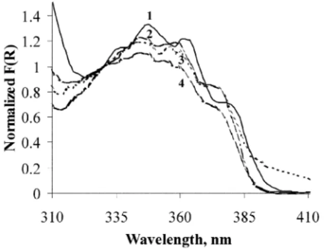
Ground-state diffuse reflectance absorption spectra for benz-ophenone included within H4CLX[4] and HnPrmCLX[4] were
obtained using an integrating sphere (further details in ref. 5), and are presented in Fig. 1 after subtraction of the remission function of the substrate.
The benzophenone n π* transition has an absorption
maximum at 348 nm for H4CLX[4] inclusion and presents a
clear vibronic structure, characteristic of this ketone absorp-tion.5a In all propylated calixarenes, the absorption spectra of
the included benzophenone is hypsochromic shifted, with an absorption maximum at 344 nm in all HnPrmCLX[4] hosts.
These data are consistent with a non-polar environment in the H4CLX[4] case and a more polar environment in the HnPr
m-Fig. 1 Remission function for BZP/H4CLX[4] (curve 1) and BZP/ HnPrmCLX[4] (n = 2, 1 and 0; m = 2, 3 and 4, respectively) inclusion
complexes, normalised to unity at 330 nm (curves 4, 3 and 2 respectively). The guest–host molar ratio is 1 : 2.5 in all samples.
CLX[4] cases, suggesting endo- and exo-calix inclusion,
respectively.
9aThe n π* absorption region of benzophenone within
HnPrmCLX[4] does not present the very clear vibronic structure
seen for BZP/H4CLX[4], but rather, a less structured and broad
band in these hosts. This broadening effect increases in the order Pr4CLX[4] < HPr3CLX[4] < H2Pr2CLX[4], suggesting
conformational behaviour as a result of increased flexibility in the above-mentioned order, possibly due to the greater mobility of the less propylated calixarenes.
The other important information regarding the geometry of the benzophenone molecule within the calixarenes comes from the FTIR spectra. Table 1 presents the carbonyl stretching frequencies obtained from FTIR absorption spectra for microcrystalline samples of benzophenone, benzophenone/ H4CLX[4] and benzophenone/HnPrmCLX[4].
The C᎐᎐O stretching band of microcrystalline benzophenone is located at 1651 cm⫺1 and the C᎐᎐C skeletal breathing mode at 1593 cm⫺1.4c For the planar conformations of benzophenone
(microcrystals), an important resonance interaction exists. Inclusion into calix[4]arene and HnPrmCLX[4] results in a clear
increase in the energy of the C᎐᎐O stretching mode due to enhancement of C᎐᎐O double bond character (inclusion of the calixarenes into cavities promotes deviations from planarity in the phenyl–carbonyl conjugated system, thereby decreasing resonance). A similar argument applies to the C᎐᎐C skeletal breathing mode. Data from Table 1 clearly show that benzo-phenone is included into the calixarenes cavities in all cases,
although it does not distinguish between H4CLX[4] and
HnPrmCLX[4] inclusion.
As indicated in the Experimental, the FTIR spectra have a Fig. 2 FTIR absorption spectra of microcrystalline samples of BZP (1), and BZP/H4CLX[4] (2) and BZP/HnPrmCLX[4] (n = 2, 1 and 0; m =
2, 3 and 4, respectively) inclusion complexes (3–5, respectively). All spectra are normalised in order to have a common baseline with a 1 : 2.5 guest–host molar ratio (except for BZP/H4CLX[4], where a 1 : 1 molar ratio was used) in a KBr matrix.
Table 1 Carbonyl stretching and skeletal breathing mode bands from FTIR absorption spectra of microcrystalline samples of BZP, and BZP/ H4CLX[4] and BZP/HnPrmCLX[4] (n = 2, 1 and 0; m = 2, 3 and 4,
respectively) inclusion complexes (1 : 2.5 guest–host molar ratio in all cases, except for H4CLX[4], where a 1 : 1 molar ratio was used)
νC᎐᎐Oa/cm⫺1 νC ᎐᎐Ca/cm⫺1 Microcrystals of BZP 1651 1593 BZP/H4CLX[4] 1655 1599 BZP/Pr4CLX[4] 1655 1598 BZP/HPr3CLX[4] 1656 1599 BZP/H2Pr2CLX[4] 1660 1598 aError ±0.5 cm⫺1.
resolution of 1.0 cm⫺1, so the fact that the peaks are not sharp in Fig. 2 reflects the heterogeneity of the environment felt by the probe, benzophenone.
Room temperature laser-induced phosphorescence
Room temperature phosphorescence spectra of the
benzo-phenone/H4CLX[4] inclusion complex were reported
pre-viously.4c These time-resolved spectra were obtained under
air-equilibrated conditions and were identical those obtained using argon-purged samples. Lifetimes of about 80 µs were found, as compared to about 50 µs for benzophenone micro-crystals,4b determined at the maximum emission wavelength
(448 nm).
Fig. 3, 4 and 5 present similar data for benzophenone as the guest in H2Pr2CLX[4], HPr3CLX[4] and Pr4CLX[4] hosts
(air-equilibrated samples). As before,4 short pulses were used as
excitation sources, either from a N2 laser pulse at 337 nm
(600 ps halfwidth, ∼1.0 mJ pulse⫺1), or from a Nd:Yag laser at 355 nm (6 ns halfwidth, 1–5 mJ pulse⫺1), both being quite suit-able for benzophenone time-resolved luminescence studies, due to their short durations when compared with the lifetime of benzophenone in all samples.
The data presented in Fig. 3, 4 and 5 were obtained using two different time gates: a small time gate (500 or 1000 ns width) to evidence the short time behaviour following the laser excitation pulse, and a very large time gate (20 ms width) to record all room temperature phosphorescence.
The small time gate width enabled us to observe the appear-ance of a new emission, which peaks at 545 nm (a few micro-seconds after the laser pulse), superimposed on the normal benzophenone phosphorescence emission. The rise time of Fig. 3 Laser-induced room temperature phosphorescence emission spectra from air-equilibrated samples of BZP/H2Pr2CLX[4] inclusion complex (guest–host molar ratio 1 : 2.5): (a) 500 ns gate width; (b) 20 ms gate width. The excitation wavelength was 337 nm in both cases.
the new emission is quite clear in Fig. 5(a) (tetrapropylated calixarene host), when compared with Fig. 3(a) (dipropylated calixarene), which shows no new emission superimposed on the benzophenone phosphorescence. This new emission in Fig. 5(a) does not appear in trace 1, but can be clearly seen in traces 2–5 (about 3–10 ms timescale). The new emission could not be observed either on shorter timescales with the short time gate width (500 ns). Only with the use of larger gate widths could the emission of the photoproduct (PhPD) be recorded, which is the predominant emission for the tri- and tetrapropylated hosts It is also important to note that the relative importance of the new emission at 545 nm increases with the degree of propylation, as Fig. 3(b), 4(b) and 5(b) clearly show. This means that this pro-poxy group plays a decisive role in the formation of the new emissive species, because no new emission was detected in the case of the benzophenone/H4CLX[4] inclusion complex.
These samples were also argon purged and their spectra recorded. In all cases, the air-equilibrated and argon-purged spectra are identical, within experimental error, as are the life-times, showing that inclusion into propylated calixarenes prevents oxygen reaching the excited benzophenone molecule during the lifetime of its triplet excited state. Table 2 summar-ises the lifetimes obtained for all samples, for benzophenone and for the new emission. Clearly, there is a quenching of benzophenone triplet state as the degree of propylation increases, with a consequent decrease in the benzophenone lifetime.
The assignment of this new emission was difficult. Two main hypotheses were assumed as starting points: emission from transient species derived from benzophenone alone or from new molecules resulting from photochemical reaction of benzo-phenone with the host. The second hypothesis seems to be the Fig. 4 Laser-induced room temperature phosphorescence emission spectra from air-equilibrated samples of BZP/HPr3CLX[4] inclusion complex (guest–host molar ratio 1 : 2.5): (a) 500 ns gate width; (b) 20 ms gate width. The excitation wavelength was 337 nm in both cases.
most logical. The simple fact that the new emission peaks at 545 nm, and also its lifetime, excludes emission from the ketyl radi-cal of benzophenone, which peaks at about 570 nm as the result of a two-photon excitation process (fluorescence emission).12
It also excludes emissions from aryl alkyl ketones which
phos-phoresce around 420 nm.6b Good candidates for the new
emission could be diketones, like benzil, resulting from an α-cleavage of benzophenone, leading to benzoyl radicals (which requires a two-photon absorption process13), which could
com-bine to form benzil. However, the phosphorescence of this ketone within calix[6]- and -[8]arenes peaks at about 565 nm.4a
A second good candidate is 1-phenyl-1,2-propanedione (PhPD). A parallel experiment was done with PhPD included
within Pr4CLX[4]: the phosphorescence emission maximum
was observed at 545 nm, with a similar phosphorescence decay, suggesting that the new emitting species was PhPD because the spectra were identical within experimental error. We also tried to prepare a sample of benzil/Pr4CLX[4] complex; we
con-cluded that there is no inclusion at all in this case because benzil Fig. 5 Laser-induced room temperature phosphorescence emission spectra from air-equilibrated samples of BZP/Pr4CLX[4] inclusion complex (guest–host molar ratio 1 : 2.5): (a) 500 ns gate width; (b) 20 ms gate width. The excitation wavelength was 337 nm in both cases. Table 2 Phosphorescence emission lifetimes (longer components, air-equilibrated samples) for samples of BZP/H4CLX[4] and BZP/ HnPrmCLX[4] (n = 2, 1 and 0; m = 2, 3 and 4, respectively) inclusion
complexes, compared with equivalent data for microcrystals of benzophenone τPa/µs At 448 nm At 545 nm BZP microcrystals 50 50 BZP/H4CLX[4] 70 70 BZP/H2Pr2CLX[4] 6 80 BZP/HPr3CLX[4] 3 580 BZP/Pr4CLX[4] 2 580 aEstimated error ±5%.
emits as if it were in the form of microcrystals, with a maximum at 522 nm from an s-cis conformation, and not as a guest, as in ref. 4a, for a benzil/H4CLX[4] sample.
Previous work4 indicates that calixarenes are good hydrogen
atom donors, so it seems reasonable to assume that efficient formation of the ketyl radical of benzophenone may also occur here.
Diffuse reflectance laser flash photolysis
Time-resolved absorption spectra of samples of benzophenone/ HnPrmCLX[4] inclusion complexes were obtained using the
dif-fuse reflectance laser flash photolysis technique, developed by Wilkinson and co-workers.2 In this study, the use of an
intensi-fied charge-coupled device as the detector allowed us to obtain time-resolved absorption spectra with nanometer spectral reso-lution.1,4
Fig. 6 shows the time-resolved absorption spectra of these inclusion complexes with a guest–host molar ratio of 1 : 2.5. All spectra were obtained with air-equilibrated samples, exciting at 355 nm.
Diffuse reflectance laser flash photolysis provided us with further relevant experimental information to help solve the
Fig. 6 Time-resolved absorption spectra of BZP/H2Pr2CLX[4] (a), BZP/HPr3CLX[4] (b) and BZP/Pr4CLX[4] (c). The guest–host molar ratio is 1 : 2.5 and the excitation wavelength 355 nm in all cases.
problem of the identification of the species responsible for the new emission peaking at 545 nm.
Transient absorption spectra of benzophenone/H4CLX[4]
samples have revealed the simultaneous formation of triplet benzophenone and also of the hydroxybenzophenone radical (BZPOHⴢ),4c as Fig. 7 shows. This time-resolved absorption
spectra relate to a 1 : 10 guest–host molar ratio sample, i.e. a rather dilute sample.
The triplet–triplet absorption spectra of benzophenone were easily identified by comparison with that published by Wilkin-son and Willsher.14 Two absorption maxima at 322 and 535 nm
can be seen in the 1 µs spectrum, corresponding to the T1 T3
and T1 T2 absorption transitions of benzophenone. The
fact that these maxima are retained at longer timescales (10 and 20 µs after the laser pulse) shows that no ketyl radical of benzophenone is formed by inclusion of benzophenone into H4CLX[4], as reported in ref. 4c for more concentrated samples
(1 : 2.5 guest–host molar ratio). The transient absorption which peaks at 395 nm can be assigned to the BZPOHⴢ radical by comparison with previously reported spectra in solution.15
Another very important feature regarding the transient absorption spectra relates to the scattered radiation at the excit-ation wavelength, 355 nm. This scattered radiexcit-ation originates from the Nd:YAG laser (third harmonic) used in the diffuse reflectance laser flash photolysis set-up.1b The original pulse
width is only 6–7 ns and, in spite of this, scattered radiation is detected in the ICCD, using its time-gated detector, at least 20 µs after the laser pulse. This shows that many photons may remain available and could possibly excite the solid powdered sample a long time after the initial laser pulse, possibly due to multiple internal reflections trapping light within the non-Fig. 7 (a) Time-resolved absorption spectra of a BZP/H4CLX[4] sample (guest–host molar ratio 1 : 10). Traces 1–4 were recorded 5, 10, 20 and 100 µs after the laser pulse, respectively. (b) Scattered radiation obtained with the ICCD using a 500 ns time gate and delay times after laser pulse of 2 (1), 5 (2), 10 (3), 20 (4) and 100 µs (5). The sample is the same as in (a) and the excitation wavelength was 355 nm.
absorbing microcrystals in the powdered substrate. This could explain how the photoproduct formed in the microsecond time-scale could be excited following a single nanosecond laser pulse, and is in accordance with our product formation hypothesis.
Scattered radiation (at 355 nm) was detected with our ICCD for low [see Fig. 7(b)] and high loadings of benzophenone included into the HnPrmCLX[4] samples, although its
import-ance decreases with increasing F(R) of benzophenone at the excitation wavelength (i.e. with increasing probe concen-tration), because the more excitation radiation is absorbed, the less scattered radiation is detected. Diffuse reflectance transient absorption spectra are corrected for emission (and scattered) radiation. Scattered radiation affects only a small and localised spectral region, while the phosphorescence emission is broader in spectral terms. It is therefore reasonable to correct for emis-sion in the first place. Whenever the intensity of the scattered radiation is three or four times more intense than the emission itself, to completely correct the emission, the scatter is over-corrected, as is the case of Fig. 7(a). This is probably due to the occurrence of some degradation of the probe, which occurs because each spectrum is an average of several laser shots (depending on the sample).
Transient absorption spectra of benzophenone included into HnPrmCLX[4] are presented in Fig. 6 (peaking at 535 nm, about
1 µs after the laser pulse). In these spectra, a new species appears and becomes more evident at longer times (∼2–10 µs after the laser pulse), which appears as a shoulder at about 555 nm, characteristic of the benzophenone ketyl radical absorp-tion.5a,4c,16 So the H
nPrmCLX[4] compounds behave as hydrogen
atom donors towards the excited aromatic ketone. The same behaviour was detected for the benzophenone/CLX[6] and CLX[8] inclusion complexes.4c The fact that no benzophenone
ketyl radicals were formed in the H4CLX[4] samples, but
evidence for ketyl radical formation was obtained in the HnPrmCLX[4] cases (and in the CLX[6] and CLX[8] cases),
4c
reflects the fact that only in the latter are spatial arrangements suitable for the hydrogen atom abstraction reaction to occur possible, whereas, due to the reduced internal space of the H4CLX[4] cavity, no reaction occurs with this host.
4c
This again suggests exo-calix inclusion of benzophenone within the propylated calixarenes.
It is important to point out that at very long times (≥1 ms), a residual transient absorption is still detected [see trace 4 of Fig. 6(a) and (c)]. This transient peaks at about 395 nm (BZPOHⴢ radical) and also shows a kind of long absorption ‘tail’ which ends at about 500 nm. This tail may be due to the formation of an α-diketone, namely 1-phenyl-1,2-propanedione. Benzo-phenone/HPr3CLX[4] does not show such strong evidence for
BZPOHⴢ radical formation, and only the superimposed triplet– triplet and ketyl radical absorptions of benzophenone could be detected.
The fact that the photoproduct is formed with a single pulse could also be explained by assuming that PhPD was formed in an excited state. However, to form a PhPD molecule, a benzoyl radical and an acetyl radical have to come into contact. From the combination of the two radicals, a ground-state PhPD mol-ecule is most probably formed. However, if an excited state was formed—and this is not a common hypothesis in literature—it could also explain the PhPD phosphorescence.
Photodegradation products studies
Initially, lamp irradiation conditions (15 W mercury lamp) were used. The analytical results showed that 2-hydroxybenzo-phenone is one of the major degradation products of all the samples under study. A typical GC-MS chromatogram of the irradiated extracts is shown in Fig. 8(a). However, this is a well-known and very stable and non-luminescent compound due to the very rapid intramolecular non-radiative mechanism of de-excitation, involving hydrogen bond formation between the
carbonyl and the nearby hydroxy groups.17a Analysis of the
extracts of the benzophenone/Pr4CLX[4] sample after
pro-longed irradiation (4 h), at a distance of 5 cm from the lamp housing, showed only traces of benzil and PhPD. However, the
analyses of the BZP/H2Pr2CLX[4] and BZP/HPr3CLX[4]
samples, irradiated under the same conditions, showed no traces of these compounds. Since all the samples showed the same emission results, it is clear that another process must be present.
Assuming that the formation of an α-diketone is the process that leads to the observed new emission, the failure to detect this compound in significant amounts could be due to differ-ences in the experimental conditions used to obtain the phosphorescence emission spectra and the photodegradation products. The same luminescence results were obtained with 337, 355 and 266 nm laser excitation; therefore, the observed result cannot be attributed to differences in the excitation wave-length used (the mercury lamp employed here irradiates mainly
at 254 nm) but could due to the high fluency of the laser
Fig. 8 Typical chromatograms (GC-MS) of: (a) an extract of a sample of BZP/Pr4CLX[4] irradiated with a mercury lamp at 254 nm; (b) a laser-irradiated (at 337 nm) sample of benzophenone within Pr4CLX[4]; (c) a laser-irradiated (at 337 nm) sample of benzophenone within H2Pr2CLX[4].
excitation and the possibility of a two-photon process occur-ring.12b,13 Therefore, the same analyses were conducted on
sam-ples irradiated with a laser at 355 nm [see Fig. 8(b) and (c)]. The samples were irradiated for 1 h and shaken every 5 min.
1-Phenyl-1,2-propanedione was the main degradation product of benzophenone in HnPrmCLX[4] hosts, as detected
by GC-MS, in all cases. This result agrees with the assign-ment made in the time-resolved luminescence as an α-diketone phosphorescence emission, and clearly indicates significant differences depending on whether laser or lamp irradiation is employed. Extracts of samples containing H2Pr2CLX[4]
showed lower levels of PhPD, whereas increased amounts were formed with HPr3CLX[4] and the Pr4CLX[4]. This result is in
agreement with the luminescence results, since the emission increases with the degree of propylation of the host. As expected, no PhPD was detected when the non-propylated calixarene was used. 2-Hydroxybenzophenone was detected in all cases. Other products found include biphenyl, benzoic acid, methyl benzophenone and benzhydrol, the latter detected by HPLC. Also, traces of acetophenone and benzene were found. All identifications were based on comparison with authentic samples and/or GC-MS spectra.
The solution photochemical reactions of carbonyl com-pounds have been extensively studied and include photo-reduction (eqn. 3), α-cleavage (eqn. 4), intramolecular hydrogen abstraction and dimerisation.13,8a,16 The solution
photo-chemistry of benzophenone was studied by flash photolysis a long time ago,8a,17a and may be briefly described by the
follow-ing processes:
Solution photochemical studies of benzophenone have shown that the hydrogen abstraction reaction with formation of the benzophenone ketyl radical (intermolecular hydrogen atom abstraction from suitable H-donating solvents) clearly pre-dominates over the formation of the benzoyl radical (Norrish type I cleavage).13,17
A two-step photon absorption process is necessary for benzophenone to produce a highly excited triplet state from which the benzoyl and phenyl radicals, and decarb-onylation may occur.12b,13 As we said, this process occurs within
a single laser pulse, probably because of the diffuse reflected excitation radiation (355, 337 or 266 nm) detected long after the laser initial pulse (depending on the concentration of benzophenone in the sample).
The new emission shown in Fig. 3, 4 and 5 for benzophenone encapsulated in H2Pr2CLX[4], HPr3CLX[4] and Pr4CLX[4],
respectively, was obtained by irradiating either with a Nd:YAG laser (at 355 nm, 1–30 mJ pulse⫺1, irradiating an area of about 1 cm2
) or with a N2 laser (at 337 nm, 0.3–1.0 mJ pulse⫺1,
irradi-ating approximately the same sample area). Due to the rather large difference in fluence and to the fact that the new emission appears under both types of excitation, it is logical to assume that the two-photon absorption is sequential and not simul-taneous, and therefore depends on the pulse length, as described in the literature for the formation of highly excited states of benzophenone.13
As we described, product analysis and identification was attempted following laser excitation (355 nm). HPLC and GC-MS analysis clearly show that the main degradation product of
the benzophenone/H4CLX[4] samples was
2-hydroxybenzo-(1) (2)
(3)
(4)
Scheme 2 phenone and, for the BZP/H2Pr2CLX[4] and BZP/Pr4CLX[4]
pairs, the main photoproducts were 1-phenyl-1,2-propanedione and 2-hydroxybenzophenone. In contrast with the solution behaviour, the α-cleavage of benzophenone occurs and becames a very important degradation pathway in solid powdered samples.
All the experimental observations described herein, which lead to the PhPD phosphorescence emission and to the appear-ance of the trace compounds, are summarised in Scheme 2. In this scheme, the crucial reaction is the benzophenone hydrogen abstraction from the methylene group bound to the oxygen of the ether linkage. Further α-cleavage of this intermediate generates a methyl radical and an aryl enolether. The methyl radical thus formed could further react with several of the intermediate radicals resulting from the α-cleavage of BZP**, and thus accounting for the products formed: mainly PhPD, from which the new phosphorescence emission originates.
4
Conclusions
We can therefore conclude that inclusion of benzophenone into H4CLX[4] and HnPrmCLX[4] (n = 2, 1 and 0; m = 2, 3 and 4,
respectively) results in significant changes in both the ground-state absorption spectra and the time-resolved emission and transient absorption spectra. Product analysis and identi-fication showed that the two main degradation products detected in the latter case were 1-phenyl-1,2-propanedione and 2-hydroxybenzophenone. The α-diketone is efficiently excited by scattered laser radiation long after the initial nanosecond laser pulse and a new phosphorescence emission occurs, which becomes the predominant luminescence on the millisecond timescale.
The fact that benzil is not formed as a result of the recombin-ation of two benzoyl radicals suggests that stereochemical con-strainments play a decisive role inside the calixarene cavity, i.e. the acetyl radical and the benzoyl radical are able to recombine, while two benzoyl radicals are not.
Comparing the results presented in this paper with those
reported by Barra and Scaiano18 for benzophenone within
p-tert-butylhexaethoxycalix[6]arene, very important differences are apparent. In the case of ref. 18, no photochemistry was reported. The simple difference in the phenolic substituent group (a propyl group in our case) prompts some remarkable photochemical behaviour of benzophenone, with the formation and emission of a new α-diketone, 1-phenyl-1,2-propanedione.
Acknowledgements
The authors thank ICCTI/CAPES for financial support.
A. S. O. and J. P. S. thank FCT for Post-Doctoral Fellowships SFRH/BPD/36500/2000 and SFRH/BPD/15589/2001.
References
1 A. M. Botelho do Rego and L. F. Vieira Ferreira, in Handbook of Surfaces and Interfaces of Materials, Vol. 2,ed. H. S. Nalwa, Academic Press, New York, 2001, ch. 7, pp. 275–313.
2 (a) F. Wilkinson and G. P. Kelly, in Photochemistry on Solid Surfaces, ed. M. Anpo and T. Matsuara, Elsevier, Amsterdam, 1989, pp. 31–47; (b) F. WilkinsonG. P. Kelly, in Handbook of Organic Photochemistry, Vol. 1, ed. J. C. Scaiano, CRC Press, Boca Raton, FL, 1989, ch. 12, pp. 293–314.
3 J. H. Hurtubise, Solid-matrix luminescence analysis: photophysics, physicochemical interactions and applications, Anal. Chim. Acta, 1997, 351, 1–22.
4 (a) L. F. Vieira Ferreira, I. Ferreira Machado, A. S. Oliveira, M. R. Vieira Ferreira, J. P. Da Silva and J. C. Moreira, A diffuse reflectance comparative study of benzil inclusion within p-tert-butylcalix[n]arenes (n = 4, 6 and 8) and silicalite, J. Phys. Chem. B, 2002, 106, 12 584–12 593; (b) L. F. Vieira Ferreira, M. R. Vieira Ferreira, A. S. Oliveira and J. C. Moreira, Potentialities of diffuse reflectance laser-induced techniques in solid phase: a comparative study of benzophenone inclusion within p-tert-butylcalixarenes, silicalite and microcrystalline cellulose, J. Photochem. Photobiol., A, 2002, 153, 11–18; (c) L. F. Vieira Ferreira, M. R. Vieira Ferreira, A. S. Oliveira, T. J. F. Branco, J. V. Prata and J. C. Moreira, Diffuse reflectance studies of β-phenylpropiophenone and benzophenone inclusion complexes with calix[4], [6] and [8] arenes, Phys. Chem. Chem. Phys., 2002, 4, 204–210.
5 (a) L. F. Vieira Ferreira, J. C. Netto-Ferreira, I. Khmelinskii, A. R. Garcia and S. M. B. Costa, Photochemistry on surfaces: matrix isolation mechanisms for study of interactions of
benzophenone adsorbed on microcrystalline cellulose investigated by diffuse reflectance and luminescence techniques, Langmuir, 1995, 11, 231–236; (b) L. F. Vieira Ferreira, M. R. Freixo, A. R. Garcia and F. Wilkinson, Photochemistry on surfaces: fluorescence quantum yield determination of dyes adsorbed on microcrystalline cellulose, J. Chem. Soc., Faraday Trans., 1992, 88, 15–22; (c) L. F. Vieira Ferreira, A. R. Garcia, M. R. Freixo and S. M. B. Costa, Photochemistry on surfaces: the solvent-matrix effect on the swelling of cellulose. An emission and absorption study of adsorbed auramine-O, J. Chem. Soc., Faraday Trans., 1993, 89, 1937–1944. 6 (a) J. P. Da Silva, L. F. Vieira Ferreira, A. M. Da Silva and A. S.
Oliveira, A comparative study of the photophysics and photochemistry of 4-chlorophenol adsorbed on silicalite and b-cyclodextrin, J. Photochem. Photobiol., A, 2002, 151, 157–164; (b) L. F. Vieira Ferreira, and A. S. Oliveira J. C. Netto-Ferreira, in Fluorescence Microscopy and Fluorescent Probes 3, ed. A. Kotyc, Espero Publishing, Prague, 1999, pp. 199–208.
7 L. F. Vieira Ferreira, M. J. Lemos, M. J. Reis and A. M. Botelho do Rego, UV/Vis absorption and luminescence and X-ray photoelectron spectroscopic studies of rhodamine dyes adsorbed onto different pore size silicas, Langmuir, 2000, 16, 5673–5680. 8 (a) N. J. Turro, Modern Molecular Photochemistry, Benjamin
Cummings, Menlo Park, CA, 1978; (b) N. J. Turro, A. Masayuki and I. R. Gould, The laser vs. the lamp. A novel laser-induced adiabatic reaction and luminescence of benzophenone, J. Am. Chem. Soc., 1982, 104, 856–860.
9 (a) C. D. Gutshe, Calixarenes, Royal Society of Chemistry, Cambridge, 1989; (b) I. Alam and C. D. Gutsche, Calixarenes. 24. Complexation by water soluble calixarenes, J. Org. Chem., 1990, 55, 4487–4489; (c) C. D. Gutsche and I. Alam, Calixarenes. 23. The complexation and catalytic properties of water soluble calixarenes, Tetrahedron, 1988, 44, 4689–4694.
10 D. M. Rudkevich, Nanoscale molecular containers, Bull. Chem. Soc. Jpn., 2002, 75, 393.
11 (a) C. D. Gutsche and M. Iqbal, p-tert-Butylcalix[4]arene, Org. Synth., 1990, 68, 234–237; (b) K. Iwamoto, K. Araki and S. Shinkai,
Conformations and structures of tetra – O-alkyl-p-tert-butylcalix-[4]arenes. How is the conformation of calix[4]arenes immobilized?, J. Org. Chem., 1991, 56, 4955–4962.
12 (a) J. C. Netto-Ferreira and J. C. Scaiano, Photochemistry and photophysics from the excited states of diaryl ketyl radicals [1], Res. Chem. Intermed., 1989, 12, 187–201; (b) M. Barra and J. C. Scaiano, Photoinduced transient phenomena in cyclodextrin solid complexes: photochemistry of aromatic ketones, Photochem. Photobiol., 1995, 62, 60–64.
13 (a) W. C. McGimpsey and J. C. Scaiano, Chem. Phys. Lett., 1987, 138, 13; (b) Y. Kajii, T. Suzuki, Y. Takatori, K. Shibuiya and K. Obi, Photodissociation of highly-excited triplet state of benzophenone studied by a time-resolved thermal lensing technique, Bull. Chem. Soc. Jpn., 1992, 65, 1349–1355.
14 F. Wilkinson and C. J. Willsher, Detection of triplet-triplet absorption in microcrystalline benzophenone by di ffuse-reflect-ance laser flash photolysis, Chem. Phys. Lett., 1984, 104, 272– 276.
15 (a) M. B. Ledger and G. Porter, Primary photochemical processes in aromatic molecules. Part 15, J. Chem. Soc., Faraday Trans. 1, 1972, 68, 539–553; (b) R. V. Bensasson and J. C. Gramain, Benzophenone triplet properties in acetonitrile and water, J. Chem. Soc., Faraday Trans. 1, 1980, 76, 1801–1810; (c) S. B. Sharma, M. Mudaliar, B. S. M. Rao, H. Moahn and J. P. Mittal, Radiation chemical oxidation of benzaldehyde, acetophenone and benzo-phenone, J. Phys. Chem. A, 1997, 101, 8402–8408.
16 S. Monti, L. Flamigui, A. Martelli and P. Bortolus, Photochemistry of benzophenone-cyclodextrin inclusion complexes, J. Phys. Chem., 1988, 92, 4447–4451.
17 (a) P. Suppan, Chemistry and Light, Royal Society of Chemistry, Cambridge, 1994; (b) J. C. Scaiano, E. B. Abuin and L. C. Stuart, Photochemistry of benzophenone in micelles. Formation and decay of radical pairs, J. Am. Chem. Soc., 1982, 104, 5673–5679.
18 M. Barra and J. C. Scaiano, Diffuse reflectance laser-flash photolytic study of aromatic ketones within calixarene solid matrixes, Supramol. Chem., 1998, 10, 91–95.
![Table 2 Phosphorescence emission lifetimes (longer components, air-equilibrated samples) for samples of BZP/H 4 CLX[4] and BZP/](https://thumb-eu.123doks.com/thumbv2/123dok_br/18800513.925852/5.918.91.438.143.786/table-phosphorescence-emission-lifetimes-components-equilibrated-samples-samples.webp)
