Recombinant antigen-based
immuno-slot blot method for
serodiagnosis of syphilis
1Seção de Sorologia, Divisão de Biologia Médica, Instituto Adolfo Lutz, São Paulo, SP, Brasil
2Department of Chemistry and Biotechnology, Graduate School of Engineering, University of Tokyo, Tokyo, Japan
3Departamento de Análises Clínicas e Toxicológicas, Faculdade de Ciências Farmacêuticas, Universidade de São Paulo, São Paulo, SP, Brasil
N.S. Sato1, T. Suzuki2, T. Ueda2, K. Watanabe2, R.D.C. Hirata3 and M.H. Hirata3
Abstract
Three recombinant antigens of Treponema pallidum Nichols strain were fused with GST, cloned and expressed in Escherichia coli, resulting in high levels of GST-rTp47 and GST-rTp17 expression, and supplementation with arginine tRNA for the AGR codon was needed to obtain GST-rTp15 overexpression. Purified fusion pro-tein yields were 1.9, 1.7 and 5.3 mg/l of cell culture for GST-rTp47, GST-rTp17 and GST-rTp15, respectively. The identities of the anti-gens obtained were confirmed by automated DNA sequencing using ABI Prism 310 and peptide mapping by Finningan LC/MS. These recombinant antigens were evaluated by immuno-slot blot techniques applied to 137 serum samples from patients with a clinical and laboratory diagnosis of syphilis (61 samples), from healthy blood donors (50 samples), individuals with sexually trans-mitted disease other than syphilis (3 samples), and from individu-als with other spirochetal diseases such as Lyme disease (20 samples) and leptospirosis (3 samples). The assay had sensitivity of 95.1% (95% CI, 86.1 to 98.7%) and a specificity of 94.7% (95% CI, 87.0 to 98.7%); a stronger reactivity was observed with fraction rTp17. The immunoreactivity results showed that fusion recombi-nant antigens based-immuno-slot blot techniques are suitable for use in diagnostic assays for syphilis.
Correspondence
N.S. Sato Seção de Sorologia Divisão de Biologia Médica Instituto Adolfo Lutz Av. Dr. Arnaldo, 355, 10º andar 01246-902 São Paulo, SP Brasil
Fax: +55-11-3085-3505 E-mail: [email protected]
Presented at the 99th General Meeting of the American Society for Microbiology, Chicago, IL, USA, May 30-June 3, 1999.
Research supported by CAPES, JICA.
Publication supported by FAPESP.
Received July 15, 2003 Accepted April 5, 2004
Key words
•Treponema pallidum •Cloning
•Recombinant antigen •Syphilis
•GST
Treponema pallidum subsp. pallidum is the etiological agent of syphilis, a sexually transmitted disease (STD), and serology is essential for screening and for its diagnosis. However, a major problem for the improve-ment of treponemal tests is the limited avail-ability of T. pallidum, a spirochetal bacte-rium that cannot be readily cultured in vitro (1). For most research and diagnostic
T. pallidum antigen.
The major T. pallidum antigen identified in studies of humoral ontogeny of experi-mental syphilis (3) and by Western blot analysis has a molecular mass of 47 kDa, 17 kDa and 15 kDa (4,5). All three antigens have been characterized as highly immunogenic membrane lipoproteins (6-10) and as patho-gen-specific antigens (6,10). Their DNA sequence has also been reported (6,9,11).
Based on published sequence data for Tp47, Tp17 and Tp15 (6,9,11) these three proteins of T. pallidum subsp. pallidum have been cloned in fusion with GST and ex-pressed in Escherichia coli (12). In the Western blot assay the purified recombinant antigens showed reactivity specifically against the treponemal fraction of the fusion protein, as no serum sample reacted with purified GST (13). In the present study we evaluated the GST-rTp47, GST-rTp17 and GST-rTp15 antigens in an immuno-slot blot (IB) assay for the diagnosis of syphilis.
For cloning, the DNA fragments of Tp47, Tp17 and Tp15 were prepared by PCR using the chromosomal DNA of T. pallidum subsp. pallidum (Nichols strain) as template. The open reading frame (ORF) encoding the full-length mature protein was amplified by PCR with a primer designed according to a pub-lished sequence, GenBank accession num-bers M27493, M74852 and M3941 (6,9,11). The sense primer includes the BamHI site and the antisense primer includes the EcoRI site except for Tp47, which had the BamHI site in both primers. PCR cycling (Perkin-Elmer 9600, Foster City, CA, USA) condi-tions were as follows: denaturation at 94ºC for 5 min, 30 cycles of 94ºC for 1 min, T-annealing for 1 min, 72ºC for 1 min for polymerization, and a final elongation step at 72ºC for 10 min. T-annealing was carried out at 49º, 59º and 62ºC for Tp47, Tp17 and Tp15, respectively. Restriction digestion of vector pGEX4T-2 (Pharmacia) and PCR products and ligation were performed ac-cording to manufacturer instructions. E. coli
DH5α or JM109 were used for cloning.
Correct insertion of amplified DNA frag-ments into the expression vector was con-firmed by automatic sequencing using a dye terminator cycle sequencing kit and an Ap-plied Biosystems Prism 310 DNA Analyzer (Foster City, CA, USA).
Recombinant T. pallidum antigen was expressed in distinct strains of E. coli. The antigens Tp47, Tp17 and GST-Tp15 were expressed in E. coli BL-21, DH5α and BL-21/ArgU218, respectively.
Cells were grown at 37ºC in LB medium containing 200 µg/ml ampicillin for E. coli DH5α or BL-21, and in medium containing
200 µg/ml ampicillin and 30 µg/ml kanamy-cin for BL-21/ArgU218. Cell growth was monitored by absorbance at 600 nm and recombinant protein expression was induced by adding isopropyl ß-D thiogalactopyrano-side to a final concentration of 0.1 mM when absorbance at 600 nm reached about 0.600. Four hours after induction, cells were har-vested and disrupted by sonication in ice-cold 0.15 M PBS, pH 7.2, with 1 mM PMSF, 5 mM DTT, and 1% Triton X-100 and centrifuged at 10,000 g for 20 min at 4ºC. The soluble fraction of GST fusion proteins was purified using glutathione (Sigma).
Peptide mapping was performed by Finningan LC/MS (Thermoquest) using a purified rTp antigen digested with pepti-dases. Briefly, purified rTp was obtained by cleavage with thrombin, with each 10 µg protein being incubated for 16 h at 25ºC with 0.1 U thrombin in 50 mM Tris-HCl, pH 7.5, 150 mM NaCl, 2.5 mm CaCl2, and 5%
15-h incubation at 30ºC, peptides were extract-ed from the gel with 0.1% TFA in 60% acetonitrile, dried and re-dissolved in 0.1% formic acid for LC/MS analysis.
Recombinant proteins were analyzed by 12% SDS-PAGE stained with Coomassie brilliant blue R-250 and by Western blotting with serum from patients with secondary syphilis (1:100) as primary antibody and immunodetection was performed using horseradish peroxidase (HRP)-labeled sec-ondary antibody and 4-chloro-1-naphthol.
The diagnostic evaluation of purified re-combinant fusion antigen was performed by IB-rTp using 137 serum samples, 61 of which were from patients with a clinical and or laboratory diagnosis of syphilis. For IB, the antigen was dotted onto a nitrocellulose membrane using a Bio-Dot SF apparatus (Bio-Rad, Hercules, CA, USA), and the mem-brane was cut into 3.0- or 4.5-mm wide strips. After titration, the following amounts of each antigen were dotted: 750, 440, 1,670, and 1,250 ng/mm2 for rTp47,
GST-rTp17, GST-rTp15, and purified GST, re-spectively. All strips were prepared with GST, which was used as control for speci-ficity. All reaction steps were performed with shaking at room temperature. Nonspe-cific binding sites were blocked with sample buffer (0.15 M PBS, pH 7.3, 5% skim milk, and 0.1% Tween 20) for 15 min, and the strips were washed once with washing buf-fer (0.15 M PBS, pH 7.3, and 0.2% Tween 20). A human serum sample diluted 1:100 or goat anti-GST serum diluted 1:2,000 (Phar-macia, Uppsala, Sweden), used as control to identify GST bands, was added to the strips and incubated for 1 h. After washing three times for 10 min each, the strips were incu-bated for 60 min with a second antibody conjugated with HRP. Goat anti-human IgG-HRP (Sigma) at 1:2,000 dilution in sample buffer was used as the second antibody. Strips were washed as before, and the color was developed by adding 0.6 mg/ml 4-chloro-1-naphthol (Sigma) in 0.15 M PBS, pH 7.3,
with 0.02% H2O2. The final reaction was
stopped by washing the strips with distilled H2O. When at least one band of GST-rTp
was present, with reactivity equal to or stronger than the weakly positive control, the result was considered to be positive.
by Dr. Natalino Yoshinari (FM-USP), were also tested. After blood clotting, serum samples were separated by centrifugation and aliquots were stored at -20ºC. Control serum samples consisted of pooled samples from patients with syphilis, one sample strongly positive, one sample weakly posi-tive in conventional treponemal tests, and one sample pooled serum samples from healthy donors with negative serological re-sults for syphilis. All serum samples were tested by Veneral Disease Research Labora-tory (Behring, Marburg, Germany) and fluo-rescent treponemal antibody-absorption (FTA-Abs, Wama Diagnostica, São Carlos, SP, Brazil) procedures performed according to manufacturer instructions.
GST-rTp47 and GST-rTp17 were suc-cessfully expressed in E. coli, whereas GST-rTp15 was poorly expressed in E. coli. However, this effect was completely re-versed by supplementation with arginine tRNA for the AGR codon. The ORF of Tp15 contains 4 minor arginine codons including one tandem AGG-AGG codon, which was identified as the inhibitory factor for the
expression of GST-rTp15 (12, and Sato NS, Suzuki T, Hirata RDC, Hirata MH and Wata-nabe K, unpublished results). Codon use is critical for overexpression of heterologous protein by E. coli. It has been reported that the arginine tRNA for rare AGR codons is a limiting factor in the bacterial expression of several heterologous genes due to its infre-quent use in E. coli (14). Replacing the rare codons with more commonly used codons and the co-expression of rare tRNA in-creased significantly the expression level of GST-rTp15 (12, and Sato NS, Suzuki T, Hirata RDC, Hirata MH and Watanabe K, unpublished results). The GST-rTp17 pro-tein was expressed in doublet and the upper band was also specific, as shown in Figure 1B, reacting with human syphilis serum. This protein with higher molecular weight resulted from amber suppression at stop codon TAG, since this antigen was ex-pressed in E. coli DH5α, which carries the
supE44genotype responsible for this transla-tional event.
All three clones showed an exuberant expression of recombinant protein;
how-27 GST-rTp47
kDa
175 83 62 47.5
32.5
25
16.5
6.5
GST-rTp17
GST-rTp15
GST
26 25 24 23 22 21 20 19 18 17 16 15 14 13 12 11 10 9 8 7 6 5 4 3 2 1
M 1 2 3 1 2 3
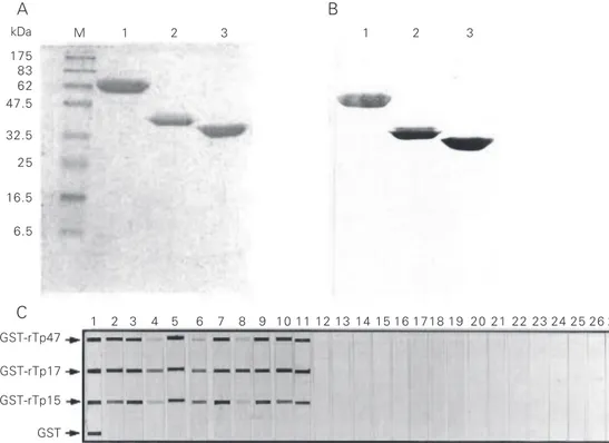
Figure 1. Purity and reactivity of recombinant antigens. Analysis of affinity chromatography-puri-fied GST-rTP fusion protein by (A) Coomassie brilliant blue (CBB)-stained SDS-PAGE and (B) Western blotting performed on serum samples from human syphilis patients and developed with horseradish peroxidase and 4-chloro-1-naphtol (4Cl1N). M = marker protein, with mo-lecular mass in kDa shown on the left. Lane 1, GST-rTp47;
lane 2, GST-rTp17; lane 3, GST-rTp15. Representative recom-binant Treponema pallidum an-tigen-based immuno-slot blot (IB-rTp) reaction (C) of recombi-nant GST-rTp antigens and puri-fied GST with goat anti-GST (lane 1); serum from patient with syphilis (lanes 2-11); serum from a healthy negative donor (lanes 12-16); serum from pa-tients with STD other than syphilis (lanes 17-19); serum from patients with leptospirosis (lanes 20-22), and serum from patients with Lyme disease (lanes 23-27).
A B
ever, a large portion of GST-rTp47 and GST-rTp15 remained in the insoluble frac-tion. The yield of purified fusion protein recovered from the soluble fraction was 1.9, 7.0 and 3.5 mg/l for rTp47, GST-rTp17 and GST-rTp15, respectively. Ex-periments are currently being conduced to improve the solubilization of expressed pro-tein in order to obtain better yields. The use of sarkosyl buffer increased GST-rTp47 solubility, although its effects on binding to affinity beads should be evaluated (data not shown). Sarkosyl buffer added to the pellet after sonication has been reported to result in a significant increase in solubility without a drastic denaturing effect on protein (15).
The purity of recombinant antigen ex-pressed in E. coli is especially important because antibodies against contaminating E. coli protein may cause false-positive reac-tions. The expression of recombinant anti-gens in fusion with GST allowed an efficient one-step purification by glutathione Sepha-rose 4B affinity chromatography. All three antigens showed high purity and reactivity with a serum sample from a patient with diagnosis of syphilis (Figure 1A,B).
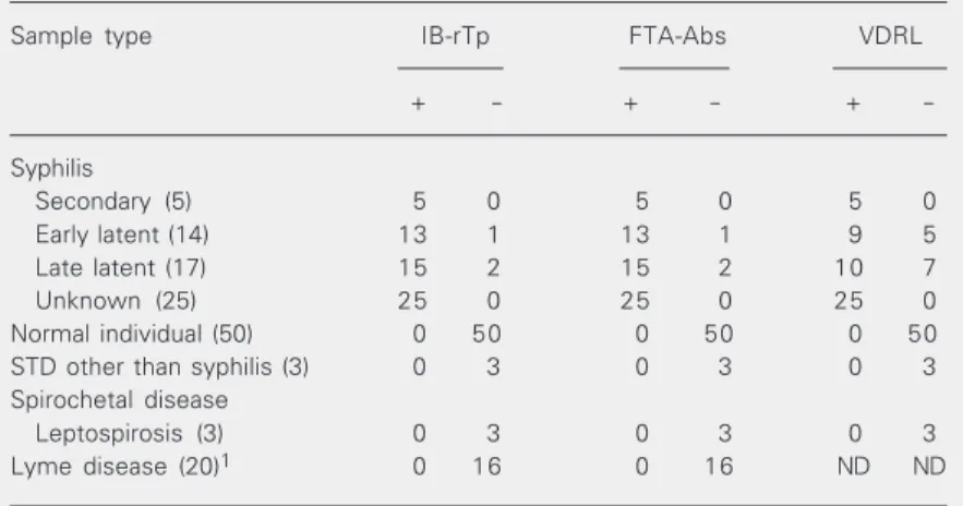
The antigenicity of purified recombinant fusion protein was evaluated by IB. Results of IB and classical serological tests for syphi-lis for 137 clinical samples are presented in Table 1. A representative reaction profile is shown in Figure 1C. Reaction with anti-GST (lane 1) in IB displays the positions of GST-rTp and GST.
Almost all of the samples from patients with syphilis reacted with the rTp17 antigen, and at a higher intensity compared to the other antigens. Some samples demonstrated weak or no reactivity against the 47-kDa and/ or 15-kDa antigens (Figure 1C, lanes 2-11). A stronger reactivity of the 17-kDa antigen was also reported by other investigators (16). Three patients with latent syphilis pre-sented a negative result in both IB-rTP and FTA-Abs tests (Table 1).
Samples from patients with other
spiro-chete infections showed no cross-reaction with samples from patients with leptospiro-sis. However, 4 samples from patients with Lyme disease had some faint and weak background reaction with at least one re-combinant antigen and/or purified GST. These four samples showed a nonspecific back-ground reaction in the FTA-Abs test also. The false-positive reaction in serum samples from patients with Lyme disease has been reported in ELISA for syphilis using a native whole antigen (17). When using whole cells as antigen, it may be speculated that cross-reaction may be due to the lipid component present in the native antigen, since the major antigens are lipoproteins in both T. pallidum and Borrelia burgdorferi (18-20). The back-ground reaction observed in the IB assay was unexpected, since the recombinant antigen has no lipid modification. The specificity of these T. pallidum recombinant antigens cross-reacting withantibodies can be further evaluated by Western blotting techniques using whole cell antigens of B. burgdorferi. Serum samples from blood donors and from patients with STD other than syphilis were all negative by IB-rTp, indicating a
Table 1. Results of IB-rTp and other serological tests for syphilis (FTA-Abs and VDRL) for different clinical serum samples analyzed.
Sample type IB-rTp FTA-Abs VDRL
+ - + - +
-Syphilis
Secondary (5) 5 0 5 0 5 0
Early latent (14) 13 1 13 1 9 5
Late latent (17) 15 2 15 2 10 7
Unknown (25) 25 0 25 0 25 0
Normal individual (50) 0 50 0 50 0 50
STD other than syphilis (3) 0 3 0 3 0 3
Spirochetal disease
Leptospirosis (3) 0 3 0 3 0 3
Lyme disease (20)1 0 16 0 16 ND ND
1Four samples showed a faint background reaction in IB-rTP with at least one of the
highly specific reactivity of these tested re-combinant antigens. Similar results were obtained by Western blotting (13).
Analysis by the IB technique showed that none of the samples studied reacted with GST, indicating that the reaction to the tre-ponemal portion of the fusion protein is specific. Although the treponemal portion (rTp) might be purified after cleavage of fusion protein (GST-rTp) by treatment with thrombin, this step could be excluded from the protocol in view of the specificity ob-served in the present study.
The advantage of the IB test is that it is of lower cost and is less cumbersome to per-form, since it does not require SDS-PAGE or electrophoretic transfer to nitrocellulose membranes. Furthermore, the amount of antigen dotted on the nitrocellulose mem-brane is minimal when compared with SDS-PAGE/Western blotting. The IB-rTp mem-brane for the detection of anti-T. pallidum IgG antibodies was dotted with recombinant protein amounts as low as 23, 13, 50, and 38 ng per strip for GST-rTp47, GST-rTp17, GST-rTp15, and GST, respectively. Fusion recombinant antigen-based IB techniques are suitable for use in diagnostic assays for syphilis. Further studies on the application of recombinant antigen-based IB for detection of different anti-T. pallidum antibody iso-types (IgM and IgA), as well as ELISA for screening tests are now being conducted.
Acknowledgments
The authors are grateful to Dr. Zila Rosa Belém (Biolab, São Paulo, SP, Brazil) for providing T. pallidum Nichols strain, to Dr. Kazunori Yamada, PhD (Molecular Medicine Laboratory, Yokohama Researcher Center, Mitsubishi Chemical Corporation, Japan), for donating BL-21/pArgU218, to Dr. Luiz Jorge Fagundes from the Health Center “Geraldo de Paula Souza”, School of Public Health, USP, SP, for providing serum samples from patients with a clinical diagnosis of syphilis and STD other than syphilis, to Dr. Amadeo Saez Alquézar from Blood Bank Center of São Paulo, SP, for seronegative samples from blood donors, and to Dr. Natalino Yoshinari, Department of Reuma-thology, Faculty of Medicine, USP, SP, for providing serum samples from patients with a diagnosis of Lyme disease. We are grateful to Dr. Chie Takemoto and Akio Inouye, University of Tokyo, for helpful technical advice, and also to the group of the Labora-tory of Serology, Adolfo Lutz Institute, São Paulo, Brazil, for collaboration. Neuza Satomi Sato was a participant in the Training Pro-gram for Researchers of Japanese Descent sponsored by the Japan International Coop-eration Agency (JICA), from April 1997 to April 1998. She thanks Dr. Elsa M. Mamizuka and Dr. Mirthes Ueda for encouragement while participating in this program.
References
1. Fieldsteel AH, Cox DL & Moeckli RA (1981). Cultivation of virulent
Treponema pallidum in tissue culture. Infection and Immunity, 32: 908-915.
2. Norris SJ & Stell S (1994). Antigenic complexity of Treponemas pallidum: antigenicity and surface localization of major polypep-tides. Journal of Immunology, 133: 2686-2692.
3. Hanff PA, Bishop NH, Miller JN & Lovett MA (1983). Humoral response in experimental syphilis to polypeptides of Treponema pallidum. Journal of Immunology, 131: 1973-1977.
4. Byrne RE, Laska S, Bell M, Larson D, Philips J & Todd J (1992). Evaluation of a Treponema pallidum Western immunoblot assay as
a confirmatory test for syphilis. Journal of Clinical Microbiology, 30: 115-122.
5. Hanff PA, Fehninger TE, Miller JN & Lovett MA (1982). Humoral immune response in human syphilis to polypeptides of Treponema pallidum. Journal of Immunology, 129: 1287-1291.
6. Akins DR, Purcell BK, Mitra MM, Norgard MV & Radolf JD (1993). Lipid modification of the 17-kilodalton membrane immunogen of
Treponema pallidum determines macrophage activation as well as amphiphilicity. Infection and Immunity, 61: 1202-1210.
Tre-ponema pallidum are proteolipids. Infection and Immunity, 57: 2872-2877.
8. Norgard MV, Chamberlain NR, Swancutt MA & Goldberg MS (1986). Cloning and expression of the major 47-kilodalton surface immu-nogen of Treponema pallidum in Escherichia coli. Infection and Immunity, 54: 500-506.
9. Weigel LM, Brandt ME & Norgard MV (1992). Analysis of the N-terminal region of the 47-kilodalton integral membrane lipoprotein of Treponema pallidum. Infection and Immunity, 60: 1568-1576. 10. Centurion-Lara A, Arrool T, Castilho R, Shaffer JM, Castro C, van
Voorhis WC & Lukehart SA (1997). Conservation of the 15-kilodal-ton lipoprotein among Treponema pallidum subspecies and strains and other pathogenic treponemes: genetic and antigenic analyses.
Infection and Immunity, 65: 1440-1444.
11. Purcell BK, Chamberlain NR, Goldberg MS, Andrews LP, Robinson EJ, Norgard MV & Radolf JD (1989). Molecular cloning and charac-terization of the 15-kilodalton major immunogen of Treponema pallidum. Infection and Immunity, 57: 3708-3714.
12. Sato NS (1999). Clonagem, expressão e caracterização de antíge-nos recombinantes do Treponema pallidum com potencial imuno-diagnóstico. Doctoral thesis, Faculty of Pharmaceutical Sciences, University of São Paulo, São Paulo, SP, Brazil.
13. Sato NS, Hirata MH, Hirata RDC, Zerbini LCMS, Silveira EPR, Melo CS & Ueda M (1999). Analysis of Treponema pallidum recombinant antigens for diagnosis of syphilis by Western blotting technique.
Revista do Instituto de Medicina Tropical de São Paulo, 41:
115-118.
14. Brinkmann U, Mattes RE & Buckel P (1989). High-level expression of recombinant genes in Escherichia coli is dependent on the availability of the dnaY gene product. Gene,85: 91-114. 15. Grieco F, Hay JM & Hull RI (1992). An improved procedure for the
purification of protein fused with glutathione S-transferase. Bio-Techniques, 13: 856-858.
16. Fujimura K, Ise N, Ueno E, Hori T, Fujii N & Okada M (1997). Reactivity of recombinant Treponema pallidum (r-Tp) antigens with anti-Tp antibodies in human syphilitic sera evaluated by ELISA.
Journal of Clinical and Laboratory Analysis, 11: 315-322. 17. Lefevre JC, Bertrand MA, Bauriaud R & Lareng MB (1992). False
positive reactions occurring with Captia Syphilis G EIA in sera from patients with Lyme disease. Genitourinary Medicine, 68: 142 (letter).
18. Radolf JD, Chamberlain NL, Clausell A & Norgard MV (1988). Identification and localization of integral membrane proteins of virulent Treponema pallidum subsp. pallidum by phase partitioning with nonionic detergent Triton X-114. Infection and Immunity, 56: 490-498.
19. Belisle JT, Brandt ME, Radolf JD & Norgard MV (1994). Fatty acids of Treponema pallidum and Borrelia burgdorferi lipoproteins. Jour-nal of Bacteriology, 176: 2151-2157.