Érica Nair André de Freitas
Degree in Biochemistry
Cloning of FUT8 gene and characterization of
its expression in human cell lines
Dissertation to obtain a M
aster’s Degree in
Biotechnology
Supervisor: Paula Videira, PhD
Supervisor: Margarida Castro Caldas, PhD
III
Érica Nair André de Freitas
Degree in Biochemistry
Cloning of FUT8 gene and characterization of
its expression in human cell lines
Dissertation to obtain a M
aster’s Degree in
Biotechnology
Supervisor: Paula Videira, PhD
Supervisor: Margarida Castro Caldas, PhD
V Copyright Direitos de Autor
Cloning of FUT8 gene and characterization of
its expression in human cell lines
Érica Nair André de Freitas, FCT-UNL,UNL
The Faculty of Sciences and Technology and the NOVA University of Lisbon have the right, forever and without geographical limits, to file and publish this dissertation through printed copies reproduced in paper or by digital means, or by any other mean known or that is invented, and to disclose it through scientific repositories and to allow its copying and distribution for non-commercial educational or research purposes, provided that the author and editor are credited.
VII
Acknowledgments
Chegando ao fim desta jornada tao desafiadora e ao mesmo tempo tão exaustiva, chega a hora de agradecer a todos os que contribuíram de forma direta e indireta.
Antes de mais, quero expressar a minha maior gratidão pelas minhas orientadoras, Paula Videira e Margarida Castro Caldas, pela oportunidade que me proporcionaram, pela confiança em iniciar este projeto e pela liberdade de investigação que me deram ao longo deste ano. Tal liberdade foi extremamente importante no meu desenvolvimento científico e serviu para talvez de uma vez por todas sucumbir qualquer dúvida que tinha em relação ao caminho que quero seguir. Muito obrigada!
Quero também agradecer ao José Ramalho por me ter recebido no seu laboratório de braços abertos, pelo tempo despendido, pelos constantes desafios de superação e pela partilha de conhecimento. Agradeço também a todo o grupo Glycoimmunology por me ter recebido tão bem e por todos, sem exceção, me terem ajudado sempre que precisei: Fanny Deschepper, Roberta Zoppi, Tiago Ferro, Zélia Silva, Tiago Costa, Rita Loios, Carlota Pascoal e Gonçalo Mineiro.
Um obrigado especial a ti Gonçalo Mineiro, meu companheiro de laboratório, das noitadas na câmara de fluxo, dos inúmeros planos e estratégias, das horas infinitas de trabalho. Muito obrigada!
Obrigada também à Mariana Pais, amiga de todas as horas! Obrigada pela amizade, sinceridade e companheirismo. Do início do mestrado até ao fim, sempre juntas, sempre a ajudarmo-nos! Obrigada. Aikaterini Manolakelli, a amiga que este projeto me presenteou. A tua amizade, suporte, carinho e doçura foram fundamentais no período mais crítico desta jornada. As gargalhadas, as conversas intermináveis, o trabalho de equipa, a força…. Foram fundamentais. Obrigada!
Ao Isaac, pela atenção, pela paciência, pelo interesse nas minhas experiências sem pouco perceber de ciência e pela perspicácia de tentar comigo perceber o que estaria a correr mal quando uma experiência falhava. Pela palavra certa no momento certo e pelo incentivo, o meu muito obrigada!
Agora às pessoas mais importantes desta jornada. Às pessoas que me proporcionaram a oportunidade de lutar por um futuro melhor, abdicando das suas vidas, da sua família e da sua profissão. Às pessoas que me formaram como ser humano e me transmitiram os valores que vou levar para a vida toda. Muito obrigada! Obrigada pelo amor incondicional, pela amizade, pelo respeito, pelo carinho.
IX
Abstract
Altered glycosylation is a universal feature of cancer cells, and certain glycans are well-known markers of tumor progression. Glycans are involved in fundamental molecular and cell biology processes occurring in cancer, such as tumor cell dissociation and invasion, cell–matrix interactions, immune modulation, tumor angiogenesis, and metastasis formation.
Fucosylation, which comprises the transfer of a fucose residue to oligosaccharides and proteins, is regulated by many types of molecules, including fucosyltransferases. Fucosylation levels in normal colon is relatively low but increases during carcinogenesis.
In the present work, we developed a molecular tool, through an adenoviral expression system to express the fucosyltransferase FUT8. FUT8 enzyme is responsible for core alpha-(1,6)-fucosylation, which is reported to be increased in cancer, but its role in colorectal cancer (CRC) is still unknown. The FUT8-adenoviral system reported here was used to transduce CRC cell lines, and the resultant transduced cell lines were evaluated for the FUT8 expression, for alpha-(1,6)-fucosylation and tested for implications on the hallmarks of cancer: proliferation, invasion and metastasis.
Our data show that the overexpression of FUT8 increases the proliferation rate and increases the migration capacity of CRC cells. We also show that increased expression of FUT8 leads to changes of the expression of growth factors like TGF-β.
Our results suggest that increased FUT8 expression in CRC is associated with increased malignancy and further studies should be envisaged to understand the underlying mechanisms.
Keywords:
XI
Resumo
A glicosilação aberrante é uma característica universal das células cancerígenas, sendo certos glicanos marcadores bem conhecidos da progressão tumoral. Os glicanos estão envolvidos em processos fundamentais da biologia celular e molecular que ocorrem no cancro, como a dissociação e invasão de células tumorais, interações célula-matriz, modulação imunológica, angiogénese tumoral e formação de metástases.
A fucosilação, que compreende a transferência de um resíduo de fucose para oligossacáridos e proteínas, é regulada por vários tipos de moléculas, incluindo as fucosiltransferases. Os níveis de fucosilação nas células de cólon são relativamente baixos, mas aumentam durante a carcinogénese. No presente trabalho, desenvolvemos uma ferramenta molecular, através de um sistema de expressão adenoviral para expressar a fucosiltransferase FUT8. A enzima FUT8 é responsável pela alfa-(1,6)-fucosilação nuclear, que é relatada como estando aumentada no cancro, sendo o seu papel no cancro colorretal (CRC) ainda desconhecido. O sistema FUT8-adenoviral descrito neste trabalho foi utilizado para transduzir linhas celulares de CRC, e as resultantes linhas celulares transduzidas foram avaliadas quanto à expressão de FUT8, quanto à alfa-(1,6)-fucosilação e testadas quanto a implicações nas características cancerígenas : proliferação, invasão e metastisação. Os nossos resultados mostram que a sobreexpressão de FUT8 aumenta a taxa de proliferação e aumenta a capacidade de migração nas células CRC. Também mostra que o aumento da expressão de FUT8 altera
a expressão de fatores de crescimento como TGF-β.
Os nossos resultados sugerem que o aumento da expressão de FUT8 no CRC está associado ao aumento da malignidade, no entanto outros estudos devem ser considerados para entender os mecanismos subjacentes.
Keywords:
XIII
Table of Contents
1. Introduction ... 1
1.1. Cancer ... 1
1.1.1. Hallmarks of cancer ... 1
1.1.2. Colorectal cancer (CRC) ... 3
1.2. Carbohydrates- The sweet side of the life ... 4
1.3. Glycosylation ... 4
1.3.1. N-glycosylation ... 5
1.3.2. O-glycosylation ... 6
1.3.3. Glycan’s biological function ... 6
1.3.4. Hallmarks of glycosylation in cancer ... 7
1.3.5. Glycosylation characteristics of colorectal cancer ... 8
1.4. Fucosylation ... 8
1.4.1. FUT8 ... 8
1.4.1.1.FUT8 structure and localization in cells ... 8
1.4.1.2.FUT8’s implications in cancer ... 9
1.4.1.2.1. FUT8 and CRC... 10
1.4.1.3.FUT8 in anti-cancer therapy ... 10
2. Aims of this thesis work ... 12
3. Materials and Methods ... 13
3.1. Adenoviral system- General introduction ... 13
3.1.1. Adenovirus structure ... 13
A. FUT8 overexpression-encoding plasmid ... 14
3.2. Cloning techniques ... 15
3.2.1. Plasmids isolation and purification from bacterial culture ... 15
3.2.1.1.Miniprep ... 15
3.2.1.2.Midiprep ... 16
3.2.2. Agarose gel electrophoresis ... 16
3.2.3. Enzymatic restriction digestion ... 16
3.2.4. Cloning of DNA fragments using the Gateway Cloning Technology ... 16
3.2.4.1.BP reaction ... 17
3.2.5. Bacterial Strain ... 17
3.2.6. Polymerase Chain Reaction (PCR) ... 17
3.2.6.1.LR reaction ... 18
XIV
3.2.8. Transfection ... 18
3.2.9. Adenoviral production ... 18
3.2.10. Mini scale production and purification of FUT8 ... 18
3.3. Analytical test- HEK 293A ... 19
3.3.1. Western Blot ... 19
3.4. Cell culture ... 20
3.4.1. Transduction of colorectal cancer cell line ... 21
3.5. Biological assays ... 21
3.5.1. Assessment of cell migration capacity ... 21
3.5.2. Assessment of cell proliferation capacity ... 21
3.5.3. Immunofluorescence assay ... 21
3.6. Analytical tests ... 22
3.6.1. Real-Time Quantitative Polymerase Chain Reaction Protocol ... 22
3.6.1.1.Gene Expression Analysis- HT29 cell line ... 22
3.6.2. Flow cytometry ... 23
4. Results and Discussion ... 25
4.1. General introduction ... 25
4.2. Molecular Cloning of FUT8 ... 25
4.3. Adenovirus production ... 27
4.4. Validation of FUT8 overexpression using adenoviral system in HEK293A cell line ... 27
4.5. FUT8 purification from transduced HEK293A cell extracts... 28
4.6. Validation of FUT8 overexpression using adenoviral system in HT29 cell line ... 28
4.7. Evaluation of the abundance of core fucosylation ... 31
4.8. Cell morphology of HT29 cell line overexpressing FUT8 ... 32
4.9. Impact of overexpression of FUT8 on CRC cells phenotype ... 33
4.9.1. Proliferation properties of CRC cells overexpressing FUT8 ... 33
4.9.2. Migration properties of CRC cells overexpressing FUT8 ... 35
5. Final remarks and future perspectives ... 36
XV
Figure index
XIX
Table index
Table 3- List of antibodies used in Western Blot assay. ... 20
ble 1- List of antibodies used in Immunofluorescence assay. ... 22
Table 2- RT-PCR reaction conditions. ... 23
Table 4- List of antibodies used in Flow Cytometry assay. ... 24
XXI Abreviations
ADCC- Antibody-dependent cellular cytotoxity AFP- Alpha-fetoprotein
Asn- Asparagine
BSA- Bovine serum albumin
CAR- Coxsackie and adenovirus receptor CFSE- Carboxyfluorescein succinimidyl ester CmR- Chloramphenicol resistance
CMV- Cytomegalovirus CRC- Colorectal cancer
DMEM- Dulbecco’s Modified Eagle Medium DMSO- Dimethyl sulfoxide
DNA- Deoxyribonucleic acid Dol-P-P- Dolichyl pyrophosphate EGFR- Epidermal growth receptor ECL- Enhanced chemiluminescence ECM- Extracellular matrix
ER- Endoplasmic reticulum FBS- Fetal bovine serum Fuc- Fucose
FUT- Fucosyltransferase Gal- Galactose
GAPDH- Glyceraldehyde 3-phosphate dehydrogenase Glc- Glucose GalNAc- N-Acetylgalactosamine GlcNAc- N-Acetylglucosamine GnT-V- N-acetylglucosaminyltransferase GPI- Glycosylphosphatidylinositol IgG- Immunoglubulin IL- Interleukin
ITRs- Right Inverted Terminal Repeats LEF-1- Lymphoid enhancer-binding factor-1 Man- Mannose
MUC1- Mucin 1
PBS- Phosphate-buffered saline PCR- Polymerase Chain Reaction RB- Retinoblastoma
RT- Room temperature Ser- Serine
Singlec-9- Sialic acid-binding immunoglobulin-like lectin 9 TE- Trypsin-EDTA
TGF- β- Transforming growth factor-β
TGF- βR- Transforming growth factor-β receptor Thr- Threonine
1 1. Introduction
1.1. Cancer
In the last decade cancer caused 20% of deaths in Europe. It is one of the most important causes of death and morbidity in Europe after cardiovascular diseases and each year it is responsible for 1.7 million deaths and 3 million of new cases1.
Cancer is the uncontrolled division of cells derived from genetic changes in a single cell or set of cells2. The changes can be triggered by inherent genetic factors and/or external agents like alcohol3, radiation3 or tobacco3, and can affect almost any organ of the body. A cancer cell often invades surrounding tissue and can metastasize to distant sites.
1.1.1.Hallmarks of cancer
Cancer hallmarks can be defined as acquired evolutionary-advantageous characteristics that complementarily promote transformation of phenotypically normal cells into malignant ones. In cancer it is promoted the progression of malignant cells while sacrificing/exploiting host tissue4.
Cancer hallmarks was a way fomented by scientists to organize principle for rationalizing the complexities of neoplastic disease and summarize the most common characteristics that malignant cells acquired to sustain their development.
Initially, six hallmarks were defined by Hanahan and Weinberg: sustain proliferative signaling, evading growth suppressors, resisting cell death, enabling replicative immortality, inducing angiogenesis, and activating invasion and metastasis5. One decade after the same researches did an updating of the hallmarks and added more two: the reprogramming of energy metabolism and evading immune destruction6.
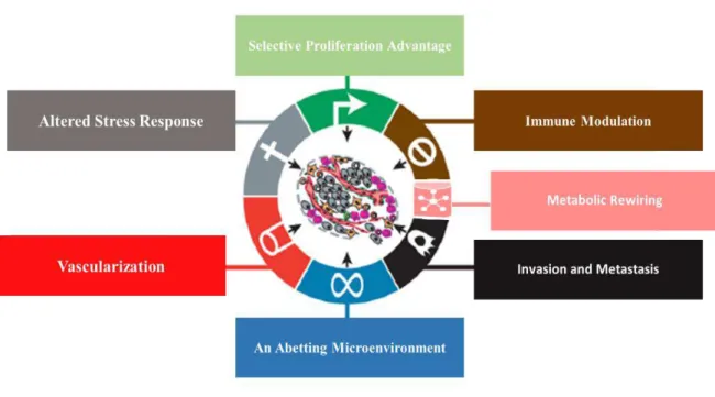
Nowadays the hallmarks of cancer can be summarized in a more organized and clearly way being individualized by Fouad and Aanei as: selective growth and proliferative advantage, altered stress response favoring overall survival, vascularization, invasion and metastasis, metabolic rewiring, an abetting microenvironment, and immune modulation4 (figure 1).
Indeed, the most fundamental characteristic of cancer cells is their capability to sustain chronic proliferation and evading growth suppressors. They acquired this ability using different ways of signaling: autocrine, paracrine and endocrine.
In the autocrine signaling, the tumor cells produce their own growth factor ligands and in response they overexpress the cognate receptors. In a paracrine signaling they produce growth factors and they release them to the adjacent target cells. Finally in an endocrine signaling the cancer cells can release the growth factors to the blood vessels in order to achieve distant cells6.
Others strategies that cancer cells have to sustain a chronic proliferation could be downstream alterations of intracellular circuits resulting in constitutive receptor activation and recruitment of normal cells within the supporting tumor-associated stroma, to release growth factors to them6.
Towards an uncontrolled proliferation, DNA damage, hypoxia and nutrient scarcity, the ability of the cancer cells to modify the stress response is considered another hallmark.
Cancer cells can resist to apoptosis, either by the overexpression of anti-apoptotic proteins, namely by the loss of function of an important protein like the p53 protein4.
Cancer cells also can escape from other mechanisms of defense against stress, as DNA repair and senescence. The senescence mechanism is a process that stops the cell cycle in an irreversible way7. A successive shortening of the telomeres in consequence of consecutive divisions induces the senescence to prevent a genomic instability and accumulation of mutations7,8. To overcome this obstacle the cancer cells upregulate the telomerase enzyme, that it is responsible for the reconstruction of the telomeres and allow a further replication8.
Tumors need sustenance in the form of nutrients and oxygen as well a way to evacuate metabolic wastes and carbon dioxide. The tumor-associated neovasculature, created by the process of vascularization supply these necessities and allows a growth above 2-3 mm3 and the ability to form metastasis that would not be possible without new vasculature8.
2 downregulation of important molecules in cell-cell adhesion, e.g. e-cadherin and degradation of the ECM for metalloproteinases and cysteine cathepsin proteases9.
In the multistage process of development and progression of a neoplastic disease there is not only alterations on the control of the proliferation but there are also adjustments of energetic metabolism. Normal cells under aerobic conditions process glucose into pyruvate via glycolysis in the cytosol and thereafter to carbon dioxide in the mitochondria.
On other hand, tumor cells even under aerobic conditions, limited their energy metabolism and process glucose into lactate. The reprogrammed metabolism, limiting energy metabolism largely to glycolysis, facilitates the biosynthesis of the macromolecules and organelles required for assembling new cells by the diversion of glycolytic intermediates into various biosynthetic pathways4.
To understand all the mechanisms involved in the oncogenesis process it is crucial to study the individual specialized cell types within the tumor as well as the “tumor environment” that surround them during the course of the process. Cancer cells and stromal cells have a continuous paracrine communication that creates a rich and dynamic microenvironment during all the stages of carcinogenesis6.
The immune system can have a great influence during the oncogenesis process by its interaction with tumor cells. Under normal conditions, the immune system can activate the “cancer immunoediting”, process by which the immune system eliminates and shapes malignant cells. However, during the development of cancer, tumor cells gain the capability to evade the immune system and multiple cellular and molecular mechanisms by which tumor cells can escape the immune system have been identified. In this way, it becomes extremely challenging in the face of so many variables, to achieve the goal of fully elucidating the all mechanisms of cancer pathogenesis and to develop novel therapies able to successfully target both primary and metastatic tumors.
3 1.1.2.Colorectal cancer (CRC)
Colorectal cancer (CRC) is a cancer that begins in the colon or in the rectum. Generally, colon and rectal cancer (depending on where they start) can be grouped because of many features that they share10. Both are cancers of the large intestine, which is the final part of the digestive tract. Most of the cases begin as small, noncancerous clumps of cells called adenomatous polyps. Polyps are small and produce few, if any, symptoms, making very difficult an early diagnosis.
For this reason, it is recommended regular screening tests to prevent colorectal cancer by the identification and removal of polyps before they became CRC.
In most cases, the causes of the disease are not clear, but some studies showed an association between diet and cancer. In diets riches in fat and with a low content of fibers were verified an increase of the risk of these cancers11.
Other factors that may increase the risk of CRC include an older age, inflammatory intestinal conditions, inherited syndromes that increase colon cancer risk, like hereditary nonpolyposis colorectal cancer, a sedentary lifestyle, diabetes, smoking and alcohol12.
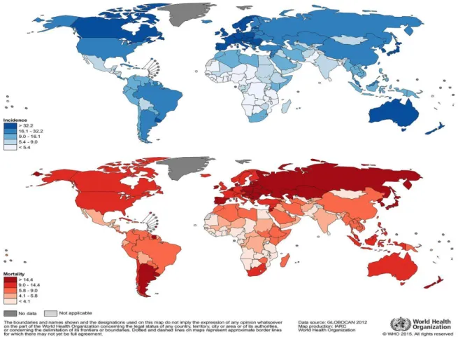
In 2012, the estimated incidence rates in males varied from <5 (per 100 000) in several African countries to over 40 in certain countries in Europe, Northern America and Oceania (figure 2)13.
So, it is crucial to find new methods of diagnosis for a disease that in most of the cases, as was mentioned before, do not present symptoms and it is one of the most common causes of cancer-related death in women and men14.
4 1.2. Carbohydrates- The sweet side of the life
Carbohydrates are the most abundant among the major classes of biomolecules belonging to a class of organic compounds found in living organisms on earth.
They can be represented by the stoichiometric formula (CH2O)n, where n is the number of carbons in the molecule. This formula also explains the origin of the term carbohydrate, that means “hydrates of carbon”15.
Monosaccharides (mono– = “one”; sacchar– = “sweet”) are simple sugars in which the number of carbons usually ranges from three to seven. The sugars can be classified as aldose or ketose, depending if the functional group is an aldehyde or a ketone, respectively. The monosaccharides also can be known as trioses (three carbons), pentoses (five carbons), and or hexoses (six carbons), according with the number of carbons.
Disaccharides (di– = “two”) are composed by two monosaccharides units and oligosaccharides (oligo- = few) are composed by 3 to 10 monosaccharides.
A long chain of monosaccharides linked by glyosidic bonds is known as a polysaccharide (poly– = “many”). The chain may be branched or unbranched, and it may contain different types of monosaccharides16.
Glycosylation is most abundant protein post-translational modification, which is essential for many biological processes. It is a complex process including numerous functional proteins and resulting in a great diversity of carbohydrate–protein bonds and glycan structures.
Glycosylation of some proteins has a great impact on their structures and functions, and interactions of protein‐linked glycans with carbohydrate‐specific proteins (lectins) modulate many important biological processes17.
1.3. Glycosylation
Glycosylation is one of the most common post-translational modifications mediated by the enzymatic addition of glycans to proteins or lipids. Transmembrane receptors, organelle-resident proteins, secreted proteins and surface proteins, as well as many others are modified by glycosylation to regulate their structure, stability and function.
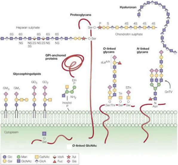
The glycans can be found in a free form or linked by covalent bonds to proteins (glycoproteins and proteoglycans) or lipids (glycolipids), identified as glycoconjugates (figure 3). The glycoconjugates can be classified by the identity of the carrier molecule and by the type of the glycosidic linkage17.
The biosynthesis of glycoproteins occurs in in the lumen of the Endoplasmic Reticulum (ER) and/or Golgi Apparatus, but also in the Cytoplasm and Plasma Membrane. Proteins can be N-glycosylated, O-glycosylated, and modified with glycosylphosphatidylinositol (GPI) anchors, and some called proteoglycans are modified with glycosaminoglycan chains.
N-glycans, in which the glycans are attached to the amine group of an asparagine (Asn) residue of a protein side chain, especially to the motif Asn-X-Ser/Thr18.
O-glycans in which glycans are attached to serine (Ser) or threonine (Thr) residues on glycoproteins, mainly on secreted and membrane bound mucins; and glycosaminoglycans (or proteoglycans) in which the glycan structure is also attached to Ser or Thr but differs from O-glycans by consisting of linear molecules which are often sulfated19. The glycolipids are the result of the lipid glycosylation and can be represented by glycoglycerolipids and glycosphingolipids. The main role of the glycolipids is to maintain the stability of the cell membrane and to facilitate cellular recognition, which is crucial to the immune response20.
5 1.3.1.N-glycosylation
Glycosylation is considered to be one of the most prevalent mechanisms of protein post-translational modifications. It is proposed that more than half of all eukaryotic proteins are glycoproteins and 90% of these proteins carry N-linked glycans21.
N-glycosylation takes place in the ER and in Golgi apparatus22. The first stage of the N-glycosylation biosynthetic pathway in mammalian cells involves the assembly of a 14-oligosaccharide “core” unit as a membrane bound dolichyl pyrophosphate (Dol-P-P) lipid precursor by enzymes located on both sides of the ER membrane22,23.
This core oligosaccharide is common to most eukaryotes and has a defined structure containing three glucoses (Glc), nine mannoses (Man) and two GlcNAc residues24.
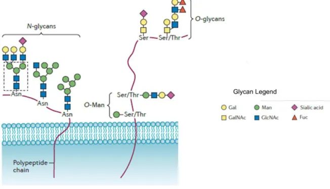
After the biosynthesis of core oligosaccharide, it is transferred to the nascent protein by the oligosaccharyltransferase, a complex enzyme with its active site in the ER lumen, via a N-glycosidic linkage of a GlcNac to an Asn residue (figure 4). The Asn residue must be part of the consensus amino acid sequence Asn-X-Ser/Thr, where X can be any amino acid, except proline25. Before exiting the ER,
6 takes place a removal process of the two outermost Glc residues and mannose residues by the activities of α-glucosidases (I and II) and mannosidases. When the glycoprotein moves to the Golgi complex, the glycan chains undergo further trimming of mannoses before further extension of the glycan branches. Following step-wise adding of sugars to different positions of the extending glycan is catalyzed by many glycosyltransferases, each adds a specific monosaccharide through a glycosidic bond.
1.3.2.O-glycosylation
The O-linked glycosylation takes place in the Golgi apparatus and consist on the sequentially addition of monosaccharaides residues or glycan structures to Ser or Thr residues of the protein backbone (figure 4). There is no consensus sequence and there are multiple types of O-linked glycans (e.g. O-GalNAc, O-GlcNAc, O-α-Fuc, O-β-Glc, O-β-Gal)26. The main forms of O-glycans on higher eukaryotic secretory proteins are mucin-type glycans27. The synthesis of the mucins begins with the addition of an GalNAc residue to a Ser/Thr side chain, followed by the construction of one of the four distinct core structures by the addition of Galactose (Gal) and GlcNAc residues in many linkages27.
1.3.3.Glycan’s biological function
Glycosylation creates a huge repertoire of glycan structures whose variety derives from the cell-specific expression levels and intracellular location of the enzymes involved and the availability of sugar substrates.
Glycans play different roles according to the molecule to which it is attached, the development stage and the biological context and its location. They are involved in many different biological functions and their significance varied cellular interactions depends on their extracellular location.
The functions of glycans comprise the specific recognition by other molecules- most commonly, glycan-binding proteins; the regulation of protein stability, solubility and vulnerability to enzymatic degradation; intracellular and intercellular communication; regulation of receptor activation and signal transduction28. They also present structural and modulatory properties29.
7 Given such important functions, alterations on the structure of the oligosaccharides leads to changes in aspects like proliferation, differentiation, migration, tumor invasion, cell-trafficking and trans-membrane signaling30.
1.3.4.Hallmarks of glycosylation in cancer
It is well known that fundamental changes in the glycosylation pattern of cell surface occur during malignant transformation and cancer development. The significance of glycosylation in cancer is further highlighted by the fact of most of the Food and Drugs Administration (FDA)- approved tumor markers are glycoproteins or glycan antigens30,31.
Glycosylation is itself a hallmark of cancer and recent studies have shown that glycans play a role in many hallmarks of cancer (figure 5).
The principal characteristic of cancer is their ability to maintain chronic proliferation. It was verified that the expression of N-glycans and their branching degree can control the activity and signaling of different growth factor receptors and has a significant role in the proliferative signaling32. Many growth factor receptors, including TGF-beta, EGFR, FGFR, PDGF, MET and IGFR33 are known to be regulated by glycosylation.
Invasion and metastasis are features of a lot of tumors and evidences propose that certain glycans are involved in these processes. E-cadherin, a transmembrane glycoprotein, which connects epithelial cells together at adherent junctions plays an essential role in the suppression of cancer cell migration and metastasis. This protein has four potential N-glycosylation sites and many studies have hypothesized that different expression profiles of E-cadherin-linked N-glycans may be related to the stability of adherent junctions. Thus, modifications by N-glycosylation have been proposed to underlie E-cadherin downregulation or inactivation in cancer34.
Apoptosis represents energy-requiring, spontaneous single cell death, with specific morphological and biochemical features, and the capability of cancer cells to evade that process is considered another hallmark of cancer. The glycosylation of death receptors and their ligands may critically control apoptosis by disrupting ligand-receptor interactions, influencing ligand secretion from effector cells and modulating the formation of signaling complexes35.
Cancer cells have a higher need of nutrients and oxygen as well as a way to remove metabolic waste like e.g. carbon dioxide. Different glycosylation associated genes has been linked to the angiogenesis process by the vascular endothelial growth factor receptor-2 (VEGFR-2), that is an important receptor tyrosine kinase (RTK)36.
The extracellular domain of VEGFR-2 is composed of seven immunoglobulin-like domains, each with multiple potential N-glycosylation sites. N-glycosylation plays a central role in RTK ligand binding, trafficking, and stability and altered glycosylation may affect its function36.
8 1.3.5.Glycosylation characteristics of colorectal cancer
As was mentioned before, an aberrant glycosylation has many functional consequences, contributing to the progression, severity and dissemination of CRC.
Glycans have been found to contribute in many fundamental biological processes involved in cancer, such as tumorigenesis and metastasis.
Tumorigenesis is a multistep process, the progression of which depends on a sequential accumulation of mutations within tissue cells. The glycosylation plays an important role of the multiple cell survival pathways and consequently in tumorigenesis.
It was found an overexpression of N-acetylglucosaminyltransferase, GnT-V, in CRC cells37. The increase of the expression of this glycosyltransferase plays a crucial role in the regulation of these oncogenic processes.
GnT-V is an enzyme that catalyzes beta 1-6 branching of N-acetylglucosamine on N-linked oligosaccharides. Its product, the beta 1–6 branching structure, is further elongated forming poly- N-acetyllactosamine and the level of this residue is increased in highly metastatic colon cancer cells38. The increase of polylactosaminylation on N-glycans of surface receptors such as growth-, arrest-, and angiogenesis promoting receptors, may has an effect in the tumor invasion and angiogenesis, because the galectin 3 with these modified residues induces the formation of molecular lattices, which delay the endocytosis of them and maintain their responsiveness to the ligand39.
The O-glycosylation also plays an important role in the regulation of CRC cell growth40.
The tumor infiltrating cells express the sialic acid-binding immunoglobulin-like lectin 9 (singlec-9) and its interaction with sialylated O-glycans of the cancer-associated transmembrane mucin protein MUC1 on CRC cells was shown to induce the recruitment of β-catenin and to promote tumor growth40,41. GnT-V also plays an important role in the formation of metastasis. The increase of its product is associated with cancer invasion and metastasis in CRC cause the GnT-V-mediated glycosylation changes on, e.g. matriptase, β1-integrin, and N-cadherin, proteins involved in cell adhesion, regulate tumor cell motility by decreasing cell-cell adhesion and increasing the interaction between cells37.
1.4. Fucosylation
Fucose (6-deoxy-L-galactose) is a monosaccharide that is found in glycoproteins and glycolipids present in vertebrates, invertebrates, plants, and bacteria.
The attachment of fucose residues to oligosaccharides and proteins by the activity of innumerous glycosyltransferases comprises one of the most common modifications of glycans on proteins or lipids. This process is regulated by different types of molecules, as glycosyltransferases, in particularly fucosyltransferases, guanosine diphosphate (GDP)-fucose synthetic enzymes, and GDP-fucose transporters.
The proteins that catalyzed this reaction, fucosyltrasnferases, can be classified based on the sites of fucose addition into α1,2-(FUT1 and FUT2), α1,3/4-(FUT3 to 7 and FUT9 to 11), α1,6-(FUT8), and O-(POFUT1 and POFUT2) FUTs42.
In the present work we will focus on fucosyltransferase 8 (FUT8). This enzyme is the only fucosyltransferase involved in core fucosylation (addition of fucose in α-1,6-linkage to the innermost N-acetyl glucosamine of N-glycans). Core fucosylation of glycoproteins has significant regulatory
functions for adhesion molecules and growth factor receptors, such as α3β1 integrin and epidermal
growth factor receptors43.
1.4.1.FUT8
1.4.1.1. FUT8 structure and localization in cells
The gene encoding fucosyltransferase 8 protein is located on chromosome 14q23.3 and encompasses approximately 333 kb. Its sequence contains nine exons with coding regions and three 5'-untranslated exons43.
FUT8 was first purified and cloned from the porcine brain and from a human gastric cancer line and it consists of 575 amino acids and it has a molecular weight of 66516 Da43. The protein contains no N-glycosylation sites and belongs to the GT23 family of the CAZy classification44.
9 The enzyme catalyzes the transference of a fucose residue from GDP-fucose to the reducing terminal GlcNAc of Asn-linked oligosaccharide (N-glycan) via an α1,6-linkage and the resultant fucosyl residue is referred to as a core fucose (figure 6). The donor substrate and the acceptor substrate for FUT8 are GDP-fucose and a biantennary N-glycan, respectively. The GDP-fucose is synthesized in the cytoplasm through both de novo pathway and the salvage pathway. In de novo pathways, the GDP-mannose is transformed to GDP-fucose, via three enzymatic reactions carried out by GDP-mannose 4,6-deoxymannose (GMD) and GDP-keo-6-4,6-deoxymannose 3,5-epimerase, 4-reductase. The savage pathway synthesizes GDP-fucose from free L-fucose derived from extracellular or lysosomal sources45. FUT8 was detected on cytosol and Golgi Aparatus46 and its physiological significance has been confirmed by genetic ablation of the fut8 gene: 80% of these mice die three days after birth and the survivors present severe growth retardation47.
1.4.1.2. FUT8’s implications in cancer
Over the time various studies were conducted to study and understand the significance and impact of FUT8 on cell growth and differentiation, since was reported that the enzyme is widely expressed in mammalian tissues and there is an increase in its expression in different cancerous tissues.
FUT8 mRNA expression and activity are highly elevated in cases of ovarian serous adenocarcinoma, in comparison with normal ovary. The elevated expression of FUT8 and the subsequent modifications of N‐glycans constitute a significant characteristic of this kind of ovarian cancer48.
In thyroid cancer was found that 33,3% of the cases of papillary carcinoma present an overexpression of FUT8 and the enhancement of the expression of this fucosyltransferase was associated with tumor size and lymph node metastasis49.
It is known and well study that the alpha-fetoprotein (AFP), the major fetal plasma protein, plays an important role in the development of hepatocellular carcinoma (HCC)50.
A core-fucosylated isoform of AFP, AFP-L3, was identified and was found that the expression of this protein it is highly in HCC tissue51.
The expression of FUT8 is high in HCC tissue and in liver cirrhosis, but surprisingly, was not observed a concomitant elevated expression of AFP-L3 in liver cirrhosis. This inconsistency can be explained by the difference in the synthesis of GDP-fucose, the donor substrate for fucosyltransferases, as FUT8, because the expression of GDP-fucose is higher in HCC, as compared to a liver cirrhosis. Thus, it appears that AFP-L3 could be used as a marker for HCC51.
Another evidence that shows that FUT8 play an important role in cancer is the elevate expression of the haptoglobin, a highly fucosylated glycoprotein in serum of patients with pancreatic cancer.
Structural analyses using mass spectrometry and lectin blotting showed that core fucosylation is increased in haptoglobin from serum of these patients. Thus, Fucosylated haptoglobin could be a novel marker for pancreatic cancer52.
10 1.4.1.2.1. FUT8 and CRC
A specific increase in FUT8 expression and its enzyme activity have been reported in innumerous human tumor processes such as ovarian serous adenocarcinoma, thyroid cancer, HCC and pancreatic cancer, as mention before.
In CRC the behavior and significance of FUT8 remain almost unknown, but some studies have shown that FUT8 activity is considerably elevated in some colorectal tumors as compared with the surrounding healthy tissues53.
The activity of FUT8 and standard clinicopathological features exhibited significant correlations between the protein activity and lymph node infiltration, tumor stage and type of growth54.
Studies revealed an increase in the FUT8 activity in polypoid tumors, more localized and less invasive than non-polypoid tumors and corresponded to the increase observed in tumors without lymph node metastasis. Likewise, was detected a progressive decrease in FUT8 activity as the degree of infiltration in the intestinal wall progressed54.
Therefore, considering these evidences, was postulated a hypothesis that the increase in the enzyme activity, in combination with different cell alterations produced during the malignant process, would be involved in the initial development of the tumors, since the initial steps of this development are characterized by the uncontrolled division of the transformed cells54.
In other hand, the decrease of the FUT8 activity in advanced stages could the linked to the acquisition of the invasive potential of the tumor cells.
1.4.1.3. FUT8 in anti-cancer therapy
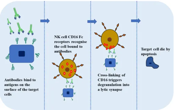
The antibodies have multiple therapeutic functions like e.g. antigen biding, induction of apoptosis, complement-dependent cellular cytotoxicity but the antibody-dependent cellular cytotoxity (ADCC) is considered one of the most important function of some antibodies used for therapy.
ADCC is the mechanism that comprehends the killing of an antibody‐coated target cell by a cytotoxic effector cell through a nonphagocytic process, characterized by the release of the content of cytotoxic granules or by the expression of cell death‐inducing molecules. This mechanism is triggered upon biding of lymphocytes receptors (FcγRs) to the constant region (Fc) of the antibodies55 (figure 7). So, it is crucial to find ways to improve and enhance the mechanism of the antibody-dependent cellular cytotoxicity. Recent studies have shown that engineering the oligosaccharides of IgGs may yield optimized ADCC. Analysis of different studies showed that recombinant IgG1 produced by rat hybridoma YB2/0 cells presents at least 50-fold higher ADCC that that produced by CHO cells, one of the most used host cell lines for production of recombinant antibodies56.
YB2/0 cells expressed a lower level of FUT8 mRNA than CHO cells, and overexpression of FUT8 in YB2/0 led the increase of fucosylation of IgG1 and decrease of ADCC56.
In the study conducted by Shields, they demonstrated that nonfucosylated anti-Her2 humanized IgG1 and anti-IgE humanized IgG1 produced by a variant of CHO cells, Lec13, had enhanced ADCC relative to fucosylated IgG1s produced by normal CHO cells57.
These results suggest that Fuc-deficient IgG1 may need a lower concentration of antibody on the surface of the target cell to activate an effector cell and start the ADCC mechanism. There are a few possible explanations of why the antibodies with nonfucosylated oligosaccharides give rise to stronger binding to FcγRIIIa than those in which the glycoforms are absent56. A core Fuc has been shown to influence the conformational flexibility of biantennary oligosaccharides56.
11
12 2. Aims of this thesis work
Bearing in mind, that an increase in FUT8 expression and activity has been associated with many human tumors such as ovarian serous adenocarcinoma, thyroid cancer, HCC, pancreatic cancer and CRC, it is vital to get supplementary understandings on the role of FUT8 in cancer.
The FUT8 behavior and its implication in CRC remains almost unknown, but there are some reports linking the activity of the protein and standard clinicopathological features as tumor stage and type of growth. Thus, it is important to develop molecular tools enabling the control of FUT8 expression in order to try to reveal its implications in this particular type of cancer.
According to this, the main goals of this project were the development of a molecular system with the objective to modify FUT8 expression; validate this system in HEK293A and HT29 cell lines and study the impact of FUT8 modulated expression in CRC cells phenotype as well in the expression of different growth factors.
The first major step of this work was the construction of the FUT8-adenoviral system, in collaboration with Dr. José Ramalho, from CEDOC, FCM/UNL, using the Gateway Technology. In this first phase of the project several cloning techniques were used.
After the expression system obtained, the final plasmid was transfected into the HEK293A cell line. This cell line was also chosen to produce the adenovirus particles and then to perform a mini-scale production of FUT8 with the aim of purifying the protein.
The purification of the protein was carried out with the purpose of evaluating its activity in order to verify if the purified protein could be used in a partnership with the group led by Dr. Daniel Varon Silva, from Max Planck Institute, which works on glycoprotein synthesis. In this particular case, FUT8 would be used to catalyze the transfer of α1,6-linked fucose to the first N-acetylglucosamine in N-linked glycans.
13 3. Materials and Methods
3.1. Adenoviral system- General introduction
The Adenoviral Expression System (InvitrogenTM, Life Technologies, USA) is an adaptation of the Gateway® System 58 that allows the creation of a replication incompetent adenovirus that can be used for delivery and transiently express a gene of interest in dividing and non-dividing mammalian cells. Choosing an adenoviral vector as a destination can be achieved, high efficiency generation of recombinant adenovirus containing our gene of interest under the control of the human cytomegalovirus
(CMV) promoter.
The use of the Gateway® Technology bring advantages like a highly efficient, rapid cloning of the gene of interest and create efficiently delivers the gene to mammalian cells in culture.
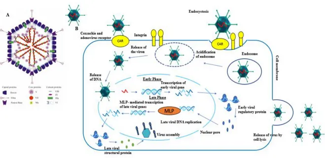
3.1.1.Adenovirus structure
The adenovirus are non-enveloped virus with linear, double-stranded DNA genome of 26-45 kb. Its structure is comprised by a nucleocapsid in the form of 3 major types: fiber, peton and hexon based proteins. The basis of the adenovirus structure is conferred by the hexon and it composes most of the viral capsid. The peton based proteins and fiber are for receptor binding and internalization of the adenovirus into host cells 59.
The adenoviral system basically comprehends the entry of the adenovirus to the cells by binding the Coxsackie and adenovirus receptor (CAR) 60. After the binding of the adenovirus, the internalization is conducted via integrin-mediated endocytosis and the transport to the nucleus it is achieved by active transport 61. By the time that the adenovirus reaches the nucleus the early phase starts take place the transcription and translation of the early genes, followed by the expression of the adenoviral late genes and viral replication (figure 8). The viral replication is dependent of the expression of the E1 protein and as the adenovirus used in the present study are replication incompetent particles the E1 is supplied by the HEK293A producer cells.
14 A. FUT8 overexpression-encoding plasmid
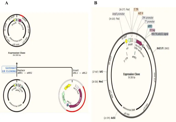
One plasmid was constructed with the aim to produce adenovirus particles to overexpress FUT8. The pAd/CMV/V5-DEST was the plasmid that was chosen as a destination vector and contain the following elements: human adenovirus type 5 sequences encoding genes (Right Inverted Terminal Repeats (ITRs), encapsidation signal sequence, late genes) and the elements required for proper packaging and production of adenovirus. A Chloramphenicol resistance gene (CmR) located between the two attR sites for counterselection, two recombination sites, attR1 and attR2 for recombinational cloning of the DNA sequence of interest from an entry clone, and a human cytomegalovirus (CMV) immediate early promoter for high-level constitutive expression of the gene of interest in a wide range of mammalian cells. An Ampicillin resistance gene for selection in E. coli and pUC origin for high-copy replication and maintenance of the plasmid in E. coli (figure 9).
A
B
15 3.2. Cloning techniques
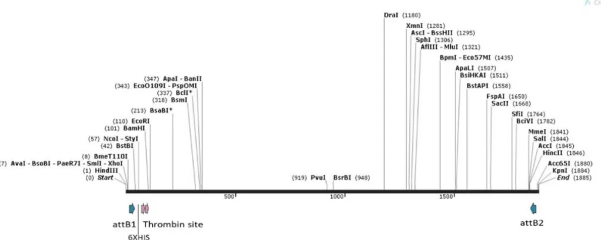
The first step of this work was the construction of the recombinant protein. The gene was synthesized by GeneCust company (Luxembourg) and we decided to add a Histidine tail (His tag) in the N-terminal position of the sequence in order to facilitate the purification process. We also decided to add a thrombin site between a His tag to simplify a possible separation of His tag from the protein coding sequence of interest (figure 10).
After obtained the plasmid with the synthesized FUT8 encoding gene, different cloning techniques were used, first with the aim of obtaining the Gateway Technology pENTR vector and afterwards with the aim to get the final vector, the adenoviral plasmid which we will use to overexpress the fucosyltransferase 8.
The plasmid was assembled using consecutive cloning strategies. Briefly, all the cloning was executed as follows: isolation and purification of the obtained plasmid DNA with the synthesized FUT8 encoding gene, enzymatic restriction digestion, Gateway reaction (BP reaction), transformation of competent cells with the resulting plasmid, PCR screening of the colonies, isolation and purification of the Gateway plasmid, confirmation of the construct by digestion with restriction enzymes, Gateway Technology reaction (LR reaction), transformation of competent cells with the resulting plasmids, PCR screening of the colonies, isolation and purification of the Gateway plasmid, confirmation of the construct by digestion with restriction enzymes and sequencing of the final plasmid.
3.2.1.Plasmids isolation and purification from bacterial culture
The plasmids were purified with mini and midi kits (Omega Bio-Tek, USA and Zymo Research, USA, respectively) and both rely on the same principle: centrifugation to obtain a pellet of the bacteria; resuspension and lyse to expose the genetic material and proteins, removal of RNA and protein, biding the DNA to a specific column, wash and elution of the DNA from the biding column.
3.2.1.1. Miniprep
The E.Z.N.A® Plasmid DNA Mini Kit I (Omega Bio-Tek, USA) was used to isolate and purify plasmid DNA in accordance with the manufacturer’s recommendations. Cells from 10ml bacterial culture were harvested and the plasmid DNA was eluted in 100 μL of Tris (pH 7,4).
16 3.2.1.2. Midiprep
Plasmid DNA isolation and purification from 50 mL cultures were done with the Zymo Pure Plasmid Midiprep Kit (Zymo Research, USA) according to the manual.
3.2.2.Agarose gel electrophoresis
DNA was separated according to size and evaluated on 0,8% agarose gel by electrophoresis. Before electrophoresis, appropriate volume of loading buffer was added to each sample.
The size standard GeneRuler™ 1kb DNA ladder (Thermo Fisher Scientific, USA) was used to determine the size of the migrated DNA fragments. The gel was run in 1x TAE buffer (40mM Tris-acetate, 1mM EDTA) for 20-30 minutes.
3.2.3.Enzymatic restriction digestion
PCR products and the vectors were digested with restriction enzymes to prepare them for the Gateway reactions and to confirm the constructions through horizontal agarose gel electrophoresis.
To digest the vectors and inserts were used NheI and PacI enzymes (Takara Bio, USA) and the digestions were performed at 37 ºC during 1 h, as described by the manufacturer.
3.2.4.Cloning of DNA fragments using the Gateway Cloning Technology
The Gateway® Technology is based on the bacteriophage lambda site-specific recombination system which allows the integration of lambda into E. coli chromosome and switch between the lytic and lysogenic pathways62. This technology has some modifications of the components of the lambda recombination system to improve the specificity and efficiency of the system63.
The Gateway® Technology has to main reactions:
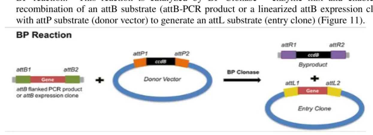
- BP reaction: This reaction is catalyzed by BP Clonase™ enzyme mix and enable the recombination of an attB substrate (attB-PCR product or a linearized attB expression clone) with attP substrate (donor vector) to generate an attL substrate (entry clone) (Figure 11).
Figure 11- Schematic representation of BP reaction from the Gateway® Technology. The reaction comprehends the recombination of an attB- PCR product and a Donor vector (attP substrate) in order to create an Entry Vector containing the attL sites.
17 3.2.4.1. BP reaction
This reaction was performed by mixing the PCR product containing our gene of interest and flanked by the attB sequences with the pDONR vector, being catalyzed by the BP Clonase™ enzyme mix. The purpose of this reaction was to obtain an entry vector that would serve as a substrate for the LR reaction.
3.2.5.Bacterial Strain
Escherichia coli (E. coli) HB101 (Promega, USA) competent cells were used to produce the DNA plasmids.
HB101 is a hybrid K12 X B strain bacterium containing the recA13 mutation that minimizes recombination and aids in insert stability. Additionally, this strain carries the hsdS20(rB- mB-) restriction minus genotype which prevents cleavage of cloned DNA by endogenous restriction enzymes. The bacteria transformation was performed following the manufacturer's instructions and working banks of transformed bacteria for each used plasmid were created. The bacteria suspension was mixed with 50% of glycerol, aliquoted and frozen at -80 ºC.
Briefly, plasmid DNA was placed in contact with HB101 cells for 30 minutes on ice, after that the cells were incubated at 42ᵒC for 1 minute. Following the previous steps, the HB101 cells were incubated for 3-4 minutes at 0ᵒC and afterwards, LB medium was added to the cells and incubate for 1,5 h at 37ᵒC while shaking with the require antibiotic in order to allow them to acquire the antibiotic resistance and to grow.
After transformation, bacteria were spread on agar plates. The agar plates were prepared with Agar (NZYTech, Portugal) accordingly to the manufacturer’s instructions.
Following the inoculation, bacteria were grown at 37ºC for 16- 17 hours. Single and well individualized colonies were picked from the previously inoculated agar plates and each colony was grown in a small volume LB medium (5 mL in 50 mL tubes) at 37 ºC for 16-17 h in an orbital shaker at 150 rpm.
3.2.6.Polymerase Chain Reaction (PCR)
Polymerase Chain Reaction (PCR) was used to amplify desired segments of DNA.
Following three steps, the target sequence is amplified. First, denaturation of the template occurs by heating, second, primers flanking the target sequence anneal to their complementary sequences, and during the last step the annealed primers are extended by DNA polymerase. The cycle is repeated and leads to an exponential amplification of the DNA segment (figure 10).
We amplify our sequence and flanked them with gateway sequences (attB sites) to obtain expression clones. We selected the bacteria colony containing the vector of interest, a screening colony PCR was performed with GoTaq DNA Polymerase (Promega, USA) using specific primers for each construct enabling to infer the successful cloning.
After the screening, one single positive colony was incubated and grown in LB medium supplemented with appropriated antibiotic for 24 h at 37ᵒC.
18 3.2.6.1. LR reaction
The LR reaction took place between the attL sites of the generated entry clone that was obtained by the BP reaction and the attR sites of the destination vector. The reaction was catalyzed by the LR Clonase enzyme mix and as a result, an expression clone with the DNA of interest flanked by attB sites was generated.
As a destination vector was choose an adenoviral vector, pAd/CMV/V5-DEST™, where our sequence of interest was controlled by the human cytomegalovirus (CMV) promoter.
3.2.7.Mammalian cell lines- HEK293A
In the present work we choose the HEK293A cells to produce the adenovirus. This cell line is a subclone of the HEK cell line that is a permanent line established from primary embryonal human kidney and exhibits a relatively flat morphology, that enable an easier visualization on the plaques.
The genes E1a and E1b are expressed in these cells and participate in transactivation of some viral promoters, allowing very high levels of protein production. The protein E1 also complements the E1- deletion in recombinant adenoviral vectors, allowing viral replication.
3.2.8.Transfection
The cells were seeded in 6-well plates 24h prior to transfection. The transfection mix was prepared according to the manufacturer's protocol. We diluted 2 µg of DNA into 500 µl jetPRIME buffer (Polyplus Transfection, USA), was added 5 µl of jetPRIME (Polyplus Transfection, USA) and the mix was incubated for 30 minutes.
After the cells reached 60% confluence we added the transfection mix into the plates and we waited for 9 days. After that time, we collected the medium and we reinfected new cells with the aim to amplify the adenovirus.
3.2.9.Adenoviral production
Adenoviral plasmid was purified as described in chapter 3.1.1.1 and 3.1.1.2.
Adenoviral production was performed by transient transfection. HEK293A cells, cultured with DMEM high glucose (Gibco®, Life Technologies, USA) at 50-80% confluence in 25 cm2 polylisin-coated flask, were transfected as described in the chapter 3.4.
Nine days after the transfection, the HEK293A cells released the produced adenovirus particles by exocytosis and the supernatants containing these viral particles were collected and centrifuged to clean debris. The adenovirus production was evaluated by the transduction.
3.2.10. Mini scale production and purification of FUT8
To express a greater amount of protein than the one expressed by the tests mentioned before, a mini
scale production was performed.
The cells were seeded in 170 cm3 roller bottles, incubated at 37ᵒC, 5% CO2 until reaching a confluence
Figure 10- Diagram representing the settings used for pcr amplification with Promega GoTaq DNA Polymerase.
19 of 50-80%. Then, 160 mL of adenovirus containing supernatant was added to each roller bottle and after approximately 48 hours the supernatant was collected and centrifuged at low speed. Afterward centrifugation, 200 μl of the supernatant was aliquoted and stored at -80°C, given the name "Total" at this aliquot. The pellet was resuspended in 50 mL lysis buffer and vortexed and sonicated in 4 cycles of 20 seconds, always keeping the samples on ice. Then, the lysate was centrifuged at 100,000 g for 1 hour. Next, a 200 μl sample of the supernatant which was given the name "Sup" was collected and again aliquoted and stored at -80°C. The remaining supernatant was diluted in 100 mL of the wash buffer and maintained on ice. The pellet was resuspended in lysis buffer and was aliquoted and stored at -80°C, given the name "pellet". The Hight Density Nickel column (Agarose Bead Technologies (ABT), USA) was equilibrated by washing with the wash buffer and the supernatant was added to the column. The flow thought was collected and aliquoted as the other aliquots mentioned before. Th elution was done with the addition of 2 mL of elusion buffer and several samples were collected and aliquots at -80.
3.3. Analytical test- HEK 293A cell line 3.3.1.Western Blot
To separate proteins based upon their molecular size, we performed 12% polyacrylamide gels. Gels were poured into pre-made gel chambers, and well-forming combs were immediately added, and the gel was left to polymerize. After polymerization, combs were removed, and the wells were filled with running. The samples were prepared for running on polyacrylamide gels by adding an equal amount (500 µl) of loading buffer 3x. Once loading buffer had been added, the samples were heated to 95°C for 5 minutes, loaded into the polyacrylamide gels along with a molecular weight marker for reference at 30mA for 30-40 minutes. Following separation of proteins by SDS-PAGE, proteins were transferred into nitrocellulose membrane (GE Healthcare Life Science, USA) using the following method. Transfer cassettes contained the following layers: a sponge, Whatman 3MM blotting paper, nitrocellulose membrane, SDS-PAGE gel, Whatman blotting paper and a sponge (all equipment was pre-immersed in transfer buffer containing 20% v/v methanol, 0.19M glycine and 0.05M Tris). The transfer cassette was then placed in a transfer tank filled with transfer buffer for 1 hour at 250mA. Following the transfer, nitrocellulose membranes were washed for 5 times each time and the Nitrocellulose membranes were kept in 2% egg albumin solution in PSB Tween for 1 hour in order to block non-specific binding sites. Following blocking, primary antibodies were diluted in PBS Tween 1x solution and incubated with the nitrocellulose membrane overnight at 4°C on a rocking table.
20 The antibodies used for Western blot are listed in table 3.
Table 3- List of antibodies used in Western Blot assay.
Primary Antibody
Host Specificity Dilution Origin
Monoclonal
anti-FUT8 Mouse Human FUT8 1:1000 Biotechnology Santa Cruz Policlonal
anti-GAPDH Goat Human 1:2500 Sicgene
Secondary Antibody
Anti-goat HRP
conjugated Donkey Goat IgG 1:5000 Bio-Rad
Anti-mouse HRP
conjugated Donkey Mouse IgG 1:5000 Bio-Rad
Mammalian cell lines- HT29 cell line
The HT29 CRC cell line (ATCC®HTB-38TM) derived from an adenocarcinoma of colon from a 44-year-old Caucasian female, was used and transduced with adenoviral vector pAd/CMV/V5-DEST™ containing the cDNA of the gene that encodes for human FUT8. Similarly, the wild type cell line was transduced with an adenoviral empty vector (MOCK) to mimic some effects that the transduction could bring to the cells.
3.4. Cell culture
HEK293A (figure 13A) and CRC (figure 13B) cell lines were cultured in Dulbecco’s Modified Eagle Medium (DMEM) (Gibco®, Life Technologies, USA) supplemented with 10% (v/v) FBS (Gibco®, Life Technologies, USA), 1% (v/v) Penicillin/Steptomycin (Gibco®, Life Technologies, USA) and 1% (v/v) Glutamine (Gibco®, Life Technologies, USA), in an incubator (Panasonic, Japan) at 37ºC with a humidified atmosphere and 5% CO2. The medium was changed every three days. To split the cells at a confluence of 80-90%, they were incubated with trypsin ethylenediaminetetraacetic acid (Trypsin-EDTA) (Gibco®, Life Technologies, USA) for 5-10 minutes, followed by the addition of 3 times the volume of trypsin of pre-warmed medium and a centrifugation at 200g, for 5 minutes at RT.
Routine cell culture manipulations were made on Class II Biosafety Cell Culture Flow Chambers, using sterile techniques. Cell stocks were maintained in cryotubes at -80 ºC, resuspended in medium supplemented with 10% sterile DMSO.
21 3.4.1.Transduction of colorectal cancer cell line
The cells (HT29 cell line) were seeded in a 6-well plate, 10,5x10⁶ cells per well and incubated for 2 days at 37 ºC in a 5% CO2 atmosphere. After that incubation time, the cells were washed with PBS 1X and incubated with the adenoviral supernatants containing the adenovirus in DMEM (Gibco®, Life Technologies, USA). In the next day the medium was replaced with a fresh culture medium.
3.5. Biological assays
3.5.1.Assessment of cell migration capacity
Cells were grown in monolayer 12-well plates until 100% confluence. Afterwards a scratch was made in the monolayer and to remove the debris, the detached cells and smooth the edge of the scratch, cells were washed with 1X PBS and then incubated with fresh culture medium at 37ºC with 5% CO2 during
the experiment.
Image acquisition of each wound along the time was performed using an inverted microscope (Motic® AE31E, China). The first image was acquired instantly after the scratch (T0) followed by periodic acquisitions, until 96 h after T0, in the same area of the scratch.
To calculate the migration area (in µm2) for each wound was subtracted the scratched area at different times to the initial scratched area (T0).
3.5.2.Assessment of cell proliferation capacity
To evaluate the proliferation capacity of the cells while overexpressing FUT8 we used the CellTraceTM carboxyfluorescein succinimidyl ester (CFSE) Cell Proliferation Kit (InvitrogenTM, Life Technologies, USA).
Following trypsin -EDTA detachment, 1×10⁶ cells were resuspended in 2 mL of PBS and 0.2 μM CFSE solution was added in a 1:1 proportion, slowly and directly into the tube walls, for a final concentration of 0.1 μM. The solution was gently mixed, and the suspension was incubated for 10 minutes at 37ºC in the dark. To remove the dye that did not bind to the cells, 10 ml of pre-warmed PBS with 2% FBS was added to cells and incubated for 5 minutes at 37ºC, in the dark. Following the incubation time, the solution was centrifuged, and the cells were resuspended and cultured in pre-warmed DMEM media and incubated for 18 h before any other procedure to allow acetate hydrolysis to occur. Cells were collected after 1 (T1), 2 (T2), 3 (T3) and 4 (T4) days.
3.5.3.Immunofluorescence assay
Cells were cultured in round coverslips in 24 well plates (Greiner CELLSTAR®, Austria) overnight, fixed and permeabilized with 200 µL of wash solution (Fixation/Permeabilization Solution Kit from BD Biosciences, USA) for 20 minutes at 4ᵒC.
After blocking with 1% bovine serum albumin (BSA), cells were stained with different antibodies followed by the respective secondary antibodies labelled with different fluorophores. The list of antibodies used can be found in Table 1.
22
ble 1- List of antibodies used in Immunofluorescence assay.
Primary Antibody and lectin
Host Specificity Dilution Origin
Monoclonal
anti-FUT8 mouse Human FUT8 1:100 Technology Santa Cruz Biotinylated
Aleuria Aurantia Lectin (AAL)
(α -1,6) linked
fucose residue 1:100 Laboratories VECTOR
Secondary Antibody
Anti-mouse FITC Goat Mouse IgG 1:100 Southern Biotech
Streptavidin
FITC Biotin 1:100 BD Pharmingen
3.6. Analytical tests
3.6.1. Real-Time Quantitative Polymerase Chain Reaction Protocol 3.6.1.1. Gene Expression Analysis- HT29 cell line
To evaluate the genetic expression of FUT8 and TGF-beta genes, total RNA was extracted and then converted to cDNA by RT-PCR. RNA extraction was performed using the GenElute Mammalian Total RNA Miniprep Kit (Sigma Aldrich, USA).
The lysis buffer was first prepared by the addiction of β-mercaptoethanol (Sigma, USA) to the commercial lysis solution in a 1:100 proportion.
We added 250 µL of the lysis buffer and cells were lysed by throughout pipetting. To remove cellular debris and shears DNA the lysed cells were filtered in filtration column by centrifugation at 12000 g for 2 minutes at 4ᵒC. The filtration column was discarded and 250 µL of 70% ethanol were added to the filtered lysate, followed of a mix by vortex. This mixture was placed to the biding column and centrifuged at maximum.
The DNAse I solution was prepared by mixing DNAse I with digestion buffer in a 1:8 proportion and 80 μL of this solution were added to each column. After incubation for 15 minutes at RT and the columns were washed 250 µL of Wash Solution I and centrifuge.
The flow-through liquid was discarded, and the biding column was washed by the addiction of 500 µL of Wash Solution I and centrifugation.
The columns were washed twice with 500µL of Wash Solution II. To remove any residual ethanol the empty biding column was centrifuged for 2 minutes at maximum speed (12 000 g) at 4ᵒC.
To elute the RNA, 50 µL of Elute Buffer was added to the biding column, already in a fresh 2 mL collection tube and centrifuged at maximum speed.
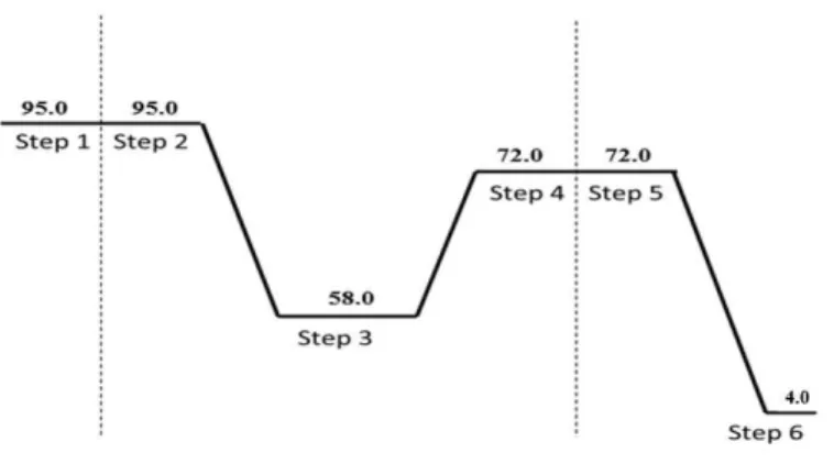
23 In a PCR tube (VWR, USA) we added 50 µL of the master mix and 50 µL of the purified RNA. The conversion was achieved using the program described in the figure 14 on a Programmable Thermal Controller PTC-100TM (MJ Research, USA). The cDNA was stored at -20ᵒC.
For the qRT-PCR analysis, we used the TaqMan Chemistry and the Rotor-Gene 6000 Series (Corbett Research, USA) with the conditions described in Table 2. To perform the reaction, we added 5 µL of TaqMan Fast Universal PCR Master Mix 2X, 3 µL of cDNA and 2 µL of diluted probe 1:4 (Applied Biosystems). All the experiments were conducted in duplicate and as endogenous controls housekeeping genes we used β-actin and glyceraldehyde 3-phosphate dehydrogenase (GAPDH).
The Rotor- Gene 6000 series software (version 1.7) was used to determine the CT values and the gene expression was assessed by the CT method. The relative quantification of mRNA was achieved by the normalization of the average of the gene expression of the gene in study against the average of the expression of the endogenous genes using the adapted equation 2-ΔCT*1000.
3.6.2.Flow cytometry
The HT29 CRC cells were detached with TE from the culture flasks and washed with 1X PBS by centrifugation at 200xg, for 5 minutes. The supernatant was discarded, and the resultant pellet was resuspended in 1 mL of medium and the cells were counted in a 1:10 dilution in a Newbauer chamber (Paul Marienfeld, Germany). 3x10⁵ cells per condition were collected in a centrifuge tube and washed again. The desired concentration of cells was then washed with 500 μl of PBS 1X and centrifuged at 1500xg for 2 minutes. The supernatant was discarded, and the preceding step was repeated but with 990 μl of 1X PBS. The pellets were resuspended in the desired volume of 1X PBS to be divided as 100 μL per condition. The primary antibodies were added, and the samples were incubated at 4ºC for 30 minutes. Another wash step was performed with 500 μl of 1X PBS and centrifuged at 1500xg for 2 minutes. The supernatant was discarded, and the pellets were resuspended in 100 μL of 1X PBS. The secondary antibody was added according to the primary antibody used and the samples were incubated at room temperature for 15 minutes in the dark. At the end of this step the cells were again washed with 500 μl of 1X PBS by centrifugation at 1500xg for 2 minutes. The supernatant was discarded, and the pellets were resuspended in 1 ml of 1X PBS to by analyzed by Flow Cytometry. If required, the cells were fixed in 4% Paraformaldehyde (PFA) to be analyzed later.
At least 1x10⁴ events were acquired in the Attune Cytometric Software (version 2.1) and all data was analyzed in FlowJo (version10). Upon the flow cytometry acquisition, the strategy adopted to gate cell population was as described on figure 15.
Figure 14- Schematic representation of the program the thermal cycler conditions- cDNA synthesis.