EFFECT OF PROPOLIS EXTRACT ON GUINEA PIG LUNG MAST CELL
ORSI R. O. (1), SFORCIN J. M. (2), FUNARI S. R. C. (1), GOMES J. C. (3)
(1) Department of Animal Production, School of Veterinary Medicine and Animal
Husbrandy, FMVZ, UNESP, Botucatu, SP, 18618-000, Brazil; (2) Department of
Microbiology and Immunology, Institute of Biosciences, UNESP, Botucatu, SP,
18618-000, Brazil; (3) Department of Pharmacology, Institute of Biosciences,
UNESP, Botucatu, SP, 18618-000, Brazil.
ABSTRACT: The direct effect of ethanolic extract of propolis on guinea pig lung cell
suspension containing mast cells, as well as its influence on the histamine release induced by antigen (ovoalbumin 10 Pg/ml) and ionophore A 23187 (3 PM) were investigated. Propolis ethanolic extract (300 Pg/ml) increased the histamine release
in guinea pig lung suspension containing mast cells by a cytotoxic effect. Lower
concentrations of propolis had no effect on histamine release. Our results demonstrated that propolis (3, 10, 30, and 100 Pg/ml) shows no significant effect on the histamine release induced by ionophore and antigen. Based on these results, we
suggest that propolis could directly activate mast cells, promoting inflammatory
mediators release by cytotoxic mechanisms, what could be related to allergic
processes in propolis sensitive people.
KEY WORDS: mast cells, allergy, histamine, propolis.
CORRESPONDENCE TO:
J. M. SFORCIN, Departamento de Microbiologia e Imunologia, Instituto de
INTRODUCTION
Propolis has been widely used in folk medicine as an anti-inflammatory agent,
attracting the researchers’ interest to elucidate its biological properties. Propolis
inhibits platelet aggregation, eicosanoid synthesis, rat paw edema, and
adjuvant-induced arthritis, showing a potent anti-inflammatory action (3, 4, 10).
The ethanolic extract of propolis showed an anti-inflamatory action on both chronic
and acute inflammation (14). It also inhibited the histamine release induced by the
compound 48/80 and concanavalin A, suggesting the presence of an unknown
compound, non-flavonoid and with anti-inflammatory activity (11).
In some cases, after propolis use, few side effects, such as cutaneous eruption, lung
dysfunction, allergy, and contact dermatitis, have been reported (1, 19, 20, 22).
Isoprenyl caffeat present in propolis composition was indicated as a pro-hapten,
which can be enzymatically oxidized in the skin cells and presented as an allergen
for T cells (7).
Mast cells play a major role in the pathogenesis of allergy process by the
elaboration of proinflammatory mediators. Mast cells secretion may be induced by
immunological mechanisms or chemical agents, releasing some mediators such as
histamine, heparin, serine proteases, prostaglandin, leukotrienes, platelet activating
factor, and cytokines. These mediators are responsible for the allergic reactions,
well known as “immediate hypersensivity reactions” (5, 15, 18).
The aim of this work was to investigate the direct effect of propolis ethanolic extract
on the guinea pig lung cell suspension containing mast cells, as well as its influence on the histamine release induced by antigen (ovoalbumin 10 Pg/ml) and ionophore A 23187 (3 PM), in order to provide evidences about the possible mechanisms involved in the hypersensivity observed in some people after propolis use.
MATERIAL AND METHODS
Propolis hydroalcoholic solution
Propolis was collected by Africanized honeybees and obtained from the apiary
located in Lageado Farm (FMVZ – UNESP). A 30% propolis ethanolic solution was
prepared; after a week, this solution was filtered, the final concentration was
calculated, and its dry weight (120 mg/ml) was obtained. Specific dilutions of this
Animals and sensitizations
Thirty Dunkin-Hartley guinea pigs (250 g) received a single dose of ovalbumin (100
mg/kg, intraperitonially) dissolved in saline. Animals were used after 21 days of the
sensitization.
Cell suspension
The cellular suspensions containing mast cells were obtained by enzymatic
dispersion with collagenase IA (6). Briefly, small pieces (approximately 1 mm3) of
lung tissue were incubated (90 min, with continuous agitation at 37oC) in Tyrode’s
solution containing bovine serum albumin (1 mg/ml), collagenase (160 units/ml), and
Hepes (10 mM). A mixture of oxygen (95%) and CO2 (5%) was bubbled in the
incubation medium every 30 min. After the enzymatic dispersion, the remaining
tissue was mechanically disrupted by expression through a syringe and filtered
through gauze moistened with Tyrode’s solution. Cells were recovered by
centrifugation (150 g, 5 min at 4oC), washed twice, first in Hepes-Tyrode containing
bovine serum albumin (0.5 mg/ml) and after only in Tyrode’s solution. Cells were
ressuspended in Tyrode’s solution.
Histamine release and fluorometric assay
After 5 min at 37oC, cells (0.5 ml) received 20 Pl of different concentrations of
propolis (3, 10, 30, 100, and 300 Pg/ml) and were incubated during 10 min at 37oC. After this period, the histamine release inducers were added. The secretion process
was interrupted by adding cold Tyrode (4oC – 1.0 ml). After centrifugation (5 min, 150
g at 4oC), the supernatants were taken and equal volumes of Tyrode were added to
the cell pellets. All the samples received perchloric acid at a final concentration of 0.4
N.
Histamine extraction and fluorometric assay were performed by continuous flow
using an automatic apparatus (17). The histamine release was expressed as a
percentage of the total content (supernatant plus pellets) discounting the
spontaneous release.
Statistical Analysis
Data were analyzed using the analysis of variance (ANOVA), followed by multiple
RESULTS AND DISCUSSION
Some cases of allergy due to propolis components have been reported, but the
possible mechanisms and the compounds responsible for such allergic reaction still
deserve further investigation.
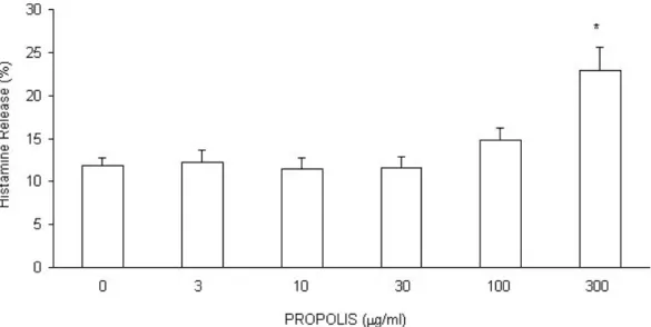
Propolis ethanolic extract (300 Pg/ml) increased the histamine release in guinea pig lung suspension containing mast cells. However, it can be seen that this effect is not
related to selective stimuli but due to cytotoxic effects, since the histamine release
induced by propolis was not inhibited by sodium cyanide (data not shown). Moreover, this effect was observed only with a high concentration of propolis (300 Pg/ml).
Propolis had no effect on histamine release in lower concentrations.
Reports have pointed out that ethanol can induce urticaria without showing evidence
of cytotoxic histamine release after oral ingestion in high concentration, and low
concentrations can cause urticaria by specific histamine release (12, 21). Previous
works in our laboratory showed that ethanol (in concentrations equal to those used to
prepare the ethanolic extract of propolis) does not have any effect on the
spontaneous histamine release (16), suggesting that the effect observed was due to
the propolis components.
Although propolis shows anti-inflammatory properties, there are few reports of its
side effects, such as allergic contact dermatitis (1). Propolis is characterized by a
complex chemical composition, with both stimulant and inhibitory compounds, as
well as synergistic ones. More than 180 compounds (such as flavonoids, phenolic
acid and their derivatives, among others) were identified in propolis composition (2).
Some cases of contact allergy due to propolis are not related to one main allergen,
but to several ones, depending on the source plant of collection by bees (9). Propolis
constituents are strong contact sensitizers when administered in guinea pig skin,
being the 1,1-dimethylallyl caffeic acid ester the main agent responsible for the
allergic process (8).
In this work, we observed that propolis induced mast cell histamine release,
suggesting that this stimulant action may occur due to its constituents concentration
in the hydroalcoholic solutions, depending either on the predominance of one
compound or on the synergism of several propolis constituents. Further
investigations with isolated constituents of propolis would permit to elucidate their
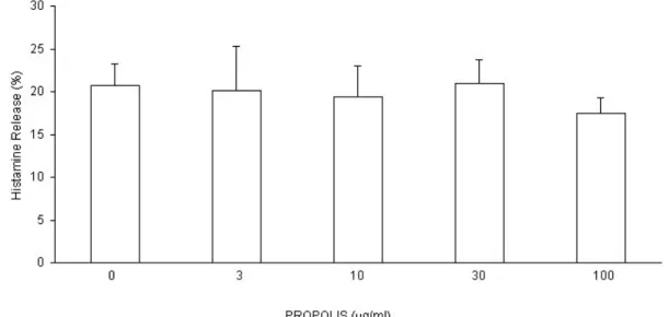
Histamine release induced by antigen and ionophore A 23187 was analyzed using propolis concentrations lower than 300 Pg/ml, which had no direct effect on
spontaneous histamine release. Our results demonstrated that propolis (3, 10, 30, and 100 Pg/ml) does not show a significant effect on the histamine release induced
by ionophore. This result indicated that propolis does not interfere with the ionic flow
promoted by the ionophore in the cell. Other authors observed that both ethanolic
and water extracts of propolis inhibited the histamine release induced by compound
48/80, suggesting that these extracts possess an anti-allergic action (11).
With regards to the histamine release induced by antigen, propolis had also no
significant effect, suggesting that this product does not interfere with the immunologic
process of mast cells degranulation.
In view of these results, we suggest that only high concentrations of propolis may
directly activate mast cells, promoting the inflammatory mediators release by
cytotoxic mechanisms. These results may be related to the allergic process in
propolis sensitive people. We encourage propolis use to non-allergic people because
of its several biological and therapeutical properties.
Figure 1. Effect of hydroalcoholic extract of propolis on the guinea pig lung cell suspension containing mast cells. Results are means r standard error of mean (SEM) of 7 similar assays.
* Statistically different from histamine release in the absence of propolis (p0.05).
Figure 2. Effect of hydroalcoholic extract of propolis on the histamine release induced
by ionophore A 23187 on the guinea pig lung cell suspensions containing mast cells. Results are means r standard error of mean (SEM) of 7 similar assays.
Figure 3. Effect of hydroalcoholic extract of propolis on the histamine release induced
by antigen on the guinea pig lung cell suspensions containing mast cells. Results are
REFERENCES
1 ANGELINI G., VENA GA., MENEGHINI CL. Psoriasis and contact allergy to
propolis. Contact Dermatitis, 1987, 17, 251-3
2 BANKOVA V., DYULGEROV A., POPOV S., EVSTATIEVA L., KULEVA L., PUREB
O., ZAMJANSAN Z. Propolis produced in Bulgaria and Mongolia: phenolic
compounds and plant origin. Apidologie,1992, 23, 79-85
3 BANSKOTA AH., TEZUKA Y., KADOTA S. Recent progress in pharmacological
research of propolis. Phytother. Res., 2001, 15, 561-71
4 BURDOCK GA. Review of the biological properties and toxicity of bee propolis
(propolis). Food Chem. Toxicol., 1998, 36, 347-63
5 CHURCH MK., HOLGATE ST., SHUTE JK., WALLS AF., SAMPSON AP. Mast cell
– derived mediators. In: MIDDLETON E., ELLIS EF., YUNGINGER JW., REED
CE., ADKINSON NF., BUSSE WW. Allergy: principles and practice. 5.ed.
Saint Louis: Mosby, 1998: 146-67
6 GOMES JC., DI STASI LC., SGARBOSA F., BARATA LES. Pharmacological
evaluation of the inhibitory effect of extracts from Anchieta salutaris on the
histamine release induced in the rat and the guinea pig. Int. Arch. Allergy
Immunol., 1994, 103, 188-93
7 HANSSON C., EZZELARAB M., STERNER O. Oxidative activation of the propolis
hapten isoprenyl caffeate. Acta Derm. Venereol., 1995, 75, 34-6
8 HAUSEN BM., WOLLENWEBER E., SENFF H., POST B. Propolis allergy. I.
Origin, properties, usage and literature review. Contact Dermatitis, 1987, 17,
163 -70
9 HEGYI E., SUCHY V., NAGY M. Propolis contact allergy. Hautarzt, 1990, 41,
675-9
10 KHAYAL MT., EL-GHAZALY MA., EL-KHATIB AS. Drugs Exp. Clin. Res., 1993,
19, 197
11 MIYATAKA H., NISHIKI M., MATSUMOTO H., FUJIMOTO T., MATSUKA M.,
ISOBE A., SATOH T. Evaluation of propolis (II): effects of brazilian and chinese
propolis on histamine release from rat peritoneal mast cells induced by
compound 48/80 and concanavalin A. Biol. Pharm. Bull., 1998, 21, 723-9
12 ORMEROD AD., HOLT PJA. Acute urticaria due to alcohol. Br. J. Dermatol.,
13 ORSI RO., FUNARI SRC., SOARES AMVC., CALVI SA., OLIVEIRA SL.,
SFORCIN JM., BANKOVA V. Immunomodulatory action of propolis on
macrophage activation. J. Venom. Anim. Toxins, 2000, 6, 205-19
14 PARK EH., KAHNG JH. Supressive effects of propolis in rat adjuvant arthritis.
Arch. Pharm. Res., 1999, 22, 554-88
15 PEARCE FL. Calcium and histamine secretion from mast cells. Prog. Med.
Chem., 1982, 19, 59-109
16 RUIZ CM., GOMES JC. Effects of ethanol, acetaldehyde and acetic acid on
histamine secretion in guinea pig lung mast cell. Alcohol, 2000, 20, 133-8
17 SHORE PA., BURKHALTER A., COHN VH. A method for the fluorometric assay
of histamine in tissue. J. Pharmacol. Exp. Ther., 1959, 127, 182-6
18 SIRAGANIAN RP. Biochemical events in basophil or mast cells activations and
mediator release. In: MIDDLETON E., ELLIS EF., YUNGINGER JW., REED
CE., ADKINSON NF., BUSSE WW. Allergy: principles and practice. 5.ed.
Saint Louis: Mosby, 1998: 204-27
19 TERAKI Y., SHIOHARA T. Propolis-induced granulomatous contact dermatitis
accompanied by marked lymphadenopathy. Br. J. Dermatol., 2001, 144,
1277-8.
20 THOMAS P., HANS-CHRISTIAN K., BERNHARD P. Propolis-induced allergic
contact dermatitis mimicking pemphigus vulgaris. Arch. Dermatol., 1998, 134,
511-3
21 TING S., RAULS DO., ASHBAUGH P., MANSFIELD LE. Ethanol-induced
urticaria – a case report. Ann. Allergy, 1988, 60, 527-30
22 YOUNG E. Sensitivity to propolis. Contact Dermatitis, 1987, 16, 49-50