RESUMO.- [Parâmetros fisiológicos em fêmeas de Mu -tum do Sudeste (Crax blumenbachii) em reabilitação.] O presente artigo reporta dados inéditos de análise hemo-gasométrica, hematológica e bacteriológica de quatro fê-meas de Mutum do Sudeste, resgatadas do tráfico ilegal de animais silvestres e em processo de habilitação e reintro-dução para vida-livre pelo Projeto Centrofauna, Botucatu/ SP. Na literatura há uma falta de dados fisiológicos sobre esta espécie, endêmica da região da Mata Atlântica e amea-çada de extinção. Esta falta de informação faz com que ava-liações clínicas nesta espécie aviária sejam muito difíceis de interpretar, causando um efeito direto e negativo nestas aves para a realização de tratamento médico em hospitais, clínicas veterinárias, centros de pesquisa e/ou conserva-ção, bem como centros de reintrodução.
TERMOS DE INDEXAÇÃO: Mutum do Sudeste,Crax blumenbachii,
parâmetros fisiológicos, microbiota cloacal, saúde animal.
INTRODUCTION
The southeastern Curassow (Crax blumenbachii Spix, 1825) is a Brazilian bird belonging to the Cracídae family (Ordem Craciformes - ex Galliformes) that originally was found in the states of Rio de Janeiro, Espírito Santo, Minas Gerais, and Bahia, in woods and forests up to an altitude of 500m, whi-ch form part of the Atlantic Forest (Rodriguez-Mahecha et al. 2005, Sigrist 2009). Nowadays, C. blumenbachii is found free living only in the Sooretama Biological Reserve and in the Biological Reserve of the Companhia Vale do Rio Doce in Espirito Santo, the Ecological Reserve of Una, the APA Pra-tisi State, the Mount Pascoal National Park, and in the Dis-covery National Park in the State of Bahia. There have been some exemplars reintroduced into the Rio Doce State Park in Minas Gerais (Bencke et al. 2006), where one estimates that no more than 249 wild individuals are present (Birdlife International 2007, Birdlife International 2008).
It is estimated that the population has had an average decline of 1 to 9%, chiefly due to deforestation and ille -gal hunting (Birdlife International 2012). This bird is now considered a species under worldwide threat of extinction (Bleul et al. 2007). In the wild, C. blumenbachii, as well as other species of curassow, has an important biological function in seed scattering and plays a fundamental role in
Physiologic parameters in female Red-billed curassows
(
Crax blumenbachii
) in rehabilitation
1Guilherme A. Marietto-Gonçalves2*, Mayra T.A. Martins2, Katiane L. Souza2, Regina K. Takahira2, Roberto C. Gonçalves2 and Raphael L. Andreatti Filho2
ABSTRACT.- Marietto-GonçalvesG.A., MartinsM.T.A., SouzaK.L, TakahiraR.K., Gonçalves R.C. & Andreatti FilhoR.L. 2016. Physiologic parameters in female Red-billed curas -sows (Crax blumenbachii) in rehabilitation.Pesquisa Veterinária Brasileira 36(2):131-135. Laboratório de Ornitopatologia, Departamento de Clínica Veterinária, Faculdade de Medicina Veterinária e Zootecnia, Universidade Estadual Paulista, Cx. Postal 560, Distrito de Rubião Júnior, Botucatu, SP 18618-000, Brazil. E-mail: gmarietto@hotmail.com
The present paper reports blood gas analysis, hematologic and micro bacteriologic information on four female Red-billed curassows, rescued from illegal wild animal tra-ding, that were undergoing habilitation for reintroduction into the wild through Projeto Centrofauna/Botucatu-SP. There is a lack of physiological data on this species, endemic to the region of Atlantic Forest (Mata Atlântica), now under threat of extinction. This lack of information makes the clinical evaluation of these birds very difficult and has a direct and adverse effect on any of these birds received for medical treatment in hospitals, ve-terinary clinics and centers for research and conservation, as well as in reintroduction centers.
INDEX TERMS: Red-billed curassow,Crax blumenbachii, physiological parameters, cloacal microbiota, animal health.
1 Received on June 2, 2015.
Accepted for publication on December 29, 2015.
2 Departamento de Clínica Veterinária, Faculdade de Medicina
Veteriná-ria e Zootecnia, (FMVZ), Universidade Estadual Paulista “Júlio de Mesqui
-ta Filho” (Unesp), Cx. Pos-tal 560, Distrito de Rubião Júnior, Botucatu, SP
maintaining the tropical forest by the dispersion of its favo-rite plants,7 especially the fruit of the Mystaceae family, whi-ch are very common in the area whiwhi-ch this bird inhabits. C. blumenbachii also helps to distribute fleshy fruits, seedlin -gs, small prey and hard seeds (IBAMA 2004, Bruno 2008).
In spite of the extreme threat of extinction of these birds, biological data on normal physiological standards for them are rare in the literature. This makes veterinary examinations very difficult, what may compromise conser -vation programs aimed at recuperation of the species, in the wild and in captivity. Given the concept that to preserve something, it is necessary to know enough about, this ar-ticle presents information on body form, blood and serum values, biochemical and microbiological findings, as well as blood gas data of four captive C. blumenbachii females (Fig.1). They were apprehended by the São Paulo State En-vironmental Police, as victims of illegal wild animal trade. At the moment they are in the process of rehabilitation of the Centro-Fauna Project, Botucatu/SP.
done immediately after blood collection using a CG8+ (Abbout
La-boratories, USA) cartouche into which two drops of venous blood were instilled. The instrument evaluated the specimen and gave the result in about 3 minutes.
For the hemogram, after blood collection, a blood smear was done from the samples, without contact with anticoagulant, to give less cellular lysis, better morphological preservation and less platelet agglutination (Green & Blue McLendon 2000). As stated, we used heparin to condition the samples, as this preservative can be used for both hemogram determination as well as for genetic and biochemical analysis (Thrall 2007).
The blood smears were made using a small drop of blood wi-thout anticoagulant on one end of a microscope slide, and with the use of a slide extensor (positioned at 45°), the drop was spre-ad out. After air drying, the smears were stained using Wright stain, as per Schalm (1965), for 10 minutes. They were then co-vered with distilled water until a reverse meniscus formed, for more than 15 minutes. They were then rinsed in running water to remove excess stain and afterwards heat dried. In the blood sme-ar all cellulsme-ar types were seen, with good morphological detail: presence or not of inclusions and blood parasites was noted, and a differential leukocyte count was done.
The hematocrit was obtained using a microhematocrit.
Ca-pillary non-heparinized tubes were filled to two thirds of their
volume with the blood samples. These were heat sealed, and cen-trifuged at 15,000 rpm for 5 minutes. The capillary tubes were read on a standardized scale and the value obtained in percenta-ges (Bierer et al. 1963). The plasma was examined for color alte-rations or turbidity due to hemolysis and /or lipemia.
Additionally, with the capillary tube we obtained the total plas-ma protein levels (TP). The tube was divided and the plasplas-ma pla-ced in a refractometer for evaluation. Hemoglobin measurement was done by the cyanmethemoglobin method. The principle of this method is the oxidation of ferric iron by the action of potassium ferrocyanide present in Drabkin´s Reagent (Thrall 2007). Quanti-zation of the hemoglobin cyanide formed was done by colorimetry. Fig.1. Female red-billed curassaw (Crax blumenbachii) is different
from the male (which is totally black with a white breast and a
red fleshy structure at the base of the Bill). Female is rusty bla -ck with a showy tuft heavily barred with white. Its abdomen is rusty red, the beak grey with a black base and its feathers have a reddish tinge (Rodriguez-Mahecha et al. 2005, Bruno 2008).
MATERIALS AND METHODS
All procedures were done within 10 minutes. Firstly, the birds
were physically confined, and temperature red with an analog
thermometer. They were then weighed on a digital scale
rea-ding up to 6 kg (Toledo do Brazil Indústria de Balanças Ltda, São
Bernardo/SP). Auscultation of the lungs was done for abnormal sounds representing possible respiratory disturbances. Finally
the superficial pectoral muscle was palpated to evaluate body
mass (scoring done as shown in Figure 2).
Along with the process described above, blood samples were drawn by puncture of the brachial vein; these samples were im-mediately divided into three fractions for different examinations: heparinized blood for a hemogram, [0.7mL], unprocessed blood for blood gas determination [0.3mL], and serum for biochemical and serological measurements [1mL]. A cloacal swab was done to identify any aerobic Gram negative Enterobacteria possibly
pre-sent in the cloacal microflora of these birds.
The blood gas measurements were performed with a portable clinical analyzer I-STAT® (Abbott Laboratories, USA); they were
Fig.2. Physical score parameters used in this study. (Body score classification: Score 1: Little muscle mass and the external crest is prominent; Score 2: Muscle mass is present and ex-ternal crest is palpable; Score 3: Muscular mass more signifi -cant, leaving only the extremity of the external crest palpable;
Score 4: Muscle mass not palpable and the extreme end of
Red blood cells and the leukocyte and platelets numbers were done using a Neubauer Hemacytometer chamber. Two mL of to-luidine Blue (0.01%) was added to 20µL of blood and allowed to rest for 10 minutes to color the cells. The Neubauer chamber
was then filled, the cells were allowed to sediment for 15 minutes
and the reading was taken. The red blood cells were counted in
the same quadrants used for mammals (five quarters of a central square) at 400x magnification, and the value found was multi -plied by a factor of 5,050. White blood cells and platelets were counted separately in the same quadrants used for counting leu-kocytes in mammals (four large quadrants) and the value found was multiplied by 252.5.
The red blood cell indices MCV and MCHC were obtained using the following mathematical calculations:
MCV (fL) = Ht (%)/red blood cells (x106/uL)*10
MCHC (g/dL) = hemoglobin (g/dL)/Ht (%) *100
For biochemical evaluations, 70µL of serum was used for each analysis, and the doses were found using the Cobas Mira Plus da
Roche (Roche Diagnostics, Germany) equipment with specific kits
for each Ebram test (Ebram, Brazil).
Serological tests for Pullorum disease and Mycoplasmosis were done using the rapid serum agglutination assay (RSA). For Newcastle Disease, the beta hemaglutination -inhibition method was used as described by Santos & Silva (2000). In the RSA we used the commercially available antigen Pulor Teste® for
Salmo-nella Pullorum, the Myco-Galli Teste® for Mycoplasma
gallisepti-cum and the Synovitest® for M. synoviae (Biovet, Brazil). For each
50µL of antigen, we used 50µL of serum which was mixed with a glass rod in the SAR reading plate.
After homogenization of the serum with the antigen, gentle rotatory movements of the reading plate were made for two mi-nutes. Samples which showed agglomerations during this time were considered positive.
For each test we used known positive control samples from a domestic chicken (Gallus gallus domesticus).A live virus strain (La Sota, New vac LS®, Fort Dodge, Brazil), was used for the IH
test. One unit of hemaglutinate (UHA) =1/8 (titration ascertained using the Hemaglutination test HA). For the IH we used 4 UHA. In each round bottom well of the plate, with space for 8 samples, 50µL of phosphatase saline was distributed in a dozen linear
wells. In the first wells 50µL of serum was placed, being serially diluted from the first to the twelfth well. This last dilution was
discarded. Following this, 50µL of virus was placed in each well and, after homogenization, the plate was allowed to rest for 40 minutes at 5°C.
After this, 50µL of red blood cell solution and 1% red blood cells (of the domestic chicken Gallus gallus domesticus) were ad-ded and after homogenization, left to rest once again for 40 minu-tes at 5oC. Positive samples were considered to be those in which
the inhibition of hemaglutination was higher than the highest di-lution of the agglutinate (1/8).
The fecal samples collected from the cloacal swabs were ini-tially pre-enriched by incubation in study tubes containing 10mL of peptonated water at 40°C for 24 hours. Following this, 1mL of the pre-enriched cultures were added to tubes of Brain Heart in-fusion medium, and into another Tetrathionate broth, once again being incubated at 40°C for 24 hours .Finally, samples were see-ded onto Brilliant Green agar, MacConkey’s medium and Xylose--Lisine-Desoxicolate, all being incubated at 40°Cfor 24 hours. After these processes, the presence of distinct colony forms was assessed, and then a sample of each one was collected for
bio-chemical identification using the method described by Holt et al.
(1994).
RESULTS
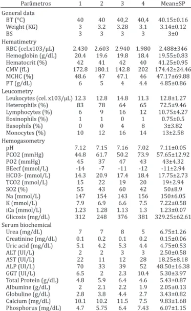
Results of the hematologic evaluations, the blood gases, the biochemical and microbiological results are presented in
Table 1. Hematologic, serum biochemical and haemogasometrics values of four female Crax blumenbachii
Parâmetros 1 2 3 4 Mean±SP
General data
BT (°C) 40 40 40,2 40,4 40.15±0.16 Weight (KG) 3 3.2 3.28 3.1 3.14±0.12
BS 3 3 3 3 3±0
Hematimetry
RBC (cel.x103/µL) 2.430 2.603 2.940 1.980 2.488±346 Hemoglobin (g/dL) 20.4 19.6 19.8 18.4 19.55±0.83 Hematocrit (%) 42 41 42 40 41.25±0.95 CMV (fL) 172.8 180.1 142.8 202 174.42±24.46
MCHC (%) 48.6 47 47.1 46 47.17±69.88 PT (g/dL) 6 5 4 4.4 4.85±0.86 Leucometry
Leukocytes (cel. x103/µL) 12.3 12.8 14.8 11.3 12.8±1.27 Heterophils (%) 83 78 64 65 72.5±9.46 Lymphocytes (%) 6 9 16 12 10.75±4.27
Eosinophils (%) 1 1 0 1 0.75±0.5
Basophils (%) 0 0 4 8 3±3.82
Monocytes (%) 10 12 16 14 13±2.58 Hemogasometry
pH 7.12 7.15 7.16 7.02 7.11±0.05
PCO2 (mmHg) 44.8 61.7 50.2 73.9 57.65±12.92
PO2 (mmHg) 45 37 47 43 43±4.32
BEecf (mmol/L) -14 -7 -11 -12 -11±2.94 HCO3- (mmol/L) 14.3 20.9 17.4 18.4 17.75±2.73 TCO2 (mmol/L) 15 22 19 20 19±2.94
SO2 (%) 55 43 60 42 50±8.9
Na (mmol/L) 147 154 143 156 150±6.05 K (mmol/L) 7.9 6.9 6.6 7.5 7.22±0.58 iCa (mmol/L) 1.23 1.28 1.13 1.3 1.23±0.07 Glicosis (mg/dL) 312 248 376 381 329.25±62.61 Serum biochemical
Urea (mg/dL) 7 7 8 5 6.75±1.26
Creatinine (mg/dL) 0.1 0.2 0.1 0.2 0.15±0.06 Uric acid (mg/dL) 5.1 4.2 5.3 4.4 4.75±0.53
ALT (UI/L) 2 2 3 3 2.50±0.58
AST (UI/L) 22 11 12 28 18.25±8.18 ALP (UI/L) 70 33 39 52 48.50±16.38 GGT (UI/L) 6.5 2 2.3 10.4 5.30±3.97 Total Protein (g/dL) 4.8 5.9 6.4 4.6 5.43±0.87 Albumine (g/dL) 2 2.1 2.2 1.9 2.05±0.13 Globuline (g/dL) 2.8 3.8 4.4 2.7 3.43±0.82 Calcium (mg/dL) 10.1 10.2 11.5 7.5 9.83±1.68 Phosphorus (mg/dL) 4.7 5.75 6.4 7.43 6.07±1.15 SD = Standart deviation, BT = Body temperature, BS = Body Score, RBC = red blood cells, MCV = Mean corpuscular volume , MCHC = Mean corpus-cular hemoglobin concentration, PT = Plasmatic proteins, PO2 = Oxygen pressure, PCO2 = Carbonic gas pressure, BE = Base excess, HCO3- = Bi-carbonate, TCO2 = Total carbon dioxide, SO2 = Oxygen saturation, iCa = Ionized calcium, ALT = Alanine aminotransferase, AST = Aspartate amino-transferase, ALP = Alkaline phosphatase.
Table 2. Aerobic Gram-negative Enterobacterias isolated by cloacal swab from four red-billed curassow females (Crax
blumenbachii)
Bacterial species Positive (T) Prevalence (%)
Escherichia coli 4(4) 100
Serratia marcensces 3(4) 75
Proteus vulgaris 4(4) 100
Proteus mirabilis 2(4) 50
Enterobacter aerogenes 1(4) 25
Citrobacter freudii 1(4) 25
Klebsiella pneumoniae 2(4) 50
Klebsiella oxytoca 1(4) 25
Table 1 and 2. There was no serological detection of any of the agents searched for in this study.
DISCUSSION
The physical evaluation showed that all the birds had a good body score, with an acceptable weight, as the mean body weight for an adult Crax blumenbachii varies from 3.0-3.5 kg (Sigrist 2009). They all had a normal temperature, which for bird species varies from 40 to 42°C. Our four bir-ds showed adult female characteristics, and we considered them to be sexually mature and at least three years of age. On respiratory examination, no abnormal sounds (such as rales, rhonchi or whistling) were heard, indicating there were no respiratory problems. For this reason, we conclu-ded that the blood gas parameters which were found are close to or normal (keeping in mind the stress to the birds caused by the procedure). There are no blood gas data avai-lable in the literature for this particular species of bird.
Blood gas measurement has a wide clinical applica-tion, as one can quantify respiratory system abnormalities based on the pH values of the blood, the partial pressure of carbon dioxide and oxygen. Additionally it is possible to deduce carbonate values, due to the acid-base organic dis-equilibrium in the blood gas concentrations, However, other parameters also may be evaluated (Sobiech et al. 2005, Zavorsky et al. 2007).
In this study, we used venous blood obtained easily due to its anatomical location. Ideally, arterial blood would be used to evaluate pulmonary function. However, venous blood still gives reasonably confident values, in spite of not allowing measurement of pulmonary oxygen carrying ca-pacity (Sobiech et al. 2005, Bleul et al. 2007, Zavorsky et al. 2007, Mota & Queiroz 2010).
We were unable to find any reference values for hema -tologic standard and biochemical serum standard for this species, to compare with our values. However, given the he-althy clinical appearance of the birds in our study, we consi-der our values to be representative of normal values. We do not know if there is a difference between the sexes in these values, as we did not evaluate any males. Similarly, we do not know if these values differ with age.
Escherichia coli were found in all (100%) of the birds in this study. Santos et al. (2010)reported this bacteria in one captive individual of C. blumenbachii. The finding of this or -ganism in the intestinal apparatus of diverse domestic bird species as well as in wild birds is considered normal (Bar-nes et al. 2008). The E. coli presence is report in captive Ortalis guttata, Penelope obscura, Pipile pipile, P. jacutinga, C. fasciolata, Pauxi tuberosa (Santos et al. 2010) and P. mitu (Saidenberg et al. 2013).
With our observations, it is possible to speculate that E. coli may be a part of the normal bacterial flora of C. blu-menbachii, at least in captivity, although there are no data available in wild birds. Proteus vulgaris was also isolated for 100% of our birds under study. The occurrence of this species as well as others which we isolated in our study has not been reported in the literature. For this reason, we do not know if these organisms are normal in C. blumenbachii, or if they represent contaminants. However, it is known
that all the species we found are commonly encountered in the industrial avian environment (Chernaki-Leffer et al. 2002, Segabinazi et al. 2005).
Due to the small number of birds available for our stu-dy, we could not do a precise statistical evaluation. As well, we were unable to obtain other birds from rehabilitation centers, breeders or zoological parks to increase the num-ber of birds evaluated, which would result in more precise analysis of the results obtained.
The few sites which have birds available are very far away from the laboratories where the analyses were per-formed. It is imperative that the blood gas analysis and the hematological studies be done immediately after collec-tion; otherwise the results cannot be relied upon.
CONCLUSION
Escherichia coli can be considered a part of the normal mi-croflora of Crax blumenbachii; however, there are few de-tails available for the species. New studies need to be done to identify the microbial flora and physiological parameters in captive members as well as in wild birds of this species. These studies are necessary to quantify measurements for the determination of standard values for C. blumenbachii, a species which is on the brink of extinction. As this species is also very important for the equilibrium of the Atlantic Forest ecosystem, it must become better understood for success in its conservation as a valuable biological entity.
REFERENCES
Barnes H.J., Nolan L.K. & Valliancourt J.P. 2008. Colibacillosis, p.775-732. In: Saif Y.M, Fadly A.M., Glisson J.R., McDougald L.R., Nolan L.K. & Swayne D.E. (Eds), Diseases of Poultry. Vol.12. Blackwell Publishing, Ames. Bencke A., Mauricio G.N., Develey P.F. & Goerck J.M. 2006. Áreas
Impor-tantes para a Conservação das Aves no Brasil. 1. Estados de Domínio da Mata Atlântica. SAVE Brasil, São Paulo, SP. 341p.
Bierer B.W., Thomas J.B., Roebuck D.E., Powell H.S. & Eleazer T.H. 1963. Hematocrit and sedimentation rate values as an aid in poultry disease diagnosis. J. Am. Vet. Med. Assoc. 143:1096-1098.
Birdlife International 2007. Threatened Birds of the World. Available at <http://www.birdlife.org> Access in July 2013.
Birdlife International. 2008. Crax blumenbachii. In: IUCN 2011. IUCN Red List of Threatened Species. Disponível em <http://www.iucnredlist. org> Access in June 2013.
Birdlife International 2012. Species factsheet: Crax blumenbachii. Avail-able at <http://www.birdlife.org> Access in June 2013.
Bleul U., Lejeune B., Schwantag S. & Kähn W. 2007. Blood gas and acid-base analysis of arterial blood in 57 newborn calves. Vet. Rec. 161:688-691. Bruno S.F. 2008. 100 animais ameaçados de extinção e o que você pode
fazer para evitar. Ediouro, Rio de Janeiro. 144p.
Chernaki-Leffer A.M., Biesdorf S.M., Almeida L.M., Leffer E.V.B. & Vigne F. 2002. Isolamento de enterobactérias em Alphitobiusdiaperinus e na cama de aviários noroeste do estado do Paraná, Brasil. Braz. J. Poult. Sci. 4(3):243-247.
Green R. & Blue McLendon A. 2000. Ratite hematology, p.1201-1206. In: Feldman B.F., Zink J.G. & Jain N.C. (Eds),Schalm’s Veterinary Hematol-ogy. Vol.5. Lippincott Williams, Philadelphia.
Holt J.G., Krieg N.R., Sneath P.H.A., Staley J.T. & Williams S.T. 1994. Bergey’s Manual of Determinative Bacteriology. Vol.9. Williams and Wilkins, Bal-timore. 787p.
IBAMA 2004. Plano de ação para conservação do Mutum do Sudeste Crax
Blumenbachii. Série Espécies Ameaçadas, Vol.1. Available at <http://
Mota I.L. & Queiroz R.S. 2010.Distúrbios do equilíbrio ácido básico e
gasometria arterial: uma revisão crítica. Revista Digital, Buenos Aires, 14. Availabale at <http://www.efdeportes.com/efd141/equilibrio-aci-do-basico-e-gasometria-arterial.htm> Access in April 2013.
Rodríguez-Mahecha J.V., Hugues N., Nieto O. & Franco A.M. 2005.Paujiles, Pavones, Pavas and Guacharacas Neotropicales. Conservatión Interna-cional, Bogotá, CO. 98p.
Saidenberg A.A.B., Allegretti L., Astolfi-Ferreira C.C.S., FerreiraJ.P.,
Almei-daM.A. & RasoT.F. 2013. Alguns genes de virulência de Escherichia coli
isoladas de mutuns-do-nordeste (Pauxi mitu) sadios no Brasil.Pesq. Vet. Bras. 33:523-527.
Santos C.H.C. & Silva E.N. 2000. Métodos de diagnósticos laboratoriais
mi-crobiológicos e sorológicos, p.173-182. In: Berchieri Júnior A. & Macari
M. (Eds).,Doenças das Aves. Fundação APINCO de Ciência e Tecnologia Avícola, Campinas, SP.
Santos H.F., Flôres M.L., Lara V.M., Sá e Silva M., Battisti L. & Lovato L.T. 2010. Microbiota cloacal aeróbia de cracídeos cativos no Rio Grande do Sul e sua susceptibilidade á antimicrobianos. Pesq. Vet. Bras. 30:1077-1082.
Segabinazi S.D., Flôres M.L., Barcelos A.S., Jacobsen G. & Eltz R.D. 2005. Bactérias da família Enterobacteriaceae em Alphitobius diaperinus
oriundos de granjas avícolas dos Estados do Rio Grande do Sul e Santa Catarina, Brasil. Acta Scient. Vet. 33:51-55.
Schalm O.W. 1965. Veterinary Hematology. Lea and Febiger, Philadelphia. 664p.
Sigrist T. 2009. The Avis Brasilis Field Guide to the Birds of Brazil: especies accounts. Avibrasilis, São Paulo, SP. 305p.
Sobiech P., Stopyra A., Kuleta Z., Zbanyszek M. & Milewski M. 2005. Acid-base balance parameters of arterial, venous and capillary blood in sheep. Bull. Vet. Inst. Pulawy49:125-127.
Thrall M.A. 2007.Hematologia e bioquímica clínica veterinária.Roca, São Paulo. 592p.